
1 of 39
Downloaded 24 times


Recommended
Mesoderm
MesodermGauri Haval
Ěý
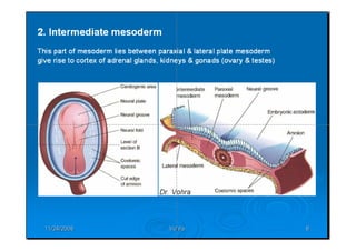
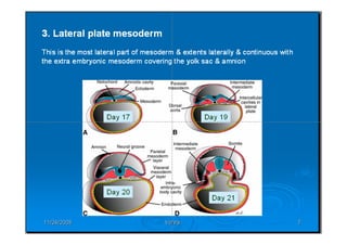
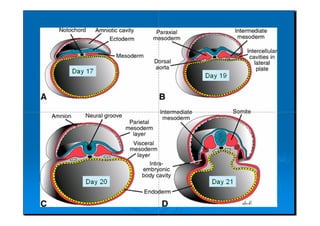
The mesoderm generates organs between the ectoderm and endoderm. It is divided into four regions - chordamesoderm, paraxial mesoderm, intermediate mesoderm, and lateral plate mesoderm. The lateral plate mesoderm splits into somatic and splanchnic layers, forming the body cavity. The heart develops from cardiogenic mesoderm along the BMP signaling pathway induced by endoderm, while the notochord and neural tube provide inhibitory signals. By 29 hours of incubation, the heart primordia have fused to form a single tube.Third week of development
Third week of developmentDr. Vibhash Kumar Vaidya
Ěý
During the third week of development, gastrulation occurs which establishes the three germ layers - ectoderm, mesoderm, and endoderm. Gastrulation begins with the formation of the primitive streak on the surface of the epiblast. Cells migrate through the primitive streak and node, some displacing the hypoblast to form endoderm, while others become mesoderm between the endoderm and remaining ectoderm. This results in the formation of the notochord, and the germ layers differentiate into various tissues and organs.3rd week of human development
3rd week of human developmentDr. Muhammad ishaque abbasi
Ěý
During the third week of development, gastrulation occurs where the three germ layers (ectoderm, mesoderm, endoderm) are formed. The notochord also begins developing from epiblast cells that ingress through the primitive streak and primitive node. These cells form the notochordal process which then fuses with endoderm and detaches to form the definitive notochord cord between the ectoderm and endoderm. The mesoderm organizes into three segments - paraxial, intermediate, and lateral plate mesoderm - which will give rise to muscles, skeleton, urinary/genital systems, and other tissues.Somite formation
Somite formationnaba al wazen
Ěý
Somites are bilaterally paired segments of paraxial mesoderm that form along the embryonic axis and give rise to important structures. Somites subdivide into sclerotomes, myotomes and dermatomes that form vertebrae, ribs, muscle, tendons and skin. Somite formation depends on a "clock mechanism" where paraxial mesoderm segments into somites according to their position in a regulated process. Within each somite, cells are specified based on location and retain flexibility before differentiating into somite-derived tissues through epithelialization and mesenchymal transformation processes.Intra-Embryonic Mesoderm (General Embryology)
Intra-Embryonic Mesoderm (General Embryology)Dr. Sherif Fahmy
Ěý
This document describes the formation of the notochord and differentiation of the intra-embryonic mesoderm in 5 steps. It explains that the intra-embryonic mesoderm forms from proliferating cells on the sides of the primitive node and streak. This mesoderm then differentiates into the paraxial, intermediate, and lateral plate mesoderm. The paraxial mesoderm forms somites which differentiate into sclerotome, dermatome, and myotome. The intermediate mesoderm forms the urogenital system, while the lateral plate mesoderm splits to form the somatic and splanchnic mesoderm separated by the intra-embryonic coelom.Neurulation developmental biology
Neurulation developmental biologyKashif Manzoor
Ěý
The document discusses the molecular regulation of neural induction and neurulation. It explains that upregulation of FGF signaling and inhibition of BMP4 activity causes ectoderm to become neural tissue by default. During neurulation, the neural plate forms the neural tube as the neural folds fuse in the midline. Neural crest cells emerge and migrate throughout the embryo to form many tissues. Precise regulation of BMP concentrations induces different cell fates in the ectoderm.Gastrulation
GastrulationAmani Riyadh
Ěý
Gastrulation is the process by which the three germ layers (ectoderm, mesoderm, endoderm) are formed in the embryo through cell movements. In amphibians, cells from the blastula's outer layer invaginate and involute to form the inner layers. The blastocoel is replaced by the archenteron. In birds, the epiblast and hypoblast layers are initially formed; the epiblast gives rise solely to the three germ layers. Gastrulation rearranges cells into layers that will develop into tissues and organs.Folding of embryo
Folding of embryoFarhan Ali
Ěý
During the 4th week of development, the embryo undergoes longitudinal and transverse folding which transforms it from a flat disc into a curved tube. This folding incorporates the yolk sac endoderm to form the gut tube and divides the coelom into the thoracic and abdominal cavities. It also repositions structures like the heart and mouth opening. By the end of the 4th week, the embryo has prominent head and tail folds, limb buds, and many organ systems are established.Gastrulation & Notochord (General Embryology)
Gastrulation & Notochord (General Embryology)Dr. Sherif Fahmy
Ěý
Gastrulation begins with the formation of the primitive streak, primitive node, buccopharyngeal membrane, and cloacal membrane. Epiblast cells migrate through the primitive streak and invaginate to form the endoderm, mesoderm, and remaining ectoderm layers. The notochord develops from the primitive pit through the stages of the notochordal process, canal, and plate. It will eventually form the primitive axial skeleton and nucleus pulposus of intervertebral discs.Alaqah 2
Alaqah 2Farhan Ali
Ěý
During the third week of development, the bilaminar germ disc differentiates into a trilaminar embryo through the process of gastrulation. Gastrulation begins with the formation of the primitive streak along the midline of the epiblast. Cells from the epiblast migrate through the primitive streak and groove to form the mesoderm and endoderm germ layers. This results in the formation of the trilaminar embryo consisting of the ectoderm, mesoderm and endoderm germ layers. Concurrently, structures like the notochord, allantois and intraembryonic coelom begin developing.1st & 2nd weeks of development
1st & 2nd weeks of developmentAbdul Ansari
Ěý
1. The document summarizes the key embryonic changes that occur during the first and second weeks of pregnancy. It describes the processes of fertilization, zygote formation, implantation, and development of the inner cell mass and outer cell mass.
2. During the second week, the inner cell mass rearranges to form two layers (the bilaminar germ disk) that will develop into the embryo, while the outer cell mass forms the trophoblasts and primary villus, which are precursors to the placenta.
3. A hormone called HCG is produced from the syncytiotrophoblast cells starting around 8 days after fertilization, and can be detected in pregnancy tests.Heart in vetrebrates
Heart in vetrebratesGovt.college,Nagda, ujjain.M.P
Ěý
Evolutionary change in heart of vertebrates
Heart is situated ventral to the oseophagus in the pericardial section of the coelom.
Heart is a highly muscular pumping organ that pumps blood into arteries and sucks it back through the veins.
In vertebrates it has undergone transformation by twisting from a straight tube to a complex multi-chambered organ.
. There has been an increase in the number of chambers in heart during evolution of vertebrates.
The heart is covered by a transparent protective covering, called pericardium. It is a single layer in fish.
Within pericardium there is a pericardial fluid, protects the heart from the external injury.
TheĚýevolution of the heart is based on the separation of oxygenated bloodĚýfrom deoxygenated blood for efficient oxygen transport.
Neurulation 
Neurulation Indian Institute of Science Education and reserach Bhopal
Ěý
The document discusses neurulation, the process of formation and closure of the neural tube. It involves primary and secondary neurulation, which are guided by molecular signaling pathways involving molecules like Shh, BMP4/7, and cadherins. Defects in neurulation can result in neural tube defects like anencephaly or spina bifida. While neurulation was once thought to be a simple one-step process, it is now understood to be more complex, with closure occurring in regions rather than entirely linearly.Paraxial and intermediate mesoderm
Paraxial and intermediate mesodermMaylowen Pescador
Ěý
Paraxial and intermediate mesoderm form important structures. Paraxial mesoderm forms somites through a "clock and wave" mechanism, with each somite giving rise to vertebrae, muscle, and dermis. Somites are specified by surrounding tissues to develop appropriately. Intermediate mesoderm forms the kidney, with the pronephros, mesonephros, and metanephros appearing sequentially in vertebrate development. The metanephros becomes the permanent kidney in amniotes.First week of development
First week of developmentDr. Vibhash Kumar Vaidya
Ěý
Hello friends..you can use these notes for your convenience as they are taken from many other standard books.. Thank you.Embryologic derivatives endoderm
Embryologic derivatives endodermMedvizz institute of medical education
Ěý
The endoderm gives rise to the epithelial lining of several internal structures including the gastrointestinal tract, respiratory tract, urinary bladder, parts of the genitourinary system, salivary glands, thyroid gland, liver, biliary tree, gallbladder, and pancreas. The endoderm also lines the branchial pouches which give rise to structures like the auditory tube, middle ear cavity, mastoid air cells, palatine tonsils, and parathyroid and thymus glands.Trilaminar germ disc (week 3 embryology)
Trilaminar germ disc (week 3 embryology)Abdoulwahab Mahde
Ěý
summary of Trilaminar Germ disc (embryology) for health science students ...
By Abdoulwahab Mahdi Ali (Student)
Development of endocrine system
Development of endocrine systemRohit Paswan
Ěý
The pituitary gland develops from an evagination of ectoderm called Rathke's pouch and a neuroectodermal diverticulum from the hypothalamus. The anterior pituitary develops from Rathke's pouch while the posterior pituitary develops from the diverticulum. Possible anomalies include ectopic posterior pituitary, pharyngeal hypophysis, agenesis of the pituitary gland, duplication, and craniopharyngioma tumor. The pineal gland develops from a diverticulum of the diencephalon. The adrenal glands develop from coelomic epithelium for the cortex and neural crest cells for the medulla. The thyroid gland is the first endocrine gland to developLeydig cell
Leydig cellkishorssawaikar
Ěý
Leydig cells are found in the testicles and produce the hormone testosterone when stimulated by luteinizing hormone from the pituitary gland. They release testosterone and other androgens which leads to male characteristics. Leydig cells have a polyhedral shape with a large nucleus and lipid-filled vesicles. They are named after Franz Leydig who discovered them in 1850.Histological structure of pituitory gland
Histological structure of pituitory glandchet08
Ěý
The pituitary gland has two main parts - the adenohypophysis and neurohypophysis. The adenohypophysis contains the pars distalis, pars tuberalis, and pars intermedia. The pars distalis is the largest part and contains chromophils like somatotrophs and mammotrophs that secrete growth hormones, as well as chromophobes that secrete ACTH. The neurohypophysis contains the pars nervosa and secretes oxytocin and anti-diuretic hormone. The pituitary gland regulates many important bodily functions through the hormones produced by its various cell types located within its distinct anatomical regions.Cleavage
CleavageAmani Riyadh
Ěý
1. Cleavage is the first phase of embryonic development after fertilization where the fertilized egg undergoes rapid, indirect cell divisions (mitosis) without an increase in overall size to form a multicellular embryo.
2. The pattern of cleavage depends on how much yolk is present in the egg. Eggs with little yolk (isolecithal) undergo total cleavage (holoblastic), while eggs with more yolk have partial cleavage (meroblastic).
3. In amphibians, which have mesoleithal eggs, cleavage is initially equal but becomes unequal with larger vegetal and smaller animal cells. This leads to the formation of a blastula with a fluid-filled cavity (Structure of Ovum
Structure of OvumSAROJ KUMAR PUJHARI
Ěý
The document summarizes the structure of an ovum or egg cell. It is round and non-motile, with a diameter of 0.15 mm in humans. Its cytoplasm is called ooplasm and its periphery contains cortical villi and granules. A vitelline membrane surrounds the plasma membrane, and a zona pellucida primary membrane surrounds that. During release from the follicle, epithelial cells attach to form the corona radiata layer.HISTOLOGY OF ADRENAL GLANDS
HISTOLOGY OF ADRENAL GLANDSDr. Ahmed Mead
Ěý
The adrenal glands secrete both steroid hormones and catecholamines. They are located above the kidneys and have two distinct regions - the cortex and medulla. The cortex lies beneath the capsule and secretes steroid hormones. The medulla lies deep to the cortex and secretes catecholamines. Although embryologically distinct, the cortex and medulla are functionally related.Development of chick embryo- structure of egg,cleavages,fate map and primitiv...
Development of chick embryo- structure of egg,cleavages,fate map and primitiv...SoniaBajaj10
Ěý
The document describes the development of a chick embryo from fertilization through 48 hours of incubation. It discusses the cleavage of the egg, formation of the blastula and blastoderm, gastrulation including primitive streak formation, and the development of key structures like the neural tube, heart, somites, and circulatory system at 24 and 33 hours. The summary provides an overview of the key stages and structures developed during early chick embryo development.First week of human development
First week of human developmentDr. Ahmed Mead
Ěý
1. Human development begins with the fertilization of an egg (oocyte) by a sperm, forming a single-celled zygote.
2. The zygote undergoes cell division and differentiation over approximately 2 weeks as it travels through the fallopian tubes to the uterus, developing into a blastocyst with an inner cell mass that will become the embryo.
3. Fertilization typically occurs as an egg released from an ovary is swept into the fallopian tubes by finger-like projections called fimbriae. Capacitated sperm that have undergone a 7-hour maturation process then fertilize the egg in the ampulla of the fallopian tubes.Spermatogenesis steps, hormonal regulation and abnormalities
Spermatogenesis steps, hormonal regulation and abnormalitiesMdNazmulIslamTanmoy
Ěý
Spermatogenesis is the process by which sperm cells are produced in males. It occurs in three stages: spermatocytogenesis where spermatogonia proliferate into primary spermatocytes, meiosis where primary spermatocytes undergo two divisions to form spermatids, and spermiogenesis where spermatids undergo changes to form spermatozoa. Hormones like testosterone, LH, FSH, growth hormone, and estrogen regulate spermatogenesis by stimulating Leydig and Sertoli cells. Abnormalities can result in conditions like azoospermia, oligozoospermia, and teratozoospermia.Gastrulation
Gastrulationamirmalekshah
Ěý
The document summarizes the process of gastrulation in humans. It discusses how the embryo develops two germ layers, the epiblast and hypoblast, just before implantation. It describes the formation of the primitive streak around day 15, which defines the body axes. Cells migrate through the primitive streak during gastrulation to form the definitive endoderm, intraembryonic mesoderm, and ectoderm. Key cellular processes in gastrulation include epithelial-to-mesenchymal transition and convergent extension.Embryological development of the nervous system and special
Embryological development of the nervous system and specialVernon Pashi
Ěý
The document summarizes key stages in the embryological development of the nervous system and special senses. It describes how the neural plate forms and folds to become the neural tube. It then discusses the formation of the three germ layers and how neural induction occurs. It provides details on neurulation and neural tube formation, as well as common defects that can arise. It also summarizes the development of the main divisions and structures of the brain and spinal cord.
VSM CLT10 Sales Production
VSM CLT10 Sales Productionhansbeyer
Ěý
This document outlines the production process for a CLT10 product from start to finish. It involves verifying parts availability, building cabinets and circuit boards, performing quality assurance testing at various stages, assembling and testing the full systems, and finally issuing certificates and shipping the completed units to customers while maintaining a safety buffer of extra units.More Related Content
What's hot (20)
Gastrulation & Notochord (General Embryology)
Gastrulation & Notochord (General Embryology)Dr. Sherif Fahmy
Ěý
Gastrulation begins with the formation of the primitive streak, primitive node, buccopharyngeal membrane, and cloacal membrane. Epiblast cells migrate through the primitive streak and invaginate to form the endoderm, mesoderm, and remaining ectoderm layers. The notochord develops from the primitive pit through the stages of the notochordal process, canal, and plate. It will eventually form the primitive axial skeleton and nucleus pulposus of intervertebral discs.Alaqah 2
Alaqah 2Farhan Ali
Ěý
During the third week of development, the bilaminar germ disc differentiates into a trilaminar embryo through the process of gastrulation. Gastrulation begins with the formation of the primitive streak along the midline of the epiblast. Cells from the epiblast migrate through the primitive streak and groove to form the mesoderm and endoderm germ layers. This results in the formation of the trilaminar embryo consisting of the ectoderm, mesoderm and endoderm germ layers. Concurrently, structures like the notochord, allantois and intraembryonic coelom begin developing.1st & 2nd weeks of development
1st & 2nd weeks of developmentAbdul Ansari
Ěý
1. The document summarizes the key embryonic changes that occur during the first and second weeks of pregnancy. It describes the processes of fertilization, zygote formation, implantation, and development of the inner cell mass and outer cell mass.
2. During the second week, the inner cell mass rearranges to form two layers (the bilaminar germ disk) that will develop into the embryo, while the outer cell mass forms the trophoblasts and primary villus, which are precursors to the placenta.
3. A hormone called HCG is produced from the syncytiotrophoblast cells starting around 8 days after fertilization, and can be detected in pregnancy tests.Heart in vetrebrates
Heart in vetrebratesGovt.college,Nagda, ujjain.M.P
Ěý
Evolutionary change in heart of vertebrates
Heart is situated ventral to the oseophagus in the pericardial section of the coelom.
Heart is a highly muscular pumping organ that pumps blood into arteries and sucks it back through the veins.
In vertebrates it has undergone transformation by twisting from a straight tube to a complex multi-chambered organ.
. There has been an increase in the number of chambers in heart during evolution of vertebrates.
The heart is covered by a transparent protective covering, called pericardium. It is a single layer in fish.
Within pericardium there is a pericardial fluid, protects the heart from the external injury.
TheĚýevolution of the heart is based on the separation of oxygenated bloodĚýfrom deoxygenated blood for efficient oxygen transport.
Neurulation
Neurulation Indian Institute of Science Education and reserach Bhopal
Ěý
The document discusses neurulation, the process of formation and closure of the neural tube. It involves primary and secondary neurulation, which are guided by molecular signaling pathways involving molecules like Shh, BMP4/7, and cadherins. Defects in neurulation can result in neural tube defects like anencephaly or spina bifida. While neurulation was once thought to be a simple one-step process, it is now understood to be more complex, with closure occurring in regions rather than entirely linearly.Paraxial and intermediate mesoderm
Paraxial and intermediate mesodermMaylowen Pescador
Ěý
Paraxial and intermediate mesoderm form important structures. Paraxial mesoderm forms somites through a "clock and wave" mechanism, with each somite giving rise to vertebrae, muscle, and dermis. Somites are specified by surrounding tissues to develop appropriately. Intermediate mesoderm forms the kidney, with the pronephros, mesonephros, and metanephros appearing sequentially in vertebrate development. The metanephros becomes the permanent kidney in amniotes.First week of development
First week of developmentDr. Vibhash Kumar Vaidya
Ěý
Hello friends..you can use these notes for your convenience as they are taken from many other standard books.. Thank you.Embryologic derivatives endoderm
Embryologic derivatives endodermMedvizz institute of medical education
Ěý
The endoderm gives rise to the epithelial lining of several internal structures including the gastrointestinal tract, respiratory tract, urinary bladder, parts of the genitourinary system, salivary glands, thyroid gland, liver, biliary tree, gallbladder, and pancreas. The endoderm also lines the branchial pouches which give rise to structures like the auditory tube, middle ear cavity, mastoid air cells, palatine tonsils, and parathyroid and thymus glands.Trilaminar germ disc (week 3 embryology)
Trilaminar germ disc (week 3 embryology)Abdoulwahab Mahde
Ěý
summary of Trilaminar Germ disc (embryology) for health science students ...
By Abdoulwahab Mahdi Ali (Student)
Development of endocrine system
Development of endocrine systemRohit Paswan
Ěý
The pituitary gland develops from an evagination of ectoderm called Rathke's pouch and a neuroectodermal diverticulum from the hypothalamus. The anterior pituitary develops from Rathke's pouch while the posterior pituitary develops from the diverticulum. Possible anomalies include ectopic posterior pituitary, pharyngeal hypophysis, agenesis of the pituitary gland, duplication, and craniopharyngioma tumor. The pineal gland develops from a diverticulum of the diencephalon. The adrenal glands develop from coelomic epithelium for the cortex and neural crest cells for the medulla. The thyroid gland is the first endocrine gland to developLeydig cell
Leydig cellkishorssawaikar
Ěý
Leydig cells are found in the testicles and produce the hormone testosterone when stimulated by luteinizing hormone from the pituitary gland. They release testosterone and other androgens which leads to male characteristics. Leydig cells have a polyhedral shape with a large nucleus and lipid-filled vesicles. They are named after Franz Leydig who discovered them in 1850.Histological structure of pituitory gland
Histological structure of pituitory glandchet08
Ěý
The pituitary gland has two main parts - the adenohypophysis and neurohypophysis. The adenohypophysis contains the pars distalis, pars tuberalis, and pars intermedia. The pars distalis is the largest part and contains chromophils like somatotrophs and mammotrophs that secrete growth hormones, as well as chromophobes that secrete ACTH. The neurohypophysis contains the pars nervosa and secretes oxytocin and anti-diuretic hormone. The pituitary gland regulates many important bodily functions through the hormones produced by its various cell types located within its distinct anatomical regions.Cleavage
CleavageAmani Riyadh
Ěý
1. Cleavage is the first phase of embryonic development after fertilization where the fertilized egg undergoes rapid, indirect cell divisions (mitosis) without an increase in overall size to form a multicellular embryo.
2. The pattern of cleavage depends on how much yolk is present in the egg. Eggs with little yolk (isolecithal) undergo total cleavage (holoblastic), while eggs with more yolk have partial cleavage (meroblastic).
3. In amphibians, which have mesoleithal eggs, cleavage is initially equal but becomes unequal with larger vegetal and smaller animal cells. This leads to the formation of a blastula with a fluid-filled cavity (Structure of Ovum
Structure of OvumSAROJ KUMAR PUJHARI
Ěý
The document summarizes the structure of an ovum or egg cell. It is round and non-motile, with a diameter of 0.15 mm in humans. Its cytoplasm is called ooplasm and its periphery contains cortical villi and granules. A vitelline membrane surrounds the plasma membrane, and a zona pellucida primary membrane surrounds that. During release from the follicle, epithelial cells attach to form the corona radiata layer.HISTOLOGY OF ADRENAL GLANDS
HISTOLOGY OF ADRENAL GLANDSDr. Ahmed Mead
Ěý
The adrenal glands secrete both steroid hormones and catecholamines. They are located above the kidneys and have two distinct regions - the cortex and medulla. The cortex lies beneath the capsule and secretes steroid hormones. The medulla lies deep to the cortex and secretes catecholamines. Although embryologically distinct, the cortex and medulla are functionally related.Development of chick embryo- structure of egg,cleavages,fate map and primitiv...
Development of chick embryo- structure of egg,cleavages,fate map and primitiv...SoniaBajaj10
Ěý
The document describes the development of a chick embryo from fertilization through 48 hours of incubation. It discusses the cleavage of the egg, formation of the blastula and blastoderm, gastrulation including primitive streak formation, and the development of key structures like the neural tube, heart, somites, and circulatory system at 24 and 33 hours. The summary provides an overview of the key stages and structures developed during early chick embryo development.First week of human development
First week of human developmentDr. Ahmed Mead
Ěý
1. Human development begins with the fertilization of an egg (oocyte) by a sperm, forming a single-celled zygote.
2. The zygote undergoes cell division and differentiation over approximately 2 weeks as it travels through the fallopian tubes to the uterus, developing into a blastocyst with an inner cell mass that will become the embryo.
3. Fertilization typically occurs as an egg released from an ovary is swept into the fallopian tubes by finger-like projections called fimbriae. Capacitated sperm that have undergone a 7-hour maturation process then fertilize the egg in the ampulla of the fallopian tubes.Spermatogenesis steps, hormonal regulation and abnormalities
Spermatogenesis steps, hormonal regulation and abnormalitiesMdNazmulIslamTanmoy
Ěý
Spermatogenesis is the process by which sperm cells are produced in males. It occurs in three stages: spermatocytogenesis where spermatogonia proliferate into primary spermatocytes, meiosis where primary spermatocytes undergo two divisions to form spermatids, and spermiogenesis where spermatids undergo changes to form spermatozoa. Hormones like testosterone, LH, FSH, growth hormone, and estrogen regulate spermatogenesis by stimulating Leydig and Sertoli cells. Abnormalities can result in conditions like azoospermia, oligozoospermia, and teratozoospermia.Gastrulation
Gastrulationamirmalekshah
Ěý
The document summarizes the process of gastrulation in humans. It discusses how the embryo develops two germ layers, the epiblast and hypoblast, just before implantation. It describes the formation of the primitive streak around day 15, which defines the body axes. Cells migrate through the primitive streak during gastrulation to form the definitive endoderm, intraembryonic mesoderm, and ectoderm. Key cellular processes in gastrulation include epithelial-to-mesenchymal transition and convergent extension.Embryological development of the nervous system and special
Embryological development of the nervous system and specialVernon Pashi
Ěý
The document summarizes key stages in the embryological development of the nervous system and special senses. It describes how the neural plate forms and folds to become the neural tube. It then discusses the formation of the three germ layers and how neural induction occurs. It provides details on neurulation and neural tube formation, as well as common defects that can arise. It also summarizes the development of the main divisions and structures of the brain and spinal cord.Viewers also liked (9)
VSM CLT10 Sales Production
VSM CLT10 Sales Productionhansbeyer
Ěý
This document outlines the production process for a CLT10 product from start to finish. It involves verifying parts availability, building cabinets and circuit boards, performing quality assurance testing at various stages, assembling and testing the full systems, and finally issuing certificates and shipping the completed units to customers while maintaining a safety buffer of extra units.
Content Audit - The Atomic Design of Content Strategy
Content Audit - The Atomic Design of Content StrategyMark Thomsen
Ěý
Oplæg, 10. november 2015. Klub, Linnesgade 25, København.
Frederik Svinth vilvorde - SNAKspil.dk // Frederiksvinth.com
Frederik Svinth vilvorde - SNAKspil.dk // Frederiksvinth.comFrederik Svinth
Ěý
Fra Ignite tale til Møderevolutionen i VilvordeNordisk præsentation
Nordisk præsentationTorben Dreier
Ěý
Udvikling af FO-byen, samt hvad sker der af aktiviteter i FO-byen
Morgenbrief med 23, MOC & Cope: Content Strategy for Video
Morgenbrief med 23, MOC & Cope: Content Strategy for VideoRasmus Skjoldan
Ěý
(Presentation is in Danish.)
Morgenbrief d. 19. marts 2015 om video, arrangeret af 23, MOC og Cope.
Velkommen og hvorfor video - Henrik Harder (23)
SĂĄdan arbejder Samvirke med video - Liv Mygind (Samvirke/Coop)
SĂĄdan arbejder Det Kongelige Teater med video - Peter Schaufuss (Det Kongelig Teater)
Content Strategy for video - Rasmus Skjoldan (Cope)
Cope's præsentation handlede om hvad content strategy fagfeltet byder på, når det handler om videoindhold. Content strategy ses af mange som indholdsstrategi for store tekstmængder, men principper for, f.eks. hvordan indhold genbruges i mange kanaler, er præcis de samme, når indholdstypen er de levende billeder.More from syed shahzaib (20)
Lecture 1 2014 
Lecture 1 2014 syed shahzaib
Ěý
1. The lecture provides an overview of human embryonic development from fertilization through the first three weeks. It examines how a single fertilized egg develops into a complex adult through cell differentiation, formation of the three germ layers, and stem cell formation.
2. Key events covered include cleavage, blastocyst formation, implantation, formation of the bilaminar embryonic disc, and gastrulation. Gastrulation involves the morphogenetic movements that generate the three germ layers - ectoderm, mesoderm, and endoderm - and establish the body plan.
3. The lecture discusses embryonic stem cells, embryonic induction, organization of the germ layers and early tissues, and clinical correlations of defectsLecture 10 muscle histology 
Lecture 10 muscle histology syed shahzaib
Ěý
Skeletal muscle is composed of fibers that contain myofibrils made up of actin and myosin filaments. The sliding of these filaments causes muscle contraction via the sarcomere, the basic contractile unit. There are three main types of muscle tissue - skeletal, cardiac, and smooth. Skeletal muscle is striated and voluntary, attaching to bones to enable movement. Cardiac muscle is exclusively found in the heart walls and has involuntary, rhythmic contractions. Smooth muscle lacks striations and has involuntary, sustained contractions that support functions like digestion.Intercellular junctions 
Intercellular junctions syed shahzaib
Ěý
Cell junctions allow for communication and coordination between cells. The major types of intercellular junctions in animals are tight junctions, gap junctions, desmosomes, and adherens junctions. Tight junctions form seals between cells to prevent leakage, while gap junctions allow communication through exchange of materials. Desmosomes fasten cells into sheets, and adherens junctions provide strong mechanical attachment between cells. In plants, plasmodesmata are channels that enable transport and communication between neighboring plant cells.Embryology ppt 
Embryology ppt syed shahzaib
Ěý
This document discusses the stages of human development from conception through the embryonic period. It describes the processes of gametogenesis and meiosis that produce egg and sperm cells. After fertilization, it outlines the prenatal development periods from zygote to blastocyst implantation. The major embryonic period of organ development from weeks 4-8 is examined, including the formation of germ layers and tissues. Embryonic folding processes that shape the embryo into its final form are also outlined.Basic embryology 
Basic embryology syed shahzaib
Ěý
The document provides an overview of basic human embryology. It describes how a zygote forms from the fusion of an egg and sperm, then divides and develops over successive weeks. During the first few weeks, the embryo develops from a single cell to a blastocyst stage, with inner cell mass and trophoblast layers. The inner cell mass then forms three germ layers - ectoderm, endoderm and mesoderm - which give rise to all bodily tissues. Over subsequent weeks, major organs and body structures develop from these layers, including the notochord, neural tube, somites and coelomic cavities. By 8 weeks, all major organs have formed in a rudimentary state.Nervous tissues 
Nervous tissues syed shahzaib
Ěý
Nervous tissue is specialized to react to stimuli and conduct impulses throughout the body. It consists of neurons, which are nerve cells that transmit electrical signals rapidly. The nervous system is divided into the central nervous system (CNS) of the brain and spinal cord, and peripheral nerves that connect the CNS to organs and muscles. There are three main types of neurons - sensory neurons transmit signals from receptors to the CNS, motor neurons transmit signals from the CNS to effectors like muscles, and interneurons connect sensory and motor neurons.Muscles 
Muscles syed shahzaib
Ěý
There are three main types of muscle: skeletal muscle, cardiac muscle, and smooth muscle. Skeletal muscle is voluntary and controls movement of the skeleton. It has a striated appearance due to repeating sarcomere units. Cardiac muscle makes up the heart walls and contracts involuntarily but also has a striated appearance from sarcomeres. Smooth muscle is found in blood vessels and organs and contracts involuntarily without striations, as its contractile proteins are arranged randomly rather than in sarcomeres. Skeletal and cardiac muscles are also called striated muscle due to their dark and light bands visible under a microscope.Junctions between cells 
Junctions between cells syed shahzaib
Ěý
This document discusses different types of junctions that connect animal and plant cells. It describes four main types of junctions in animal cells: tight junctions, adherens junctions, gap junctions, and desmosomes. Tight junctions seal adjacent epithelial cells and control molecular passage between cells. Adherens junctions provide strong mechanical attachment between cells. Gap junctions allow small molecules and ions to pass between cells. Desmosomes hold epithelial cells tightly together. The document also discusses plasmodesmata, which connect plant cells and allow communication between them through extensions of their plasma membranes.Histology of muscle 
Histology of muscle syed shahzaib
Ěý
This document summarizes the histology and classification of muscle tissue. There are three main types of muscle: skeletal, cardiac, and smooth muscle. Skeletal muscle is striated and voluntary. Cardiac muscle is striated and involuntary. Smooth muscle is non-striated and involuntary. Skeletal muscle is composed of elongated cells with multiple nuclei and striations due to the overlapping actin and myosin filaments. Cardiac muscle cells are branched with central nuclei and intercalated discs. Smooth muscle cells are spindle-shaped with a single central nucleus. The document focuses on the histology and structure of skeletal muscle.Cartilage 
Cartilage syed shahzaib
Ěý
There are three main types of cartilage: hyaline, fibro, and elastic. Hyaline cartilage is the most common and found in places like the ribs and nose. It has fine collagen fibers that give it strength. Fibro cartilage is found in intervertebral discs and is the strongest type, with thick collagen fibers arranged to withstand stress. Elastic cartilage contains elastic fibers and is located in places requiring flexibility like the ear.Hindlimb vascular system
Hindlimb vascular systemsyed shahzaib
Ěý
The document discusses the arterial blood supply to the hind limb of animals. It provides an overview of the major arteries including the caudal tibial, saphenous, caudal femoral, and plantar arteries. A schematic shows the external iliac, femoral, popliteal, cranial tibial, dorsal pedal, and dorsal metatarsal arteries as the major arterial supply to the hind limb. The document also notes that the dorsal metatarsal III (great metatarsal artery) is the main blood supply to the plantar digital vessels and crosses between the third and fourth metatarsals.Terminology used in livestock production 
Terminology used in livestock production syed shahzaib
Ěý
This document defines common terminology used in livestock production, including:
- Breed refers to a group of animals related by descent that share similar characteristics.
- Species is a group of individuals that can interbreed but are distinct from other groups.
- Terms like sire, dam, calf, heifer, cow, and bull refer to the male and female parents and offspring at different ages or conditions.
- Other terms describe processes like gestation, parturition, lactation, and the intervals between calving and reproductive cycles.
- Genetic terms explain concepts like genotype, phenotype, alleles, dominance, and heritability which influence animal traits.
- Additional terms identify animal groupings, products,Social media 
Social media syed shahzaib
Ěý
This document summarizes several major media platforms: Facebook is a social networking site with over 1.3 billion users that allows users to share photos, messages and activities. Google is a multinational corporation specializing in internet services like search engines and online advertising. Twitter is a microblogging site where users share 140-character messages called tweets, with over 100 million users posting 340 million tweets per day. YouTube is a video sharing website where users can upload, view and share videos, and is owned by Google.Livestock terminology 
Livestock terminology syed shahzaib
Ěý
This document defines common livestock terminology used for different animal species. It provides definitions for general terms like breed, breeding animal, dam, flock, and herd. It then lists terminology specific to popular livestock including cattle (calf, heifer, cow, steer, bull), chickens (chick, broiler, pullet, hen, cockerel, rooster), goats (kid, wether, doe, buck), pigs (barrow, boar, gilt, hog, pig, sow), sheep (ewe, lamb, ram, wether), turkeys (poult, hen, tom), horses (foal, colt, filly, gelding, pony, mare, stallpoultry terms
poultry termssyed shahzaib
Ěý
The document defines over 100 terms related to poultry anatomy, health, breeding and husbandry. It describes parts of chickens like the abdomen, beak, comb and feathers. It also defines common diseases, medications, housing structures and equipment used for raising poultry. Terms cover the lifecycle from eggs to chicks to mature chickens and include classifications of breeds.Development of embryo 
Development of embryo syed shahzaib
Ěý
The document outlines the key developmental stages of an embryo from fertilization through the 8th week. It discusses the formation of the blastocyst and attachment to the uterine lining in the 1st week. In the 2nd and 3rd weeks, the formation of the germ layers and notochord occur, along with gastrulation and neurulation. Somites begin to form in the 3rd week. Limbs and major organ systems develop through the 4th to 8th weeks as the embryo grows and differentiates.Parts of a chicken digestive tract 
Parts of a chicken digestive tract syed shahzaib
Ěý
The chicken digestive tract begins at the mouth and includes the crop, proventriculus, gizzard, small intestine, ceca, large intestine, and cloaca. Food enters the mouth and is pushed to the crop for temporary storage before passing to the proventriculus for digestion. The gizzard then grinds food using strong muscles before it moves to the small intestine for further digestion and nutrient absorption. Digestion waste is eliminated through the cloaca.ENDOCRINE SYSTEM OF A GOAT
ENDOCRINE SYSTEM OF A GOATsyed shahzaib
Ěý
The endocrine system works with the nervous system to regulate and control bodily functions through the secretion of hormones. The major endocrine glands include the hypothalamus, pituitary gland, thyroid gland, parathyroid glands, adrenal glands, pancreas, ovaries, and testes. Hormones are chemical messengers that are released into the bloodstream and target specific cells and tissues to bring about longer-term physiological adaptations in the body. Hemodynamics lect notes (1)
Hemodynamics lect notes (1)syed shahzaib
Ěý
The circulatory system transports nutrients, oxygen, hormones, blood cells, and removes waste through the cardiovascular and lymphatic systems. Blood circulates through arteries and veins, carrying oxygen to tissues and waste away. Lymph circulates through lymph vessels, returning excess fluid to blood. The cardiovascular system comprises the heart and blood vessels. The heart pumps blood which circulates through two loops - pulmonary circulation to the lungs and systemic circulation to the body. Hemodynamics is the study of blood flow and factors influencing circulation like blood pressure, volume, and vessel resistance.Lymphatic system lecture notes (1) 
Lymphatic system lecture notes (1) syed shahzaib
Ěý
The lymphatic system includes lymph vessels, lymph nodes, the spleen, thymus, tonsils, and bone marrow. It works to maintain fluid balance, absorb fatty acids, and fight infection. Lymph vessels carry lymph fluid and drain excess tissue fluid, returning it to the bloodstream. They also carry white blood cells to filter lymph in lymph nodes, removing pathogens and cancer cells. The filtered lymph then continues through the lymphatic system before draining into the blood via the thoracic duct.Recently uploaded (20)

Adventure Activities Final By H R Gohil Sir
Adventure Activities Final By H R Gohil SirGUJARATCOMMERCECOLLE
Ěý
Adventure Activities Final By H R Gohil SirQuickBooks Desktop to QuickBooks Online How to Make the Move
QuickBooks Desktop to QuickBooks Online How to Make the MoveTechSoup
Ěý
If you use QuickBooks Desktop and are stressing about moving to QuickBooks Online, in this webinar, get your questions answered and learn tips and tricks to make the process easier for you.
Key Questions:
* When is the best time to make the shift to QuickBooks Online?
* Will my current version of QuickBooks Desktop stop working?
* I have a really old version of QuickBooks. What should I do?
* I run my payroll in QuickBooks Desktop now. How is that affected?
*Does it bring over all my historical data? Are there things that don't come over?
* What are the main differences between QuickBooks Desktop and QuickBooks Online?
* And more
How to Modify Existing Web Pages in Odoo 18
How to Modify Existing Web Pages in Odoo 18Celine George
Ěý
In this slide, we’ll discuss on how to modify existing web pages in Odoo 18. Web pages in Odoo 18 can also gather user data through user-friendly forms, encourage interaction through engaging features. How to Configure Flexible Working Schedule in Odoo 18 Employee
How to Configure Flexible Working Schedule in Odoo 18 EmployeeCeline George
Ěý
In this slide, we’ll discuss on how to configure flexible working schedule in Odoo 18 Employee module. In Odoo 18, the Employee module offers powerful tools to configure and manage flexible working schedules tailored to your organization's needs.N.C. DPI's 2023 Language Diversity Briefing
N.C. DPI's 2023 Language Diversity BriefingMebane Rash
Ěý
The number of languages spoken in NC public schools.Year 10 The Senior Phase Session 3 Term 1.pptx
Year 10 The Senior Phase Session 3 Term 1.pptxmansk2
Ěý
Year 10 The Senior Phase Session 3 Term 1.pptxDatabase population in Odoo 18 - Odoo slides
Database population in Odoo 18 - Odoo slidesCeline George
Ěý
In this slide, we’ll discuss the database population in Odoo 18. In Odoo, performance analysis of the source code is more important. Database population is one of the methods used to analyze the performance of our code. APM People Interest Network Conference - Tim Lyons - The neurological levels ...
APM People Interest Network Conference - Tim Lyons - The neurological levels ...Association for Project Management
Ěý
APM People Interest Network Conference 2025
-Autonomy, Teams and Tension: Projects under stress
-Tim Lyons
-The neurological levels of
team-working: Harmony and tensions
With a background in projects spanning more than 40 years, Tim Lyons specialised in the delivery of large, complex, multi-disciplinary programmes for clients including Crossrail, Network Rail, ExxonMobil, Siemens and in patent development. His first career was in broadcasting, where he designed and built commercial radio station studios in Manchester, Cardiff and Bristol, also working as a presenter and programme producer. Tim now writes and presents extensively on matters relating to the human and neurological aspects of projects, including communication, ethics and coaching. He holds a Master’s degree in NLP, is an NLP Master Practitioner and International Coach. He is the Deputy Lead for APM’s People Interest Network.
Session | The Neurological Levels of Team-working: Harmony and Tensions
Understanding how teams really work at conscious and unconscious levels is critical to a harmonious workplace. This session uncovers what those levels are, how to use them to detect and avoid tensions and how to smooth the management of change by checking you have considered all of them.
Rass MELAI : an Internet MELA Quiz Finals - El Dorado 2025
Rass MELAI : an Internet MELA Quiz Finals - El Dorado 2025Conquiztadors- the Quiz Society of Sri Venkateswara College
Ěý
Finals of Rass MELAI : a Music, Entertainment, Literature, Arts and Internet Culture Quiz organized by Conquiztadors, the Quiz society of Sri Venkateswara College under their annual quizzing fest El Dorado 2025. 

A PPT Presentation on The Princess and the God: A tale of ancient India by A...
A PPT Presentation on The Princess and the God: A tale of ancient India by A...Beena E S
Ěý
A PPT Presentation on The Princess and the God: A tale of ancient India by Aaron ShepardHow to Configure Restaurants in Odoo 17 Point of Sale
How to Configure Restaurants in Odoo 17 Point of SaleCeline George
Ěý
Odoo, a versatile and integrated business management software, excels with its robust Point of Sale (POS) module. This guide delves into the intricacies of configuring restaurants in Odoo 17 POS, unlocking numerous possibilities for streamlined operations and enhanced customer experiences.Eng7-Q4-Lesson 1 Part 1 Understanding Discipline-Specific Words, Voice, and T...
Eng7-Q4-Lesson 1 Part 1 Understanding Discipline-Specific Words, Voice, and T...sandynavergas1
Ěý
Understanding Discipline-Specific Words, Voice, and Technical Terms


APM People Interest Network Conference - Tim Lyons - The neurological levels ...
APM People Interest Network Conference - Tim Lyons - The neurological levels ...Association for Project Management
Ěý
Rass MELAI : an Internet MELA Quiz Finals - El Dorado 2025
Rass MELAI : an Internet MELA Quiz Finals - El Dorado 2025Conquiztadors- the Quiz Society of Sri Venkateswara College
Ěý