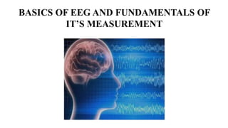
1 basics of eeg and fundamentals of its measurement
- 1. BASICS OF EEG AND FUNDAMENTALS OF IT¡¯S MEASUREMENT
- 2. Timeline of EEG invention 1875 ? Richard Caton - Presence of continuous and spontaneous electrical activity from the brain surface of rabbits and monkeys 1890 ? Adolf Beck ¨C Sensory stimulus can induce spontaneous and rhythmic oscillation 1912 ? Vladimir Pravdich Neminsky ¨C Produced first animal EEG and evoked potential of mammalian dog 1924 ? Hans Berger - Recorded the first human EEG Figure: Hans Berger and his invention
- 3. Cerebral generators of EEG potentials Figure: Neuronal structure Figure: Neuron ¨C Neuron connection
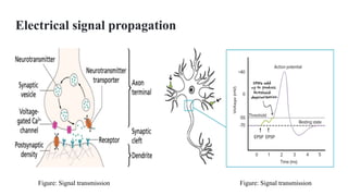
- 4. Electrical signal propagation Figure: Signal transmission Figure: Signal transmission
- 5. Figure: Pyramidal cell¡¯s alignment Figure: Single pyramidal cell
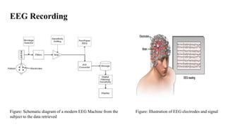
- 6. EEG Recording Figure: Schematic diagram of a modern EEG Machine from the subject to the data retrieved Figure: Illustration of EEG electrodes and signal
- 7. Figure: 10/20 System of EEG electrode placement ? Nasion ? Inion ? Left and right auricular points Figure: EEG Scalp electrodes EEG Electrode Placement
- 8. Filters Figure: Low frequency Filter Figure: Low frequency Filter characteristics
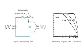
- 9. Figure: High Frequency Filter Figure: High Frequency Filter Characteristics
- 10. Figure : 60 Hz notch filter Figure : 60 Hz notch filter characteristics
- 11. Amplifier ? All EEG amplifiers are differential amplifiers. ? Differential amplifier takes two input voltages and produces an output that is an amplified version of the difference between the two inputs ? Advantage ¨C Cancels out the external noise
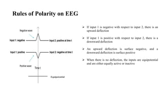
- 12. Rules of Polarity on EEG ? If input 1 is negative with respect to input 2, there is an upward deflection ? If input 1 is positive with respect to input 2, there is a downward deflection ? An upward deflection is surface negative, and a downward deflection is surface positive ? When there is no deflection, the inputs are equipotential and are either equally active or inactive Equipotential
- 13. Polarity Convention - Example
- 14. Montage ?Logical and orderly arrangement of channels/electrode pairs on the display ? Bipolar Montage ? Common electrode reference montage ? Average reference montage ? Laplacian montage
- 15. Figure : Commonly used bipolar longitudinal pattern (Double Banana) Figure : EEG of Bipolar montage
- 16. Figure: Referential montage Figure: Laplacian montage
- 17. Figure: Normal EEG in awake state
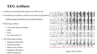
- 19. EEG Artifacts ? Artifacts are unwanted noise signals in an EEG record. ? Classification of artefacts is based on the source of generation: ?Physiological artifacts and external artifacts. ? Physiologic artifacts: ? Any minor body movements ? EMG ? ECG ? Eye movements etc. ? Non Physiologic artifacts: ? Damage of electrodes ? Cable movements ? Broken wire contacts ? Impedance fluctuation ? 60/50 H artifact etc Figure: EEG Artifacts
- 20. Advantages & Applications of EEG ? Excellent temporal resolution ? EEG can determine the relative strengths and positions of electrical activity in different brain regions. ? EEG does not involve exposure to high intensity magnetic field ? Relatively cheap and simple to operate ? Applications of the EEG in humans and animals involve: ? Research ? Clinics
- 21. ? Clinical application- EEG is one of the main diagnostic tests for epilepsy Normal EEG compared to EEG including a seizure: (A) Normal EEG of 15 seconds; (B) EEG of the same patient having an epileptic seizure visible on electrodes P8 and T8.
- 22. Clinical applications ? Monitor alertness, coma and brain death ? Locate areas of damage following head injury, stroke, tumor. ? Monitor cognitive engagement (alpha rhythm) ? Control anesthesia depth ? Investigate epilepsy and locate seizure origin ? Investigate sleep disorder and physiology. ? Etc.
- 23. References ? Teplan, M. (2002). FUNDAMENTALS OF EEG MEASUREMENT. ? Britton JW, Frey LC, Hopp JLet al., authors; St. Louis EK, Frey LC, editors. Electroencephalography (EEG): An Introductory Text and Atlas of Normal and Abnormal Findings in Adults, Children, and Infants [Internet]. Chicago: American Epilepsy Society; 2016. Available from: https://www.ncbi.nlm.nih.gov/books/NBK390354/ ? https://doi.org/10.1684/epd.2020.1217 ? Light, G. A., Williams, L. E., Minow, F., Sprock, J., Rissling, A., Sharp, R., Swerdlow, N. R., & Braff, D. L. (2010). Electroencephalography (EEG) and event-related potentials (ERPs) with human participants. Current protocols in neuroscience, Chapter 6, Unit¨C6.25.24. https://doi.org/10.1002/0471142301.ns0625s52











![References
? Teplan, M. (2002). FUNDAMENTALS OF EEG MEASUREMENT.
? Britton JW, Frey LC, Hopp JLet al., authors; St. Louis EK, Frey LC, editors. Electroencephalography (EEG):
An Introductory Text and Atlas of Normal and Abnormal Findings in Adults, Children, and Infants [Internet].
Chicago: American Epilepsy Society; 2016. Available from:
https://www.ncbi.nlm.nih.gov/books/NBK390354/
? https://doi.org/10.1684/epd.2020.1217
? Light, G. A., Williams, L. E., Minow, F., Sprock, J., Rissling, A., Sharp, R., Swerdlow, N. R., & Braff, D. L.
(2010). Electroencephalography (EEG) and event-related potentials (ERPs) with human participants. Current
protocols in neuroscience, Chapter 6, Unit¨C6.25.24. https://doi.org/10.1002/0471142301.ns0625s52](https://image.slidesharecdn.com/1basicsofeegandfundamentalsofitsmeasurement-210807063158/85/1-basics-of-eeg-and-fundamentals-of-its-measurement-23-320.jpg)
