1 basics of eeg and fundamentals of its measurement
Download as PPTX, PDF0 likes1,122 views
The document discusses the basics of EEG and its measurement. It provides a timeline of EEG invention from 1875 to 1924. It describes how EEG signals are generated from neuronal structures and propagated through electrical signals. It explains how EEG is recorded using a modern EEG machine and electrode placement systems. It discusses filters, amplifiers, polarity conventions, montages, artifacts, and clinical applications of EEG for monitoring brain activity.
1 of 24
Downloaded 39 times








![References
• Teplan, M. (2002). FUNDAMENTALS OF EEG MEASUREMENT.
• Britton JW, Frey LC, Hopp JLet al., authors; St. Louis EK, Frey LC, editors. Electroencephalography (EEG):
An Introductory Text and Atlas of Normal and Abnormal Findings in Adults, Children, and Infants [Internet].
Chicago: American Epilepsy Society; 2016. Available from:
https://www.ncbi.nlm.nih.gov/books/NBK390354/
• https://doi.org/10.1684/epd.2020.1217
• Light, G. A., Williams, L. E., Minow, F., Sprock, J., Rissling, A., Sharp, R., Swerdlow, N. R., & Braff, D. L.
(2010). Electroencephalography (EEG) and event-related potentials (ERPs) with human participants. Current
protocols in neuroscience, Chapter 6, Unit–6.25.24. https://doi.org/10.1002/0471142301.ns0625s52](https://image.slidesharecdn.com/1basicsofeegandfundamentalsofitsmeasurement-210807061846/85/1-basics-of-eeg-and-fundamentals-of-its-measurement-23-320.jpg)

Recommended
EEG artifacts


EEG artifactsSrirama Anjaneyulu
Ìý
EEG artifacts can arise from various physiological and extraphysiological sources other than brain activity. Physiological artifacts originate from the patient's own generator sources like eye movements, muscle activity, movement, and cardiac activity. Extraphysiological artifacts are externally generated, such as from medical devices, electrical equipment, or the environment. Common EEG artifacts include cardiac artifacts like ECG signals, ballistocardiographic artifacts from head or body movement, pacemaker signals, and pulse artifacts. Electrode artifacts can be transient pops or low frequency rhythms across electrodes from poor contact or movement. External artifacts include 50/60 Hz ambient noise, intravenous drips, and signals from devices like pumps and ventilators. Muscle and ocular artifactsEEG Amplifiers


EEG AmplifiersMurtaza Syed
Ìý
This document discusses different types of amplifiers:
1. Power/current amplifiers increase the power or current of an input signal, while decreasing voltage.
2. Frequency amplifiers increase the frequency component of an input signal.
3. Voltage amplifiers increase the voltage of an input signal. Voltage amplifiers are further divided into pre-amplifiers, differential amplifiers, and single-ended amplifiers.
Differential amplifiers are important in EEG machines as they can reject common mode signals and accurately amplify small voltage differences in brain waves.EEG Generators


EEG GeneratorsRahul Kumar
Ìý
This presentation discusses the basic principles governing EEG Rhythm Generation, and discusses the various circuits that generate and maintain cerebral oscillations.EEG ppt


EEG pptNeurologyKota
Ìý
The document discusses various current applications of electroencephalography (EEG) technology both within and outside of clinical settings. It outlines EEG's predominant use in epilepsy and sleep disorder diagnosis clinically. It also explores recent developments that enable portable and cheaper EEG units, allowing novel consumer and research applications. Specifically, the document examines EEG's role in investigating sleep disorders, assessing brain death, monitoring anesthesia depth, cognitive engagement, brain development, and more. It explores EEG's growing use in cognitive science, neuroscience, and other research domains. Finally, it discusses emerging areas like brain-computer interfaces, closed-loop systems, and neuromarketing.Basics of electroencephalography


Basics of electroencephalographyNeurologyKota
Ìý
- The EEG records electrical activity from the cerebral cortex which is amplified over 10 million times to be visible. It detects action potentials and post-synaptic potentials from neurons.
- Electrodes are placed on standardized locations on the scalp according to the 10-20 or 10-10 systems to allow comparison across studies. Recordings can be bipolar between adjacent electrodes or referential against a common electrode.
- Activity is recorded through amplifiers and can be displayed through different montages optimized for localization or overall brain activity. Calibration ensures consistent sensitivity and filtering removes unwanted interference.Principles of polarity in eeg


Principles of polarity in eegPramod Krishnan
Ìý
This presentation looks at EEG signal generation, pyramidal cells, recording of EEG, source localisation, polarity, analysis of dipole, derivations, montages, EEG - Montages, Equipment and Basic Physics


EEG - Montages, Equipment and Basic PhysicsRahul Kumar
Ìý
This presentation discusses the 10-20 system of electrode placement, with its modifications. Also discussed are the Equipment Specifications, basic Physics and sources of interferenceArtifacts in EEG - Recognition and differentiation


Artifacts in EEG - Recognition and differentiationRahul Kumar
Ìý
This Presentation discusses the variously commonly seen artifacts in EEG, and how to recognize them. In EEG interpretation, it is often more important to identify an artifact than to identify true pathology. Once all the artifacts are ruled out, one is sure that what one is dealing with represents disease/abnormality normal eeg 


normal eeg Sachin Adukia
Ìý
This document provides an overview of normal EEG patterns in adults. It begins with a brief history of EEG and then describes the basic electrical activity generated by the brain and how EEG recordings work. It outlines the normal frequency bands seen in EEG - delta, theta, alpha, beta and gamma. Specific normal EEG patterns like the alpha rhythm, vertex waves, sleep spindles and K-complexes are described. It also discusses benign variants and activation procedures. In summary, the document serves as a reference for the typical EEG patterns seen in healthy, awake and sleeping adults.Benign variants of eeg


Benign variants of eegNeurologyKota
Ìý
This document describes several benign EEG variants that can have an epileptiform appearance but are not epileptogenic. It discusses characteristics of alpha variants, mu rhythm, lambda waves, rhythmic mid-temporal theta discharges, wicket spikes, subclinical rhythmic electroencephalographic discharges of adults, phantom spike-wave discharges, and small sharp spikes. These benign variants can occur during drowsiness and light sleep and are seen in specific electrode sites, with features like attenuation with eye opening or movement in the case of mu rhythm. Accurate identification requires training to distinguish them from true epileptiform discharges.EEG Variants By IM


EEG Variants By IMMurtaza Syed
Ìý
EEG variants, are always to be recognized while interpreting the EEG one must be aware of these. Major and most common EEG is variants are discussed in the stated presentation.
Syed Irshad Murtaza.EEG Artifact and How to Resolve


EEG Artifact and How to ResolveLalit Bansal
Ìý
This document discusses various types of EEG artifacts including physiological artifacts generated by the body and extraphysiological artifacts from external equipment or environment. It describes common artifacts like cardiac, electrode, eye blink, muscle activity and their characteristic appearances on EEG. The key is to ensure good preparation, electrode placement and monitoring for artifacts during EEG recording to obtain clean data for accurate interpretation.Brainstem auditory evoked potentials (baep)


Brainstem auditory evoked potentials (baep)NeurologyKota
Ìý
Brainstem Auditory Evoked Potentials (BAEP) involves recording electrophysiological responses from the ear in response to auditory stimulation to assess the functioning of the auditory pathway. BAEP testing involves placing electrodes on the scalp to record waveforms representing activity in the auditory nerve and brainstem in response to click sounds. BAEP is useful for screening and monitoring conditions affecting the auditory pathway such as tumors near the cerebellopontine angle, multiple sclerosis, and coma. It can also be used for newborn hearing screening and evaluating stroke and tuberculous meningitis patients.Benign variants in eeg


Benign variants in eegNeurologyKota
Ìý
1) The document describes several benign EEG variants that can occur but are not associated with epilepsy. These include wicket waves, benign sporadic sleep spikes, 6 per second spike-waves, 14 & 6 Hz positive spikes, and more.
2) The variants are described in terms of their frequency, location, morphology, when they typically occur, and other characteristics. For example, wicket waves occur in the alpha frequency range and are seen unilaterally in the temporal region.
3) Many of the variants are considered normal variants seen in relaxed wakefulness, drowsiness, or different stages of sleep. They are commonly seen in different age groups but are not clinically significant or associated with epilepsy.Abnormal eeg


Abnormal eegrzgar hamed
Ìý
1. The document discusses various abnormal EEG patterns including slowing, spikes, sharp waves, and other abnormalities. It provides details on types of slowing such as focal, regional, and generalized slowing.
2. Different types of spikes and sharp waves are defined including their durations. Both focal and generalized spike/sharp wave abnormalities are described.
3. Specific abnormal EEG patterns are explained in detail such as frontal intermittent rhythmic delta activity (FIRDA), polymorphic delta activity (PDA), and benign focal epilepsies of childhood including rolandic and occipital epilepsy. Causes and differentiation of these patterns are provided.Eeg artifacts


Eeg artifactsNeurologyKota
Ìý
This document discusses different types of artifacts that can appear in EEG recordings. It divides artifacts into physiological artifacts, caused by body movements or electrical activity, and non-physiological artifacts, caused by external electrical interference or equipment issues. Specific artifacts covered include eye blinks and movements, muscle activity, sweat, ECG interference, ventilator and pulse artifacts, electrical interference, and electrode and lead movement issues. Proper identification of artifacts is important for interpreting EEG recordings.PLEDS


PLEDSSachin Adukia
Ìý
1. PLEDs (Periodic Lateralized Epileptiform Discharges) are a pattern seen on EEG characterized by periodic discharges that are lateralized to one hemisphere.
2. They are commonly seen in conditions involving acute brain injury or inflammation such as stroke, encephalitis, tumors, or hypoxic ischemic encephalopathy.
3. PLEDs are associated with a risk of seizures but generally indicate an unstable brain state that will improve over time as the underlying condition resolves. Prognosis depends on the specific cause.EEG artifacts


EEG artifactsSudhakar Marella
Ìý
Cardiac artifacts appear as periodic waves that are time-locked to the heartbeat as recorded by ECG. They include electrical artifacts seen as QRS complexes and mechanical artifacts seen as pulse waves. Electrode artifacts occur due to poor electrode contact or lead movement and appear as irregular waves of varying morphology and amplitude. External device artifacts are caused by electrical or mechanical devices and may appear as 50/60Hz noise, spike-like waves from IV drips, or irregular high amplitude waves from electrical motors. Artifacts must be distinguished from physiological activity and epileptiform discharges based on characteristics like distribution, morphology, and periodicity to avoid misinterpretation.EEG Variants with patterns by Murtaza Syed


EEG Variants with patterns by Murtaza SyedMurtaza Syed
Ìý
This document provides information on normal variant EEG patterns. It discusses four main types of EEG variants: rhythmic patterns, epileptiform patterns, lambda and lambdoids, and age-related variants. Six main rhythmic variant patterns are described including alpha variants, mu rhythm, rhythmic mid-temporal theta of drowsiness, subclinical rhythmic electrographic discharges in adults, midline theta rhythm, and frontal arousal rhythm. Four epileptiform variant patterns are also outlined. The document provides detailed descriptions of each variant pattern.Abnormal EEG patterns


Abnormal EEG patternsMurtaza Syed
Ìý
1. The document defines abnormal EEG patterns (AEPs) and describes two main types: non-epileptiform and epileptiform.
2. Non-epileptiform patterns include slow waves, which can be focal or diffuse, and amplitude/frequency asymmetry. Epileptiform patterns consist of spike and sharp waves that can be focal or generalized.
3. Specific AEPs are described such as benign rolandic epilepsy, 3/sec spike-wave, periodic lateralized epileptiform discharges, and others associated with various neurological conditions.Electroencephalography (eeg)


Electroencephalography (eeg)Ranjeet Singha
Ìý
Electroencephalography (eeg),electrical activity recorded via electrodes on the scalp,Neurophysiological Basis of EEG,Scalp EEG Recordings,Wearable EEG Devices,Recording EEG Signals,EEG Rhythms,EEG artefacts


EEG artefactsManchesterEEG
Ìý
EEG artefacts arise from unwanted electrical activity from sources other than the brain, such as eye movements, muscle activity, and environmental noise. Identifying artefacts can be challenging as some resemble brain activity. Methods for removing artefacts include filtering, regression-based approaches, and independent component analysis, which transforms scalp channel data into spatially independent sources that may represent brain or non-brain activity. Careful inspection of component properties like scalp maps, time courses, and spectra is needed to classify them as representing brain activity or artefacts.Mu rhythm


Mu rhythmMohibullah Kakar
Ìý
The document discusses the mu rhythm, which is a central rhythm seen on EEG with an alpha frequency band of 8-10 Hz. It has an arciform configuration and occurs in less than 5% of children under age 4 and 18-20% of children ages 8-16. The mu rhythm is not blocked by eye opening but is blocked by touch, limb movement, or thought of movement. It is usually asymmetric and independent between hemispheres. The mu rhythm is believed to originate from the sensorimotor cortex at rest and can be prominent in patients with skull defects.Electroencephalogram(EEG)


Electroencephalogram(EEG)ashikh
Ìý
Electroencephalography is the technique used to acquire electrical signals of brain through electrodes which are placed by certain montage. Different wave patterns can be observed which is useful in detecting any abnormal conditions or neurological brain disorders in human beings. There is broad future scope for medical research and creating EEG based equipments for real time applications.EEG in convulsive and non convulsive seizures in the intensive care unit


EEG in convulsive and non convulsive seizures in the intensive care unitPramod Krishnan
Ìý
Case based discussion regarding the utility of EEG in the management of convulsive and non convulsive seizures, including status epilepticus in the intensive care unitSleep EEG


Sleep EEGNeurologyKota
Ìý
The document summarizes the stages of sleep as assessed by polysomnography. It describes the key EEG patterns, eye movements, and muscle activity that characterize each stage:
Stage 1 is characterized by low voltage mixed frequency EEG activity, vertex sharp waves, and slow eye movements. Stage 2 involves sleep spindles and K complexes in the EEG along with occasional slow eye movements. Stage 3 contains 20-50% slow wave activity in the EEG. REM sleep involves rapid eye movements and EEG desynchronization resembling wakefulness, along with muscle atonia. The stages cycle throughout the night in a progression from light to deep sleep and back to light sleep.Electrocorticography


ElectrocorticographyLalit Bansal
Ìý
This document provides information about electrocorticography (ECOG). It discusses:
1. The limitations of scalp EEG and advantages of ECOG for localizing epileptic activity, including its ability to record high-quality signals close to neuronal generators with minimal attenuation.
2. Types and uses of intraoperative and extraoperative ECOG. Indications for combining ECOG with functional mapping to guide epilepsy surgery near eloquent cortex.
3. Characteristics of ECOG recordings, such as common patterns associated with cortical dysplasia, and factors that can influence recordings like anesthesia, antiepileptic drug levels, and electrical stimulation.Normal EEG patterns, frequencies, as well as patterns that may simulate disease


Normal EEG patterns, frequencies, as well as patterns that may simulate diseaseRahul Kumar
Ìý
This presentation discusses the vast range of traces that show the variations in normal EEG patterns, as well as discussing the frequency and amplitudes of various normal waveforms.EEG- CONCEPTS AND CLINICAL USES FOR NEUROLOGY RESIDENTS


EEG- CONCEPTS AND CLINICAL USES FOR NEUROLOGY RESIDENTSChirayuRegmi2
Ìý
EEG- CONCEPTS AND CLINICAL USES FOR NEUROLOGY RESIDENTSElectroenchephalography


Electroenchephalographyimabongaigaon
Ìý
This document provides a detailed overview of electroencephalography (EEG). Some key points:
- EEG was developed in the late 19th/early 20th century through studies of brain electrical activity in animals and humans. Hans Berger recorded the first human EEG in 1924.
- EEG uses electrodes placed on the scalp to detect electrical signals produced by neuron firing in the brain. It can identify abnormalities associated with conditions like epilepsy, tumors, strokes and encephalopathies.
- Quantitative EEG analysis allows measuring brain activity levels in different frequency bands to identify abnormalities in conditions like Alzheimer's, ADHD, autism, depression, OCD and schizophrenia.More Related Content
What's hot (20)
normal eeg 


normal eeg Sachin Adukia
Ìý
This document provides an overview of normal EEG patterns in adults. It begins with a brief history of EEG and then describes the basic electrical activity generated by the brain and how EEG recordings work. It outlines the normal frequency bands seen in EEG - delta, theta, alpha, beta and gamma. Specific normal EEG patterns like the alpha rhythm, vertex waves, sleep spindles and K-complexes are described. It also discusses benign variants and activation procedures. In summary, the document serves as a reference for the typical EEG patterns seen in healthy, awake and sleeping adults.Benign variants of eeg


Benign variants of eegNeurologyKota
Ìý
This document describes several benign EEG variants that can have an epileptiform appearance but are not epileptogenic. It discusses characteristics of alpha variants, mu rhythm, lambda waves, rhythmic mid-temporal theta discharges, wicket spikes, subclinical rhythmic electroencephalographic discharges of adults, phantom spike-wave discharges, and small sharp spikes. These benign variants can occur during drowsiness and light sleep and are seen in specific electrode sites, with features like attenuation with eye opening or movement in the case of mu rhythm. Accurate identification requires training to distinguish them from true epileptiform discharges.EEG Variants By IM


EEG Variants By IMMurtaza Syed
Ìý
EEG variants, are always to be recognized while interpreting the EEG one must be aware of these. Major and most common EEG is variants are discussed in the stated presentation.
Syed Irshad Murtaza.EEG Artifact and How to Resolve


EEG Artifact and How to ResolveLalit Bansal
Ìý
This document discusses various types of EEG artifacts including physiological artifacts generated by the body and extraphysiological artifacts from external equipment or environment. It describes common artifacts like cardiac, electrode, eye blink, muscle activity and their characteristic appearances on EEG. The key is to ensure good preparation, electrode placement and monitoring for artifacts during EEG recording to obtain clean data for accurate interpretation.Brainstem auditory evoked potentials (baep)


Brainstem auditory evoked potentials (baep)NeurologyKota
Ìý
Brainstem Auditory Evoked Potentials (BAEP) involves recording electrophysiological responses from the ear in response to auditory stimulation to assess the functioning of the auditory pathway. BAEP testing involves placing electrodes on the scalp to record waveforms representing activity in the auditory nerve and brainstem in response to click sounds. BAEP is useful for screening and monitoring conditions affecting the auditory pathway such as tumors near the cerebellopontine angle, multiple sclerosis, and coma. It can also be used for newborn hearing screening and evaluating stroke and tuberculous meningitis patients.Benign variants in eeg


Benign variants in eegNeurologyKota
Ìý
1) The document describes several benign EEG variants that can occur but are not associated with epilepsy. These include wicket waves, benign sporadic sleep spikes, 6 per second spike-waves, 14 & 6 Hz positive spikes, and more.
2) The variants are described in terms of their frequency, location, morphology, when they typically occur, and other characteristics. For example, wicket waves occur in the alpha frequency range and are seen unilaterally in the temporal region.
3) Many of the variants are considered normal variants seen in relaxed wakefulness, drowsiness, or different stages of sleep. They are commonly seen in different age groups but are not clinically significant or associated with epilepsy.Abnormal eeg


Abnormal eegrzgar hamed
Ìý
1. The document discusses various abnormal EEG patterns including slowing, spikes, sharp waves, and other abnormalities. It provides details on types of slowing such as focal, regional, and generalized slowing.
2. Different types of spikes and sharp waves are defined including their durations. Both focal and generalized spike/sharp wave abnormalities are described.
3. Specific abnormal EEG patterns are explained in detail such as frontal intermittent rhythmic delta activity (FIRDA), polymorphic delta activity (PDA), and benign focal epilepsies of childhood including rolandic and occipital epilepsy. Causes and differentiation of these patterns are provided.Eeg artifacts


Eeg artifactsNeurologyKota
Ìý
This document discusses different types of artifacts that can appear in EEG recordings. It divides artifacts into physiological artifacts, caused by body movements or electrical activity, and non-physiological artifacts, caused by external electrical interference or equipment issues. Specific artifacts covered include eye blinks and movements, muscle activity, sweat, ECG interference, ventilator and pulse artifacts, electrical interference, and electrode and lead movement issues. Proper identification of artifacts is important for interpreting EEG recordings.PLEDS


PLEDSSachin Adukia
Ìý
1. PLEDs (Periodic Lateralized Epileptiform Discharges) are a pattern seen on EEG characterized by periodic discharges that are lateralized to one hemisphere.
2. They are commonly seen in conditions involving acute brain injury or inflammation such as stroke, encephalitis, tumors, or hypoxic ischemic encephalopathy.
3. PLEDs are associated with a risk of seizures but generally indicate an unstable brain state that will improve over time as the underlying condition resolves. Prognosis depends on the specific cause.EEG artifacts


EEG artifactsSudhakar Marella
Ìý
Cardiac artifacts appear as periodic waves that are time-locked to the heartbeat as recorded by ECG. They include electrical artifacts seen as QRS complexes and mechanical artifacts seen as pulse waves. Electrode artifacts occur due to poor electrode contact or lead movement and appear as irregular waves of varying morphology and amplitude. External device artifacts are caused by electrical or mechanical devices and may appear as 50/60Hz noise, spike-like waves from IV drips, or irregular high amplitude waves from electrical motors. Artifacts must be distinguished from physiological activity and epileptiform discharges based on characteristics like distribution, morphology, and periodicity to avoid misinterpretation.EEG Variants with patterns by Murtaza Syed


EEG Variants with patterns by Murtaza SyedMurtaza Syed
Ìý
This document provides information on normal variant EEG patterns. It discusses four main types of EEG variants: rhythmic patterns, epileptiform patterns, lambda and lambdoids, and age-related variants. Six main rhythmic variant patterns are described including alpha variants, mu rhythm, rhythmic mid-temporal theta of drowsiness, subclinical rhythmic electrographic discharges in adults, midline theta rhythm, and frontal arousal rhythm. Four epileptiform variant patterns are also outlined. The document provides detailed descriptions of each variant pattern.Abnormal EEG patterns


Abnormal EEG patternsMurtaza Syed
Ìý
1. The document defines abnormal EEG patterns (AEPs) and describes two main types: non-epileptiform and epileptiform.
2. Non-epileptiform patterns include slow waves, which can be focal or diffuse, and amplitude/frequency asymmetry. Epileptiform patterns consist of spike and sharp waves that can be focal or generalized.
3. Specific AEPs are described such as benign rolandic epilepsy, 3/sec spike-wave, periodic lateralized epileptiform discharges, and others associated with various neurological conditions.Electroencephalography (eeg)


Electroencephalography (eeg)Ranjeet Singha
Ìý
Electroencephalography (eeg),electrical activity recorded via electrodes on the scalp,Neurophysiological Basis of EEG,Scalp EEG Recordings,Wearable EEG Devices,Recording EEG Signals,EEG Rhythms,EEG artefacts


EEG artefactsManchesterEEG
Ìý
EEG artefacts arise from unwanted electrical activity from sources other than the brain, such as eye movements, muscle activity, and environmental noise. Identifying artefacts can be challenging as some resemble brain activity. Methods for removing artefacts include filtering, regression-based approaches, and independent component analysis, which transforms scalp channel data into spatially independent sources that may represent brain or non-brain activity. Careful inspection of component properties like scalp maps, time courses, and spectra is needed to classify them as representing brain activity or artefacts.Mu rhythm


Mu rhythmMohibullah Kakar
Ìý
The document discusses the mu rhythm, which is a central rhythm seen on EEG with an alpha frequency band of 8-10 Hz. It has an arciform configuration and occurs in less than 5% of children under age 4 and 18-20% of children ages 8-16. The mu rhythm is not blocked by eye opening but is blocked by touch, limb movement, or thought of movement. It is usually asymmetric and independent between hemispheres. The mu rhythm is believed to originate from the sensorimotor cortex at rest and can be prominent in patients with skull defects.Electroencephalogram(EEG)


Electroencephalogram(EEG)ashikh
Ìý
Electroencephalography is the technique used to acquire electrical signals of brain through electrodes which are placed by certain montage. Different wave patterns can be observed which is useful in detecting any abnormal conditions or neurological brain disorders in human beings. There is broad future scope for medical research and creating EEG based equipments for real time applications.EEG in convulsive and non convulsive seizures in the intensive care unit


EEG in convulsive and non convulsive seizures in the intensive care unitPramod Krishnan
Ìý
Case based discussion regarding the utility of EEG in the management of convulsive and non convulsive seizures, including status epilepticus in the intensive care unitSleep EEG


Sleep EEGNeurologyKota
Ìý
The document summarizes the stages of sleep as assessed by polysomnography. It describes the key EEG patterns, eye movements, and muscle activity that characterize each stage:
Stage 1 is characterized by low voltage mixed frequency EEG activity, vertex sharp waves, and slow eye movements. Stage 2 involves sleep spindles and K complexes in the EEG along with occasional slow eye movements. Stage 3 contains 20-50% slow wave activity in the EEG. REM sleep involves rapid eye movements and EEG desynchronization resembling wakefulness, along with muscle atonia. The stages cycle throughout the night in a progression from light to deep sleep and back to light sleep.Electrocorticography


ElectrocorticographyLalit Bansal
Ìý
This document provides information about electrocorticography (ECOG). It discusses:
1. The limitations of scalp EEG and advantages of ECOG for localizing epileptic activity, including its ability to record high-quality signals close to neuronal generators with minimal attenuation.
2. Types and uses of intraoperative and extraoperative ECOG. Indications for combining ECOG with functional mapping to guide epilepsy surgery near eloquent cortex.
3. Characteristics of ECOG recordings, such as common patterns associated with cortical dysplasia, and factors that can influence recordings like anesthesia, antiepileptic drug levels, and electrical stimulation.Normal EEG patterns, frequencies, as well as patterns that may simulate disease


Normal EEG patterns, frequencies, as well as patterns that may simulate diseaseRahul Kumar
Ìý
This presentation discusses the vast range of traces that show the variations in normal EEG patterns, as well as discussing the frequency and amplitudes of various normal waveforms.Similar to 1 basics of eeg and fundamentals of its measurement (20)
EEG- CONCEPTS AND CLINICAL USES FOR NEUROLOGY RESIDENTS


EEG- CONCEPTS AND CLINICAL USES FOR NEUROLOGY RESIDENTSChirayuRegmi2
Ìý
EEG- CONCEPTS AND CLINICAL USES FOR NEUROLOGY RESIDENTSElectroenchephalography


Electroenchephalographyimabongaigaon
Ìý
This document provides a detailed overview of electroencephalography (EEG). Some key points:
- EEG was developed in the late 19th/early 20th century through studies of brain electrical activity in animals and humans. Hans Berger recorded the first human EEG in 1924.
- EEG uses electrodes placed on the scalp to detect electrical signals produced by neuron firing in the brain. It can identify abnormalities associated with conditions like epilepsy, tumors, strokes and encephalopathies.
- Quantitative EEG analysis allows measuring brain activity levels in different frequency bands to identify abnormalities in conditions like Alzheimer's, ADHD, autism, depression, OCD and schizophrenia.Eeg examples


Eeg examplesVijay Kumar T
Ìý
This document provides information about EEG recordings and analysis. It discusses topics like the Nyquist theorem, types of EEG recordings, artifacts in EEG like EMG and eye blinks, reviewing EEG characteristics, spectral maps of EEG under different conditions, microstates in EEG, normal and abnormal distributions in EEG data, and life span normative EEG databases.SUMSEM-2021-22_ECE6007_ETH_VL2021220701295_Reference_Material_I_04-07-2022_EE...


SUMSEM-2021-22_ECE6007_ETH_VL2021220701295_Reference_Material_I_04-07-2022_EE...BharathSrinivasG
Ìý
This document discusses electroencephalography (EEG) and evoked potentials. It begins by describing EEG as a method for recording electrical brain activity using electrodes placed on the scalp. It then discusses how EEG is used to diagnose neurological conditions like epilepsy and brain tumors. The document outlines different brain wave patterns observed in EEG like alpha, beta, theta, and delta waves. It also discusses how EEG is performed and interpreted, potential artifacts, and different electrode montages. Finally, it describes evoked potentials as the electrical response of the brain to sensory stimulation, and summarizes different types of evoked potentials including visual, somatosensory, and auditory potentials.EEG


EEGMohd. Bilal
Ìý
Electroencephalography (EEG) is a technique used to analyze neural activity in the brain. EEG measures electrical activity using electrodes placed on the scalp. It has several uses such as medical diagnosis and research. Some key points are:
- EEG was developed in the early 20th century and works by measuring voltage fluctuations resulting from ionic current flows in neurons.
- It has advantages like being noninvasive and having good temporal resolution, but disadvantages like having poor spatial resolution.
- EEG signals are classified by frequency into different wave types including alpha, beta, theta, delta, and gamma waves which correlate with different brain states.
- Applications include diagnosing brain disorders, brain-computer interfaces,Eeg


EegDr. sreeremya S
Ìý
EEG recordings measure the minute electrical potentials produced by the brain using electrodes placed on the scalp. Hans Berger first recorded human EEGs in 1924 using metal strips as electrodes. The EEG is difficult to interpret due to low amplitude brain signals and spatial mapping of brain functions. The 10-20 electrode placement system was adopted for standardization. Long-term EEG recordings require electrodes that ensure stability while avoiding infection risks. Analysis of EEGs focuses on characteristics like amplitude, frequency, and phase to understand changes in brain electrical activity.Unit 3 biomedical


Unit 3 biomedicalAnu Antony
Ìý
This document provides information about electroencephalography (EEG), electromyography (EMG), and patient monitoring. It discusses how EEG is used to measure brain activity through electrodes on the scalp. It describes the different frequency bands seen on EEG and how they relate to mental states. The document outlines the components of an EEG recording system and various EEG artifacts. It also discusses EMG and how it is used to measure muscle electrical activity. Finally, it covers patient monitoring systems, including bedside monitors, central monitoring stations, and the parameters that are measured like heart rate, blood pressure, respiration rate.EEG in neurology and psychiatry


EEG in neurology and psychiatrykkapil85
Ìý
This presentation gives basic account of characteristic EEG patterns seen in common psychiatric and neurological conditions.NEURAL ENGINEERING UNIT II ELECTROENCEPHALOGRAPHY


NEURAL ENGINEERING UNIT II ELECTROENCEPHALOGRAPHYthendralm1
Ìý
Electroencephalography (EEG): General Principles and Clinical Applications:
EEG is a non-invasive diagnostic tool that measures electrical activity in the brain. It is widely used to assess brain function, detect neurological conditions, and guide treatment in epilepsy, sleep disorders, and other conditions.
Neonatal and Paediatric EEG:
EEG in neonates and children requires specialized interpretation due to developmental differences in brain activity. It is used for diagnosing conditions such as neonatal seizures, developmental delay, and encephalopathy.
EEG Artefacts and Benign Variants:
Artefacts are unwanted signals in EEG recordings caused by non-cerebral activity (e.g., muscle movements, blinking). Benign variants are normal deviations that should not be mistaken for pathological findings.
Video EEG Monitoring for Epilepsy:
Combines EEG with synchronized video recordings to capture and analyze seizure events. This technique is critical for epilepsy diagnosis, classification, and pre-surgical evaluation.
Invasive Clinical Neurophysiology in Epilepsy and Movement Disorders:
Invasive techniques involve implanting electrodes directly into the brain to localize seizure foci or study movement disorders. This is often used in pre-surgical planning for epilepsy.
Topographic Mapping:
EEG topographic mapping provides a visual representation of brain activity, aiding in identifying localized abnormalities.
Frequency Analysis and Quantitative Techniques in EEG:
Frequency analysis helps classify brain waves (e.g., delta, theta, alpha, beta) to study brain states and conditions. Quantitative EEG (qEEG) uses mathematical techniques to analyze data for more precise diagnostics.
Intraoperative EEG Monitoring:
EEG is used during surgeries such as carotid endarterectomy and cardiac procedures to monitor brain function and reduce the risk of complications like stroke.
Magnetoencephalography (MEG):
MEG is a technique that measures the magnetic fields generated by neuronal activity. It complements EEG by providing high spatial resolution and is used in epilepsy and functional brain mapping.EEG.pptx


EEG.pptxmanjushashinde4
Ìý
The document discusses the history and procedure of electroencephalography (EEG). It describes how Hans Berger first recorded human EEG in 1924 and identified the alpha wave rhythm. An EEG involves placing electrodes on the scalp to record the brain's electrical activity with high temporal but low spatial resolution. The four main EEG wave types - alpha, beta, theta, and delta - are defined. Sleep stages are characterized by different EEG patterns, and EEG can be used to diagnose epilepsy, brain tumors, and other neurological conditions.Artifacts in eeg final


Artifacts in eeg finalNeurology resident slides
Ìý
This document discusses different types of artifacts that can appear on an EEG, including how to identify and eliminate them. It separates artifacts into physiological artifacts originating from the patient's body (e.g. eye movements, muscle activity), and extraphysiological artifacts from external sources (e.g. electrodes, equipment, environment). Specific artifact types like blinks, lateral eye movements, muscle activity are described. Guidelines provided on how to reduce artifacts include closing the eyes, relaxing muscles, ensuring good electrode contact, and shielding from environmental interference.EEG-132 Pract. (1).ppt


EEG-132 Pract. (1).pptFatmaSetyaningsih2
Ìý
The document discusses electroencephalography (EEG), which records the electrical activity of the brain from the scalp. It describes how Hans Berger first recorded EEG in 1929. It explains the different types of brain waves seen in a normal EEG - alpha, beta, theta, and delta waves. It discusses how EEG is used to study epilepsy, sleep disorders, brain tumors, and other neurological conditions. It also covers how EEG recordings are performed, including electrode placement and provocation tests.Eeg Sleep Iom Ver


Eeg Sleep Iom VerKouya71
Ìý
EEG, sleep, and evoked potentials can provide useful clinical information. EEG reflects brain electrical activity and can detect abnormal rhythms like spike and wave patterns in epilepsy. Sleep has different phases including REM and non-REM. Evoked potentials average brain responses to sensory stimuli to detect small signals against background noise and identify conduction delays or abnormalities.Artifacts in EEG.pptx


Artifacts in EEG.pptxPramod Krishnan
Ìý
This presentation reviews the common artifacts in EEG, their identification and rectification. Examples of various artifacts are provided in the presentation. EEG guest lecture_iub_eee541


EEG guest lecture_iub_eee541Md Kafiul Islam
Ìý
This was a Guest Lecture given to the Masters students of the course EE541-Biomedical Electronics on EEGEEG INTERPRETATION


EEG INTERPRETATIONRajeev Bhandari
Ìý
The document provides information about EEGs and EMGs. It defines EEG as recording electrical activity of the brain from the scalp and notes its history and applications in diagnosing conditions like epilepsy. It describes different brain waves seen in EEGs including alpha, beta, theta, and delta waves and their characteristics. It also summarizes sleep cycles and brain waves associated with each stage of sleep. The document then discusses EMG and how it records muscle activity through motor units. It notes the techniques of surface EMG and intramuscular EMG and what abnormalities in spontaneous activity or motor unit potentials can indicate.Electrocardiogram pptx


Electrocardiogram pptxSt. Xavier's college, maitighar,Kathmandu
Ìý
The Electrocardiogram document discusses:
1. An electrocardiogram (ECG) records the heart's electrical activity using electrodes placed on the skin and an ECG machine.
2. The ECG machine detects the electrical signals via electrodes and lead wires and displays or prints the output.
3. ECG leads combine electrodes in different positions to view the heart's electrical activity from different angles or planes.Recently uploaded (20)
ASP.NET Web API Interview Questions By Scholarhat


ASP.NET Web API Interview Questions By ScholarhatScholarhat
Ìý
ASP.NET Web API Interview Questions By ScholarhatEffective Product Variant Management in Odoo 18


Effective Product Variant Management in Odoo 18Celine George
Ìý
In this slide we’ll discuss on the effective product variant management in Odoo 18. Odoo concentrates on managing product variations and offers a distinct area for doing so. Product variants provide unique characteristics like size and color to single products, which can be managed at the product template level for all attributes and variants or at the variant level for individual variants.NUTRITIONAL ASSESSMENT AND EDUCATION - 5TH SEM.pdf


NUTRITIONAL ASSESSMENT AND EDUCATION - 5TH SEM.pdfDolisha Warbi
Ìý
NUTRITIONAL ASSESSMENT AND EDUCATION, Introduction, definition, types - macronutrient and micronutrient, food pyramid, meal planning, nutritional assessment of individual, family and community by using appropriate method, nutrition education, nutritional rehabilitation, nutritional deficiency disorder, law/policies regarding nutrition in India, food hygiene, food fortification, food handling and storage, food preservation, food preparation, food purchase, food consumption, food borne diseases, food poisoningOdoo 18 Accounting Access Rights - Odoo 18 ºÝºÝߣs


Odoo 18 Accounting Access Rights - Odoo 18 ºÝºÝߣsCeline George
Ìý
In this slide, we’ll discuss on accounting access rights in odoo 18. To ensure data security and maintain confidentiality, Odoo provides a robust access rights system that allows administrators to control who can access and modify accounting data. Oral exam Kenneth Bech - What is the meaning of strategic fit?


Oral exam Kenneth Bech - What is the meaning of strategic fit?MIPLM
Ìý
Presentation of the CEIPI DU IPBA oral exam of Kenneth Bech - What is the meaning of strategic fit? Full-Stack .NET Developer Interview Questions PDF By ScholarHat


Full-Stack .NET Developer Interview Questions PDF By ScholarHatScholarhat
Ìý
Full-Stack .NET Developer Interview Questions PDF By ScholarHatBISNIS BERKAH BERANGKAT KE MEKKAH ISTIKMAL SYARIAH


BISNIS BERKAH BERANGKAT KE MEKKAH ISTIKMAL SYARIAHcoacharyasetiyaki
Ìý
BISNIS BERKAH BERANGKAT KE MEKKAH ISTIKMAL SYARIAHInterim Guidelines for PMES-DM-17-2025-PPT.pptx


Interim Guidelines for PMES-DM-17-2025-PPT.pptxsirjeromemanansala
Ìý
This is the latest issuance on PMES as replacement of RPMS. Kindly message me to gain full access of the presentation. One Click RFQ Cancellation in Odoo 18 - Odoo ºÝºÝߣs


One Click RFQ Cancellation in Odoo 18 - Odoo ºÝºÝߣsCeline George
Ìý
In this slide, we’ll discuss the one click RFQ Cancellation in odoo 18. One-Click RFQ Cancellation in Odoo 18 is a feature that allows users to quickly and easily cancel Request for Quotations (RFQs) with a single click.Intellectual Honesty & Research Integrity.pptx


Intellectual Honesty & Research Integrity.pptxNidhiSharma495177
Ìý
Research Publication & Ethics contains a chapter on Intellectual Honesty and Research Integrity.
Different case studies of intellectual dishonesty and integrity were discussed.AI and Academic Writing, Short Term Course in Academic Writing and Publicatio...


AI and Academic Writing, Short Term Course in Academic Writing and Publicatio...Prof. (Dr.) Vinod Kumar Kanvaria
Ìý
AI and Academic Writing, Short Term Course in Academic Writing and Publication, UGC-MMTTC, MANUU, 25/02/2025, Prof. (Dr.) Vinod Kumar Kanvaria, University of Delhi, vinodpr111@gmail.comAzure Administrator Interview Questions By ScholarHat


Azure Administrator Interview Questions By ScholarHatScholarhat
Ìý
Azure Administrator Interview Questions By ScholarHatDr. Ansari Khurshid Ahmed- Factors affecting Validity of a Test.pptx


Dr. Ansari Khurshid Ahmed- Factors affecting Validity of a Test.pptxKhurshid Ahmed Ansari
Ìý
Validity is an important characteristic of a test. A test having low validity is of little use. Validity is the accuracy with which a test measures whatever it is supposed to measure. Validity can be low, moderate or high. There are many factors which affect the validity of a test. If these factors are controlled, then the validity of the test can be maintained to a high level. In the power point presentation, factors affecting validity are discussed with the help of concrete examples.RRB ALP CBT 2 Mechanic Motor Vehicle Question Paper (MMV Exam MCQ)


RRB ALP CBT 2 Mechanic Motor Vehicle Question Paper (MMV Exam MCQ)SONU HEETSON
Ìý
RRB ALP CBT 2 Mechanic Motor Vehicle Question Paper. MMV MCQ PDF Free Download for Railway Assistant Loco Pilot Exam.Mastering Soft Tissue Therapy & Sports Taping


Mastering Soft Tissue Therapy & Sports TapingKusal Goonewardena
Ìý
Mastering Soft Tissue Therapy & Sports Taping: Pathway to Sports Medicine Excellence
This presentation was delivered in Colombo, Sri Lanka, at the Institute of Sports Medicine to an audience of sports physiotherapists, exercise scientists, athletic trainers, and healthcare professionals. Led by Kusal Goonewardena (PhD Candidate - Muscle Fatigue, APA Titled Sports & Exercise Physiotherapist) and Gayath Jayasinghe (Sports Scientist), the session provided comprehensive training on soft tissue assessment, treatment techniques, and essential sports taping methods.
Key topics covered:
✅ Soft Tissue Therapy – The science behind muscle, fascia, and joint assessment for optimal treatment outcomes.
✅ Sports Taping Techniques – Practical applications for injury prevention and rehabilitation, including ankle, knee, shoulder, thoracic, and cervical spine taping.
✅ Sports Trainer Level 1 Course by Sports Medicine Australia – A gateway to professional development, career opportunities, and working in Australia.
This training mirrors the Elite Akademy Sports Medicine standards, ensuring evidence-based approaches to injury management and athlete care.
If you are a sports professional looking to enhance your clinical skills and open doors to global opportunities, this presentation is for you.ASP.NET Interview Questions PDF By ScholarHat


ASP.NET Interview Questions PDF By ScholarHatScholarhat
Ìý
ASP.NET Interview Questions PDF By ScholarHatInventory Reporting in Odoo 17 - Odoo 17 Inventory App


Inventory Reporting in Odoo 17 - Odoo 17 Inventory AppCeline George
Ìý
This slide will helps us to efficiently create detailed reports of different records defined in its modules, both analytical and quantitative, with Odoo 17 ERP.AI and Academic Writing, Short Term Course in Academic Writing and Publicatio...


AI and Academic Writing, Short Term Course in Academic Writing and Publicatio...Prof. (Dr.) Vinod Kumar Kanvaria
Ìý
1 basics of eeg and fundamentals of its measurement
- 1. BASICS OF EEG AND FUNDAMENTALS OF IT’S MEASUREMENT
- 2. Timeline of EEG invention 1875 • Richard Caton - Presence of continuous and spontaneous electrical activity from the brain surface of rabbits and monkeys 1890 • Adolf Beck – Sensory stimulus can induce spontaneous and rhythmic oscillation 1912 • Vladimir Pravdich Neminsky – Produced first animal EEG and evoked potential of mammalian dog 1924 • Hans Berger - Recorded the first human EEG Figure: Hans Berger and his invention
- 3. Cerebral generators of EEG potentials Figure: Neuronal structure Figure: Neuron – Neuron connection
- 4. Electrical signal propagation Figure: Signal transmission Figure: Signal transmission
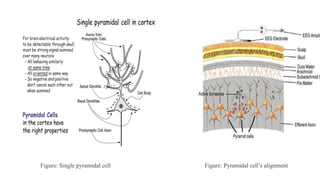
- 5. Figure: Pyramidal cell’s alignment Figure: Single pyramidal cell
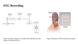
- 6. EEG Recording Figure: Schematic diagram of a modern EEG Machine from the subject to the data retrieved Figure: Illustration of EEG electrodes and signal
- 7. Figure: 10/20 System of EEG electrode placement • Nasion • Inion • Left and right auricular points Figure: EEG Scalp electrodes EEG Electrode Placement
- 8. Filters Figure: Low frequency Filter Figure: Low frequency Filter characteristics
- 9. Figure: High Frequency Filter Figure: High Frequency Filter Characteristics
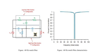
- 10. Figure : 60 Hz notch filter Figure : 60 Hz notch filter characteristics
- 11. Amplifier • All EEG amplifiers are differential amplifiers. • Differential amplifier takes two input voltages and produces an output that is an amplified version of the difference between the two inputs • Advantage – Cancels out the external noise
- 12. Rules of Polarity on EEG  If input 1 is negative with respect to input 2, there is an upward deflection  If input 1 is positive with respect to input 2, there is a downward deflection  An upward deflection is surface negative, and a downward deflection is surface positive  When there is no deflection, the inputs are equipotential and are either equally active or inactive Equipotential
- 13. Polarity Convention - Example
- 14. Montage Logical and orderly arrangement of channels/electrode pairs on the display • Bipolar Montage • Common electrode reference montage • Average reference montage • Laplacian montage
- 15. Figure : Commonly used bipolar longitudinal pattern (Double Banana) Figure : EEG of Bipolar montage
- 16. Figure: Referential montage Figure: Laplacian montage
- 17. Figure: Normal EEG in awake state
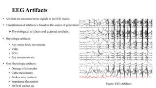
- 19. EEG Artifacts • Artifacts are unwanted noise signals in an EEG record. • Classification of artefacts is based on the source of generation: Physiological artifacts and external artifacts. • Physiologic artifacts: • Any minor body movements • EMG • ECG • Eye movements etc. • Non Physiologic artifacts: • Damage of electrodes • Cable movements • Broken wire contacts • Impedance fluctuation • 60/50 H artifact etc Figure: EEG Artifacts
- 20. Advantages & Applications of EEG • Excellent temporal resolution • EEG can determine the relative strengths and positions of electrical activity in different brain regions. • EEG does not involve exposure to high intensity magnetic field • Relatively cheap and simple to operate • Applications of the EEG in humans and animals involve:  Research  Clinics
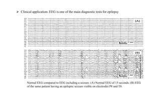
- 21.  Clinical application- EEG is one of the main diagnostic tests for epilepsy Normal EEG compared to EEG including a seizure: (A) Normal EEG of 15 seconds; (B) EEG of the same patient having an epileptic seizure visible on electrodes P8 and T8.
- 22. Clinical applications • Monitor alertness, coma and brain death • Locate areas of damage following head injury, stroke, tumor. • Monitor cognitive engagement (alpha rhythm) • Control anesthesia depth • Investigate epilepsy and locate seizure origin • Investigate sleep disorder and physiology. • Etc.
- 23. References • Teplan, M. (2002). FUNDAMENTALS OF EEG MEASUREMENT. • Britton JW, Frey LC, Hopp JLet al., authors; St. Louis EK, Frey LC, editors. Electroencephalography (EEG): An Introductory Text and Atlas of Normal and Abnormal Findings in Adults, Children, and Infants [Internet]. Chicago: American Epilepsy Society; 2016. Available from: https://www.ncbi.nlm.nih.gov/books/NBK390354/ • https://doi.org/10.1684/epd.2020.1217 • Light, G. A., Williams, L. E., Minow, F., Sprock, J., Rissling, A., Sharp, R., Swerdlow, N. R., & Braff, D. L. (2010). Electroencephalography (EEG) and event-related potentials (ERPs) with human participants. Current protocols in neuroscience, Chapter 6, Unit–6.25.24. https://doi.org/10.1002/0471142301.ns0625s52


