2015 PDA Visual Inspection Forum DS Aldrich Presentation
•
2 likes•2,067 views

The document discusses the process of forensic microscopy for particle identification. It describes a three level process: 1) detection and observation of particles, 2) separation, description and categorization of particles, and 3) identification of particles through various analytical techniques. Key techniques mentioned include microscopy, spectroscopy, X-ray diffraction and mass analysis. The goal is to methodically analyze unknown particles through increasing levels of resolution and specificity.


2015 PDA Visual Inspection Forum DS Aldrich Presentation
- 1. 2015 PDA Visual Inspection Forum | October 26-27, 2015 | Bethesda North Marriott Hotel and Conference Center | Bethesda, MD The Parenteral Drug Association presents the… 2015 PDA Visual Inspection Forum October 26-27, 2015 | Bethesda, MD Bethesda North Marriott Hotel and Conference Center Exhibition: October 26-27 | Course: October 28-29 The leading meeting and exhibition dedicated to quality assurance of injectable products
- 2. 2015 PDA Visual Inspection Forum | October 26-27, 2015 | Bethesda North Marriott Hotel and Conference Center | Bethesda, MD Forensic Microscopy – The Process of Particle Identification Scott Aldrich Ultramikro LLC info@ultramikro.com
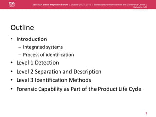
- 3. 2015 PDA Visual Inspection Forum | October 26-27, 2015 | Bethesda North Marriott Hotel and Conference Center | Bethesda, MD Outline • Introduction – Integrated systems – Process of identification • Level 1 Detection • Level 2 Separation and Description • Level 3 Identification Methods • Forensic Capability as Part of the Product Life Cycle 3
- 4. 2015 PDA Visual Inspection Forum | October 26-27, 2015 | Bethesda North Marriott Hotel and Conference Center | Bethesda, MD Introduction • Analytical Resources Supporting the Visual Inspection System are Essential • Forensic Microscopy Anchors the Process • Resources are Ideally Internal – Engineering Support of Production – Visual Inspection Training – Laboratory Investigations • When External, must be directed – Documentation of Defect – Direction of Analyses 4
- 5. 2015 PDA Visual Inspection Forum | October 26-27, 2015 | Bethesda North Marriott Hotel and Conference Center | Bethesda, MD 5 Visual Inspection Program Inspector Selection Knapp Studies Procedural Selection and Refinement Testing Inspectors With Standards Inspector Familiarization with Typical Defects Inspector Training Defect Investigation Product Inspection & Release Qualified Inspector Microscopy Lab
- 6. 2015 PDA Visual Inspection Forum | October 26-27, 2015 | Bethesda North Marriott Hotel and Conference Center | Bethesda, MD Process • Level 1: Locate, Verify • Level 2: Isolate and Characterize – Triage Process • Level 3: Identify • Life Cycle promotes remediation 6
- 7. 2015 PDA Visual Inspection Forum | October 26-27, 2015 | Bethesda North Marriott Hotel and Conference Center | Bethesda, MD Level 1 Detection • Duplicate original conditions – Verify – Observe in Situ • Initiate critical evaluation – Illumination – Dwell – Agitation 7
- 8. 2015 PDA Visual Inspection Forum | October 26-27, 2015 | Bethesda North Marriott Hotel and Conference Center | Bethesda, MD Level 1 Detection and Observations • Is photography/videography in situ possible? • By…eye, low magnification, microscopy • What observations are possible? – Size ( given lensing of container) – Color – Shape – Buoyancy – ?? 8
- 9. 2015 PDA Visual Inspection Forum | October 26-27, 2015 | Bethesda North Marriott Hotel and Conference Center | Bethesda, MD Level 2 - Separation, Description and Triage • The PM must be removed • All PM must be categorized • Categories must be explored 9
- 10. 2015 PDA Visual Inspection Forum | October 26-27, 2015 | Bethesda North Marriott Hotel and Conference Center | Bethesda, MD Level 2 Separation and Description • Removal by Capillary 10
- 11. 2015 PDA Visual Inspection Forum | October 26-27, 2015 | Bethesda North Marriott Hotel and Conference Center | Bethesda, MD 11 Isolation and Manipulation Methods • Direct removal, dry – Tungsten wire, 1-5µm tip – Cat’s whisker – Fine scalpel, cleaver (MicroTool™) – Facilitate with water or adhesive • Direct removal, wet – Capillary tube (Wiretrol) – Poly tube, drawn to fine tip – Swipe of a Membrane wedge • Filtration – Membrane selection/prep • Centrifugation • Transfers, Concentration – Dried KBr – Cleaned filter paper – Capillary tube Teetsov 1977
- 12. 2015 PDA Visual Inspection Forum | October 26-27, 2015 | Bethesda North Marriott Hotel and Conference Center | Bethesda, MD Level 2 Separation and Description • Removal by Filtration – 788 style method – Selection of media • Modified cellulose esters • PVDF • Silver • Polycarbonate/Polyester • Gold-coated media • Physical Removal – Mechanical separation with MicroTool or wires – Dry Powder, Lyo cake, Packaging – Panning: allowing foreign material to separate by gravity in a clean Petri dish - Suspensions 12
- 13. 2015 PDA Visual Inspection Forum | October 26-27, 2015 | Bethesda North Marriott Hotel and Conference Center | Bethesda, MD Level 2 – Membrane Capture <788> 2 13 Thin Flake (Poor Color Dispersal) Long Fiber + Flake (Mottled Background) 100µm in 10µm divisions
- 14. 2015 PDA Visual Inspection Forum | October 26-27, 2015 | Bethesda North Marriott Hotel and Conference Center | Bethesda, MD Triage • PM located by eye, verified at highest magnification • PM moved to work area, examined dry, examined wet (water mount) – Aha?? – Next steps directed by observations • Fundamental Testing 14
- 15. 2015 PDA Visual Inspection Forum | October 26-27, 2015 | Bethesda North Marriott Hotel and Conference Center | Bethesda, MD Microchemical • Chemical tests are ideal when attempting a general categorization of the unknown. General reference texts containing a wide variety of elemental spot tests, functional group, chemical categorization and complex material identification may be found in the following references: – Feigl: organic and inorganic spot tests – Chamot & Mason: fundamental methods and elemental tests – Stahl: practical spot tests for a variety of common materials • Further, there are selections of tests for more common types of material that may end up in the sample, especially raw materials, commodities (starches, grains, brans, etc.), oils, fibers, excreta, etc. 15
- 16. 2015 PDA Visual Inspection Forum | October 26-27, 2015 | Bethesda North Marriott Hotel and Conference Center | Bethesda, MD Wet Elemental Analysis • Microchemical testing for suspect components – Elements – Functional groups – Complex materials (Stahl, 1973) • Cellulose • Lipids/fats • Starch • Proteins 16
- 17. 2015 PDA Visual Inspection Forum | October 26-27, 2015 | Bethesda North Marriott Hotel and Conference Center | Bethesda, MD Level 2 Process • A stepwise process – Observations and tests • By Eye • By Magnifier • By Stereomicroscope • By Compound Microscope, ideally binocular PLM – Microscopy Provides Context, Association, Direct ID 17 Generally, we use the following sequence as a progression of analysis: visual → hand lens → stereomicroscopy → polarized light microscopy → SEM-EDX → spectroscopy
- 18. 2015 PDA Visual Inspection Forum | October 26-27, 2015 | Bethesda North Marriott Hotel and Conference Center | Bethesda, MD Level 2 - Diagnosis 18 • Morphology • Refractive Index • Birefringence (Michel-Levy) • Extinction • Elongation • Pleochroism Bloss 1961
- 19. 2015 PDA Visual Inspection Forum | October 26-27, 2015 | Bethesda North Marriott Hotel and Conference Center | Bethesda, MD Microscopy Unit Operations • Applied polarized light microscopy 19 McCrone 6-digit Binary code Opaque Colored Birefringent High Index (> n1.66) Flat Elongated-Needles or Rods 1 1 1 1 1 1 Value: 32 16 8 4 2 1: 0 to 63 score possible 0 0 0 0 0 0 Flat or Equant Not Flat Low Index (< n1.66) Isotropic Colorless Transparent McCrone WC, Delly JG. (1973). The Particle Atlas, Ann Arbor Science Publishers, Ann Arbor MI.
- 20. 2015 PDA Visual Inspection Forum | October 26-27, 2015 | Bethesda North Marriott Hotel and Conference Center | Bethesda, MD Particle Identity May Be Obvious 20
- 21. 2015 PDA Visual Inspection Forum | October 26-27, 2015 | Bethesda North Marriott Hotel and Conference Center | Bethesda, MD …Or Not! Mixtures and Masses 21
- 22. 2015 PDA Visual Inspection Forum | October 26-27, 2015 | Bethesda North Marriott Hotel and Conference Center | Bethesda, MD Level 3 Identification • Solid State Elemental – SEM-EDX (WDX) and X-ray fluorescence – LIBS • Micro-spectroscopy – Infrared – dispersive-Raman • X-ray diffraction • Mass Analyses 22
- 23. 2015 PDA Visual Inspection Forum | October 26-27, 2015 | Bethesda North Marriott Hotel and Conference Center | Bethesda, MD Solid State Elemental Analysis - SEM-EDX • Scanning Electron Microscopy – Energy-Dispersive X-ray Spectrometry – Robust and Non-destructive – Periodic Table coverage > Z5 (boron) – Lightest elements most difficult – Infinite thickness above 3-5 micrometers – Not Quantitative – See USP <1811> 23
- 24. 2015 PDA Visual Inspection Forum | October 26-27, 2015 | Bethesda North Marriott Hotel and Conference Center | Bethesda, MD Solid State Elemental Analysis - LIBS • Laser-Induced Breakdown Spectroscopy – Rapid analysis of the elemental constitution of the unknown. – Laser excitation produces atomic emission spectra from the (ultimately) destroyed particle. – LIBS can analyze all elements, limited by the laser power and detector efficiency (as for almost all spectroscopical techniques) – Provides relative, not highly quantitative, elemental abundance. 24
- 25. 2015 PDA Visual Inspection Forum | October 26-27, 2015 | Bethesda North Marriott Hotel and Conference Center | Bethesda, MD Microspectroscopy – Infrared • Functional groups strong in IR: C-F, Si-O, O-H, N-H, C-H; C≡N, C=O, C-Cl, NO2 – dispersive-Raman • Functional groups strong in Raman: C≡C, C=C, N=N, S- H, C=S, C-S, S-S, CH2, C≡N, C=O, C-Cl, NO2 25
- 26. 2015 PDA Visual Inspection Forum | October 26-27, 2015 | Bethesda North Marriott Hotel and Conference Center | Bethesda, MD Spectroscopy Unit Operations • Spectrometer is an Extension of the Microscope • Location of Zones/Separation by Solvents • Spatial Resolution according to Technique • Specimen Prepared by Microscopical Operations • Zone of Analysis Defined by Prior Characterization 26
- 27. 2015 PDA Visual Inspection Forum | October 26-27, 2015 | Bethesda North Marriott Hotel and Conference Center | Bethesda, MD USP 1787 - Information Chapter for Particle Analysis • Methods – Light Microscopy • Staining methods – Light Obscuration – Dynamic Image Analysis (Flow Imaging) – Electrozone-Coulter – Laser Diffraction – Infrared and Raman Microspectroscopy – Electron Microscopy – EDX, EELS – ToF-SIMS • Silicones discussed as particulate matter 27
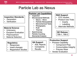
- 28. 2015 PDA Visual Inspection Forum | October 26-27, 2015 | Bethesda North Marriott Hotel and Conference Center | Bethesda, MD 28 Particle Lab as Nexus Particle Lab Capabilities • Inspection • Microscopical Methods • Macro – Micro • Particle Manipulation • Microchemical Tests • Photography • PLM • Thermal • Spectroscopy • Particle Counting • Elemental Analysis QC Release 788-1, 788-2 Production Support • Process Capability • Component Prep • Consumables Integrity • Fixtures Wear • Vendor Evaluation Regulatory • Responses • Insert changes • Registration Studies Inspection Standards • Generation • Verification QA Support • AQL Rejects • Complaints • Recalls R&D Support • CCC studies • Product Use Trials • Inserts • Labelling • Alternate Methods Material Science • Unknowns • Excipient Evaluation • Polymorphism • Material Selection
- 29. 2015 PDA Visual Inspection Forum | October 26-27, 2015 | Bethesda North Marriott Hotel and Conference Center | Bethesda, MD Thank You Questions? 29
- 30. 2015 PDA Visual Inspection Forum | October 26-27, 2015 | Bethesda North Marriott Hotel and Conference Center | Bethesda, MD References • Aldrich, D.S. and Smith, M.A. (1995). Chapter 9 - Pharmaceutical Applications of Infrared Microspectroscopy, in Practical Guide to Infrared Microspectroscopy, Howard Humecki, Editor, Marcel Dekker 1995; New York, NY, 323-375. • Aldrich, D.S. (2010). Chapter 5 - Particulate Matter: Subvisible, in Pharmaceutical Dosage Forms: Parenteral Medications, Nema S and Ludwig JD, eds. Third ed. Volume 2, Informa Healthcare, New York, pps. 114-145. • Barber, T.A. (1993). Pharmaceutical Particulate Matter - Analysis and Control, InterPharm Press, Buffalo Grove, IL. • Bloss, F.D. (1961). An Introduction to the Methods of Optical Crystallography, New York: Holt, Rinehart and Winston. • Chamot, E.M. and Mason, C.W. (1958). Handbook of Chemical Microscopy, Volume I, J. Wiley and Sons, Inc., New York, NY. • Chamot, E.M. and Mason, C.W. (1960). Handbook of Chemical Microscopy, Volume II, J. Wiley and Sons, New York, NY. • Feigl, F. and Anger, V. (1966). Spot Tests in Organic Analysis, Elsevier, New York, NY. • Feigl, F. and Anger, V. (1972). Spot Tests in Inorganic Analysis, Elsevier, New York, NY • McCrone, W.C. and Delly, J.G. (1973). The Particle Atlas, Volumes I-IV, Ann Arbor: Ann Arbor Science Publishers. • McCrone, W.C. (1982). Particle Characterization by PLM, Part I - No Polars. Microscope 30, 3, 185-196. • McCrone, W.C. (1982). Particle Characterization by PLM, Part II - Single Polars. Microscope 30, 4, 315-331. • McCrone, W.C. (1983). Particle Characterization by PLM, Part III - Crossed Polars. Microscope 31, 2, 187-206. • Stahl, E.; Ed. (1973). Drug Analysis by Chromatography and Microscopy, A Practical Supplement to Pharmacopoeias, Ann Arbor Science Publishers, Ann Arbor, MI. • Teetsov, A. S. (1977). Techniques of Small Particle Manipulation, Microscope, 25: 103. 30