際際滷

際際滷Share a Scribd company logo
IMAGING IN ABDOMINAL
TUBERCULOSIS
By Dr. Gayathri
Moderator: Dr.Anjani
 Tuberculosis can affect any organ system, particularly in
immunocompromised individuals.
 It can be divided into
1) Pulmonary
2) Extrapulmonary.
 Lungs are the primary involved organs and abdominal involvement
occurs in about 11-12% of patients with extra-pulmonary
tuberculosis.
 The abdominal presentation may involve different structures such as
gastrointestinal tract, genitourinary tract, solid organs (liver, spleen,
pancreas), gallbladder, aorta and its branches, peritoneum and lymph
nodes, frequently with concomitant involvement of those organs
ETIOLOGY
 Primary infection from M. tuberculosis
 Abdominal TB is usually secondary to pulmonary TB
 CXR normal in 2/3 of patients with abdominal TB.Only 15% have active
pulmonary disease .
 Other sources of abdominal infection with TB
1)Swallowing infected material.
2)Hematogenous spread from active or latent infection
3)Direct extension from infected tissues
CLINICAL FEATURES:
Often presents with fever, weight loss, abdominal pain , anorexia,
distension, lump, constipation and diarrhoea.
Complications include obstruction, perforation, fistula formation and
hemorrhage.
May or may not have evidence of pulmonary TB
Negative chest radiograph or negative tuberculin skin test does not
exclude extrapulmonary TB
CLASSIFICATION OF ABDOMINAL
TUBERCULOSIS
a) Peritoneal tuberculosis
b) Lymph node tuberculosis
c) Gastrointestinal tuberculosis
d) Tb of solid viscera
IMAGING FEATURES
 Often no evidence of lung disease (CXR or CT can be normal)
LYMPHADENOPATHY (TUBERCULOUS LYMPHADENITIS)
-Can range from increased number of normal-sized nodes to massively enlarged
conglomerate nodal masses,Mesenteric and peripancreatic lymph nodes most
commonly involved
-Multiple groups often affected simultaneously
-Enlarged, necrotic nodes with hypoattenuating centers and hyperattenuating
enhancing rims on CT (40-60%)Characteristic of caseous necrosis
-Mixed-attenuation nodes are also possible
-Nodes calcify with healing: TB probably most common cause of mesenteric nodal
calcification
Abdominl Tuberculosis imaging features.pptx
TUBERCULOSIS (TB) PERITONITIS :
 3 imaging patterns: Wet, dry, and fibrotic fixed
 Wet type: Large amount of free or loculated ascites, which is usually complex
with septations and fibrinous strands (and higher than water density due to
protein/cellular content)
 Dry type: Mesenteric and omental thickening, fibrous adhesions, and caseous
nodules
 Fibrotic fixed: Discrete masses in omentum with matted loops of bowel and
frequent loculated ascites Distinguishing TB peritonitis from carcinomatosis can
be difficult, but carcinomatosis more likely to demonstrate discrete implants or
omental caking (while peritoneum more often smoothly thickened in TB)
Abdominl Tuberculosis imaging features.pptx
GI TB
 Ileocecal region affected in 90% of casesCommon site due to
presence of significant lymph tissue and stasis of bowel contents in
that location (with close contact of bacilli with bowel mucosa)
 Cecum and terminal ileum are usually contracted (cone-shaped
cecum) with asymmetric wall thickening of ileocecal valve and medial
cecum and gaping ileocecal valve
 Regional inflammation is common, but prominent vasa recta
engorgement and increased vascularity associated with Crohn disease
(also on differential diagnosis) not usually present
 Over time, cecum becomes gradually smaller and irregular in shape as
result of scarring
 Regional lymphadenopathy with central caseation is frequent clue to
correct diagnosis  Involvement of stomach and proximal small bowel
is rare, but any portion of GI tract can be involved
 Stomach: Often affects antrum and distal body, simulating peptic ulcer
disease
The pathological forms of GI TB includes:
 Hypertrophic form (10 %)
 Ulcero  proliferative form (30 %)
 Ulcerative form (60 %)
SIGNS ON BARIUM STUDY
 Fleischner sign
 Stierlin sign
 String sign
 Pulled up ceacum
Abdominl Tuberculosis imaging features.pptx
PANCREATIC TB
 TB can very rarely manifest as mass, mimicking cancer (caseated
peripancreatic nodes involving pancreas)
 Hypoenhancing mass (usually pancreatic head) typically without
pancreatic duct dilatation or vascular invasion
Abdominl Tuberculosis imaging features.pptx
RENAL TB
 Usually unilateral (75%)
 Earliest finding is papillary necrosis (usually involving upper pole),
particularly evident on delayed excretory phase CT images or
intravenous urography (uncommonly utilized in modern setting)
 Over time, fibrosis and stricturing of collecting system can lead to
asymmetric caliectasis
 Involvement of parenchyma can manifest as focal, wedge-shaped
areas of low attenuation, multiple small, hypodense nodules, or
discrete renal abscess  In chronic stage, kidney appears atrophic and
markedly irregular with cortical thinning, volume loss, and
calcification (autonephrectomy or "putty" kidney)
 Kidney usually nonfunctional at this stage without contrast excretion
Abdominl Tuberculosis imaging features.pptx
URETERAL TB
 Usually secondary to renal TB
 Filling defects may be seen in ureter on delayed excretory phase
images due to sloughed papillae or debris secondary to renal
infection/papillary necrosis
 Thickened ureteral wall with strictures most common in distal 1/3 of
ureter
 Chronically can appear as corkscrew or beaded ureter due to multiple
fibrotic strictures
Abdominl Tuberculosis imaging features.pptx
URINARY BLADDER TB
 Tuberculous cystitis manifests as decreased bladder volume with wall
thickening, ulceration, filling defects and calcifications in bladder wall
 Severe cases with profuse scarring result in small, irregular, and
calcified bladder.
Abdominl Tuberculosis imaging features.pptx
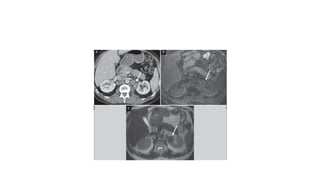
ADRENAL TB
Acute: Enlarged adrenals (often appears as discrete, centrally necrotic
adrenal mass)
Chronic: Small adrenals with dots of calcification and low signal on all
MR sequences
Abdominl Tuberculosis imaging features.pptx
FEMALE GENITAL TB
 Most commonly involves fallopian tubes (in 94% of cases) and
endometrium and can be cause of infertility
 Most often appears as bilateral hydrosalpinx (or pyosalpinx) with
fallopian tubes appearing irregular and nodular (with sites of stenosis
and scarring)
 Endometrium can appear thickened and heterogeneous with internal
calcification and distorted shape as result of adhesions and scarring
Abdominl Tuberculosis imaging features.pptx
MALE GENITAL TB
 Most often involves seminal vesicles or prostate gland with testicular
involvement much less common
 Can resemble pyogenic abscess 賊 calcification
 TB prostatitis on prostate MR resembles other causes of prostatitis
with band-like or geographic T2 hypointensity and mild restricted
diffusion and mild early
Abdominl Tuberculosis imaging features.pptx
HEPATIC AND SPLENIC TB
 Micronodular patternInnumerable tiny nodules (< 2 mm), which are most
often hypodense on CT and hyperechoic on USMay simply appear as
hepatomegaly/splenomegaly on CT (with individual lesions not visible)
 Macronodular patternFewer large nodules, which frequently demonstrate
central necrosisHypodense on CT with ill-defined margins and gradual
development of calcifications in more chronic stages
 TB and histoplasmosis are most common causes of calcified granulomasT1
hypointense and T2 hyperintense on MR with rim enhancement
Abdominl Tuberculosis imaging features.pptx
THANK YOU

More Related Content

Abdominl Tuberculosis imaging features.pptx

  • 1. IMAGING IN ABDOMINAL TUBERCULOSIS By Dr. Gayathri Moderator: Dr.Anjani
  • 2. Tuberculosis can affect any organ system, particularly in immunocompromised individuals. It can be divided into 1) Pulmonary 2) Extrapulmonary. Lungs are the primary involved organs and abdominal involvement occurs in about 11-12% of patients with extra-pulmonary tuberculosis.
  • 3. The abdominal presentation may involve different structures such as gastrointestinal tract, genitourinary tract, solid organs (liver, spleen, pancreas), gallbladder, aorta and its branches, peritoneum and lymph nodes, frequently with concomitant involvement of those organs
  • 4. ETIOLOGY Primary infection from M. tuberculosis Abdominal TB is usually secondary to pulmonary TB CXR normal in 2/3 of patients with abdominal TB.Only 15% have active pulmonary disease . Other sources of abdominal infection with TB 1)Swallowing infected material. 2)Hematogenous spread from active or latent infection 3)Direct extension from infected tissues
  • 5. CLINICAL FEATURES: Often presents with fever, weight loss, abdominal pain , anorexia, distension, lump, constipation and diarrhoea. Complications include obstruction, perforation, fistula formation and hemorrhage. May or may not have evidence of pulmonary TB Negative chest radiograph or negative tuberculin skin test does not exclude extrapulmonary TB
  • 6. CLASSIFICATION OF ABDOMINAL TUBERCULOSIS a) Peritoneal tuberculosis b) Lymph node tuberculosis c) Gastrointestinal tuberculosis d) Tb of solid viscera
  • 7. IMAGING FEATURES Often no evidence of lung disease (CXR or CT can be normal) LYMPHADENOPATHY (TUBERCULOUS LYMPHADENITIS) -Can range from increased number of normal-sized nodes to massively enlarged conglomerate nodal masses,Mesenteric and peripancreatic lymph nodes most commonly involved -Multiple groups often affected simultaneously -Enlarged, necrotic nodes with hypoattenuating centers and hyperattenuating enhancing rims on CT (40-60%)Characteristic of caseous necrosis -Mixed-attenuation nodes are also possible -Nodes calcify with healing: TB probably most common cause of mesenteric nodal calcification
  • 11. TUBERCULOSIS (TB) PERITONITIS : 3 imaging patterns: Wet, dry, and fibrotic fixed Wet type: Large amount of free or loculated ascites, which is usually complex with septations and fibrinous strands (and higher than water density due to protein/cellular content) Dry type: Mesenteric and omental thickening, fibrous adhesions, and caseous nodules Fibrotic fixed: Discrete masses in omentum with matted loops of bowel and frequent loculated ascites Distinguishing TB peritonitis from carcinomatosis can be difficult, but carcinomatosis more likely to demonstrate discrete implants or omental caking (while peritoneum more often smoothly thickened in TB)
  • 15. GI TB Ileocecal region affected in 90% of casesCommon site due to presence of significant lymph tissue and stasis of bowel contents in that location (with close contact of bacilli with bowel mucosa) Cecum and terminal ileum are usually contracted (cone-shaped cecum) with asymmetric wall thickening of ileocecal valve and medial cecum and gaping ileocecal valve
  • 16. Regional inflammation is common, but prominent vasa recta engorgement and increased vascularity associated with Crohn disease (also on differential diagnosis) not usually present Over time, cecum becomes gradually smaller and irregular in shape as result of scarring Regional lymphadenopathy with central caseation is frequent clue to correct diagnosis Involvement of stomach and proximal small bowel is rare, but any portion of GI tract can be involved Stomach: Often affects antrum and distal body, simulating peptic ulcer disease
  • 17. The pathological forms of GI TB includes: Hypertrophic form (10 %) Ulcero proliferative form (30 %) Ulcerative form (60 %)
  • 18. SIGNS ON BARIUM STUDY Fleischner sign Stierlin sign String sign Pulled up ceacum
  • 21. PANCREATIC TB TB can very rarely manifest as mass, mimicking cancer (caseated peripancreatic nodes involving pancreas) Hypoenhancing mass (usually pancreatic head) typically without pancreatic duct dilatation or vascular invasion
  • 23. RENAL TB Usually unilateral (75%) Earliest finding is papillary necrosis (usually involving upper pole), particularly evident on delayed excretory phase CT images or intravenous urography (uncommonly utilized in modern setting) Over time, fibrosis and stricturing of collecting system can lead to asymmetric caliectasis
  • 24. Involvement of parenchyma can manifest as focal, wedge-shaped areas of low attenuation, multiple small, hypodense nodules, or discrete renal abscess In chronic stage, kidney appears atrophic and markedly irregular with cortical thinning, volume loss, and calcification (autonephrectomy or "putty" kidney) Kidney usually nonfunctional at this stage without contrast excretion
  • 29. URETERAL TB Usually secondary to renal TB Filling defects may be seen in ureter on delayed excretory phase images due to sloughed papillae or debris secondary to renal infection/papillary necrosis Thickened ureteral wall with strictures most common in distal 1/3 of ureter Chronically can appear as corkscrew or beaded ureter due to multiple fibrotic strictures
  • 31. URINARY BLADDER TB Tuberculous cystitis manifests as decreased bladder volume with wall thickening, ulceration, filling defects and calcifications in bladder wall Severe cases with profuse scarring result in small, irregular, and calcified bladder.
  • 33. ADRENAL TB Acute: Enlarged adrenals (often appears as discrete, centrally necrotic adrenal mass) Chronic: Small adrenals with dots of calcification and low signal on all MR sequences
  • 35. FEMALE GENITAL TB Most commonly involves fallopian tubes (in 94% of cases) and endometrium and can be cause of infertility Most often appears as bilateral hydrosalpinx (or pyosalpinx) with fallopian tubes appearing irregular and nodular (with sites of stenosis and scarring) Endometrium can appear thickened and heterogeneous with internal calcification and distorted shape as result of adhesions and scarring
  • 37. MALE GENITAL TB Most often involves seminal vesicles or prostate gland with testicular involvement much less common Can resemble pyogenic abscess 賊 calcification TB prostatitis on prostate MR resembles other causes of prostatitis with band-like or geographic T2 hypointensity and mild restricted diffusion and mild early
  • 39. HEPATIC AND SPLENIC TB Micronodular patternInnumerable tiny nodules (< 2 mm), which are most often hypodense on CT and hyperechoic on USMay simply appear as hepatomegaly/splenomegaly on CT (with individual lesions not visible) Macronodular patternFewer large nodules, which frequently demonstrate central necrosisHypodense on CT with ill-defined margins and gradual development of calcifications in more chronic stages TB and histoplasmosis are most common causes of calcified granulomasT1 hypointense and T2 hyperintense on MR with rim enhancement