Abscess aspirate specimen analysis final
- 1. SPECIMEN ANALYSIS: ABSCESS ASPIRATE Anneka Pierzga and Yackima Saura-Welch June 16th, 2016 MLT 2010 Clinical Microbiology (Professor Tiffany Gill) ŌĆō College of Southern Maryland
- 2. THE SPECIEMN ’é© What is an abscess? ’éż Accumulation of purulent material in the dermis or subcutaneous tissue ’éż Appears as a swollen, red, tender and fluctuant mass ’ü« Often diagnosed based on history and physical exam, although studies suggest that soft-tissue ultrasonography may enhance accuracy of abscess detection, especially in cases of deeper or questionably appearing abscesses ’ü« Culture of needle-aspirated material useful for isolation of causative pathogens
- 3. SPECIMEN COLLECTION 1. Cleanse site with sterile saline or 70% alcohol 2. Aspirate area containing purulent material or fluid by needle and syringe (may need to irrigate with a small volume of non-bacteriostatic sterile saline) 3. Expel aspirated material in to sterile screw top tube 4. For anaerobic culture: Samples should be placed into oxygen free environment using anaerobic transport media
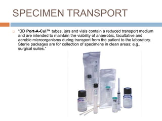
- 7. SPECIMEN TRANSPORT ’é© ŌĆ£BD Port-A-CulŌäó tubes, jars and vials contain a reduced transport medium and are intended to maintain the viability of anaerobic, facultative and aerobic microorganisms during transport from the patient to the laboratory. Sterile packages are for collection of specimens in clean areas; e.g., surgical suites.ŌĆØ
- 9. PRIMARY SET-UP ’é© Direct examination of a Gram Stained slide ’éż Determine the staining and morphological characteristics of pathogens to direct physicianŌĆÖs initial treatment plan ’é© Inoculation of media ’éż Blood Agar Plate ’éż Chocolate Agar Plate ’éż MacConkey Agar Plate ’éż CNA Anaerobic (Columbia Agar with Colistin and Nalidixic Acid) ’éż BBA (Brucella Blood Agar) ’éż LKV (Laked Blood Agar with Kanaycin and Vancomycin) ’éż BBE (Bacteroides Bile Esculin Agar)
- 11. MAJOR OFFENDERS ’é© The leading cause of subcutaneous abscesses in otherwise healthy individuals isŌĆ” Staphylococcus aureus ’é© Methicillin-Resistant Staphylococcus aureus has been found to be the most common cause of abscesses in patients presenting to the emergency department in the US, followed by methicillin-susceptible S. aureus and beta- hemolytic streptococci
- 12. About Staphyloccos aureus: ŌĆó Virulence factors ŌĆó Colony morphology ŌĆó Hemolysis ŌĆó Environmental conditions ŌĆó In vitro contamination ŌĆó Common normal flora ŌĆó Differential media ŌĆó Confirmatory testing ŌĆó Antibiotic susceptibility testing
- 13. Staphyloccos aureus ’é© Gram-positive cocci in grape-like clusters ’é© Found as part of the normal flora of the anterior nares, nasopharynx, perineal area, skin, and mucosa; may be introduced to sterile sites by traumatic introduction ’é© May also be spread from person to person by direct contact
- 14. VIRULENCE FACTORS Polysaccharide capsule inhibits phagocytosis and helps with colonization Catalase helps resist digestion by leukocytes Penicillinase provides resistance to penicillin-related antibiotics Coagulase enables the bacteria to hide within a clot, thereby escaping the
- 15. S. aureus COLONIES S. Aureus colonies typically appear to cream in color but occasionally have a yellow pigment. The golden pigmentation (staphyloxanthin) has been reported to be a virulence factor protecting the pathogen against oxidants produced by the immune system To compare in size and color S. epidermidis appears white. Medium to large (0.5-1.5 ╬╝m); smooth, entire, slightly raised, low convex, opaque; most colonies pigmented creamy yellow; most colonies beta- hemolytic
- 16. HEMOLYSIS Beta-hemolysis (╬▓-hemolysis), sometimes called complete hemolysis, is a complete lysis of red cells in the media around and under the colonies: the area appears lightened (yellow) and transparent. .
- 17. ENVIRONMENTAL CONDITIONS & INCUBATION PERIOD ’é© Grows best aerobically but are facultative anaerobic. ’é© 24 hours of incubation at 37┬░C
- 18. IN-VITRO CONTAMINATION Meaning: When something foreign and non-sterile has made contact with the plate during inoculation. Can occur by ’ü▒ Not cleaning collection site appropriately ’ü▒ Not streaking plate under the hood ’ü▒ Opening the lid and breathing/allowing any micro-contaminant to enter. ’ü▒ Not using sterile inoculating loopThe arrows indicate fungal contamination of the specimen
- 19. NORMAL FLORA Since many abscesses are located beneath the skin, it is not uncommon to have normal skin flora in the sample. Coagulase negative Staphylococcu s species and Enterococcus species are considered normal skin flora
- 20. DIFFERENTIAL/SELECTIVE MEDIA ’é© Phenylethyl alcohol agar (PEA) ’é© Mannitol Salt Agar (MSA) has a 7.5% concentration of salt and S. aureus can ferment mannitol ’é© Columbia colistin-nalidixic acid (CNA) agar ’é© Chromogenic media ’é© DNase or thermostable-endonuclease test.
- 21. CONFIRMATORY TEST ŌĆó Gram - positive cocci in grapelike clusters ŌĆó Catalase - positive ŌĆó Coagulase - positive
- 22. SUSCEPTIBILITY TESTING IF SUSCEPTIBLE: ’é© ampicillin/sulbactam ’é© amoxicillin/clavulanate ’é© oxacillin ’é© nafcillin ’é© cefazolin ’é© ceftriaxone ’é© Macrolides ’é© Clindamycin ALTERNATIVES: ’é© Trimethoprim-Sulfomethoxazole(TMP- SMX) ’é© vancomycin MRSA: ’é© vancomycin ’é© teicoplanin ’é© linezolid ’é© quinupristin/dalfopristin ’é© TMP-SMX Resistance to 0.04 U of bacitracin (Taxo A disk) and furazolidone
- 23. REFERENCES Bacteria in Photos. (2016). Beta hemolysis of Staphylococcus aureus. Growth characteristics of staph aureus on blood agar. Retrieved from http://www.bacteriainphotos.com/beta_hemolysis_on_agar.html BD (Becton, D. a. C. (2016). Diagnostic Systems: Port-A-CulŌäó Tube (for Aerobic and Anaerobic Cultures). Retrieved from http://www.bd.com/ds/productCenter/221606.asp Cardiovascular and Interventional Radiological Society of Europe. (2016). Aspiration. Microbiology in Pictures. (2016). Staphylococcus aureus colony morphology and microscopic appearance, basic characterisic and tests or identiication of S. aureus bacteria. Retrieved from http://www.microbiologyinpictures.com/staphylococcus%20aureus.html Sankaqm5 (Producer). (2012, 6/11/2016). Large Abscess Cavity Pus Aspiration With Anti Gravity Technique Retrieved from https://www.youtube.com/watch?v=xP1-ccR4O2w Singer, A. J., & Talan, D. A. (2014). Management of Skin Abscesses in the Era of Methicillin-Resistant Staphylococcus aureus. The New England Journal of Medicine, 370(11), 1039-1047. Talan, D. A., Krishnadasan, A., Gorwitz, R. J., Fosheim, G. E., Limbago, B., Albrecht, V., & Moran, G. J. (2011). Comparison of Staphylococcus aureus From Skin and Soft-Tissue Infections in US Emergency Department Patients, 2004 and 2008. Clinical Infectious Disease, 53(2). The University of Texas Medical Branch. (2016). Specimen Collection: Acceptable Specimens for Bacteriologic Analysis of Wounds. Retrieved from https://www.utmb.edu/lsg/Pages/SpecimenCollection/SpecColWounds.aspx Tille, P. M. (2014). Bailey and Scott's Diagnostic Microbiology (13 ed.): Elsevier.
Editor's Notes
- Singer, A. J., & Talan, D. A. (2014). Management of Skin Abscesses in the Era of Methicillin-Resistant Staphylococcus aureus. The New England Journal of Medicine, 370(11), 1039-1047
- Cardiovascular and Interventional Radiological Society of Europe. (2016). Aspiration. The University of Texas Medical Branch. (2016). Specimen Collection: Acceptable Specimens for Bacteriologic Analysis of Wounds. Retrieved from https://www.utmb.edu/lsg/Pages/SpecimenCollection/SpecColWounds.aspx
- Sankaqm5 (Producer). (2012, 6/11/2016). Large Abscess Cavity Pus Aspiration With Anti Gravity Technique Retrieved from https://www.youtube.com/watch?v=xP1-ccR4O2w
- BD (Becton, D. a. C. (2016). Diagnostic Systems: Port-A-CulŌäó Tube (for Aerobic and Anaerobic Cultures). Retrieved from http://www.bd.com/ds/productCenter/221606.asp
- Tille, P. M. (2014). Bailey and Scott's Diagnostic Microbiology (13 ed.): Elsevier.
- Singer, A. J., & Talan, D. A. (2014). Management of Skin Abscesses in the Era of Methicillin-Resistant Staphylococcus aureus. The New England Journal of Medicine, 370(11), 1039-1047. Talan, D. A., Krishnadasan, A., Gorwitz, R. J., Fosheim, G. E., Limbago, B., Albrecht, V., & Moran, G. J. (2011). Comparison of Staphylococcus aureus From Skin and Soft-Tissue Infections in US Emergency Department Patients, 2004 and 2008. Clinical Infectious Disease, 53(2). Tille, P. M. (2014). Bailey and Scott's Diagnostic Microbiology (13 ed.): Elsevier.
- Tille, P. M. (2014). Bailey and Scott's Diagnostic Microbiology (13 ed.): Elsevier.
- Tille, P. M. (2014). Bailey and Scott's Diagnostic Microbiology (13 ed.): Elsevier.
- Tille, P. M. (2014). Bailey and Scott's Diagnostic Microbiology (13 ed.): Elsevier.
- Bacteria in Photos. (2016). Beta hemolysis of Staphylococcus aureus. Growth characteristics of staph aureus on blood agar. Retrieved from http://www.bacteriainphotos.com/beta_hemolysis_on_agar.html
- Microbiology in Pictures. (2016). Staphylococcus aureus colony morphology and microscopic appearance, basic characterisic and tests or identiication of S. aureus bacteria. Retrieved from http://www.microbiologyinpictures.com/staphylococcus%20aureus.html
- Typical appearance of a blood agar plate resulting from contamination. (2016). Researchgate.
- S. epidermidis and S. aureus appearance and colony morphology on agar media. (2015). Microbiology in Pictures. Staphylococcus epidermidis and Staphylococcus aureus, colony morphology and hemolysis. (2016). Bacteria in Photos. Tille, P. M. (2014). Bailey and Scott's Diagnostic Microbiology (13 ed.): Elsevier.
- Tille, P. M. (2014). Bailey and Scott's Diagnostic Microbiology (13 ed.): Elsevier.
- The catalase test; postive catalase test. The catalase test result with Staphylococcus aureus. Principle of the catalase test. (2016). Bacteria in Photos. Bound coagulase (cell-bound coagulase, clumping factor of Staphylococcus aureus. (2016). Bacteria in Photos. Tille, P. M. (2014). Bailey and Scott's Diagnostic Microbiology (13 ed.): Elsevier.
- Microbiology in Pictures. (2016a). Antibiotic susceptibility test, Staphylococcus aureus and MRSA. Tille, P. M. (2014). Bailey and Scott's Diagnostic Microbiology (13 ed.): Elsevier.


