ACUTE AND CHRONIC OTITIS MEDIA notes.pptx
Download as PPTX, PDF0 likes127 views
║▌║▌▀Żs on otitis media
1 of 9
Download to read offline

Recommended
Congenital ear deformities
Congenital ear deformitiesNikhil Vaishnav
╠²
Congenital ear abnormalities can result from errors in embryonic development or intrauterine events affecting growth. They cause a variety of deformities like anotia (absence of the outer ear), microtia (underdeveloped outer ear), and macrotia (excessively large outer ear). Treatments include prosthetics, reconstructive surgery, and reducing the size of overgrown ears. Common birth defects of the ear include preauricular sinuses (nodules by the ear), Darwin's tubercles (thickened ear tissue), and ear canal stenosis (narrowed ear canal).Otosclerosis
OtosclerosisSanil Varghese
╠²
Otosclerosis is a primary metabolic bone disease that causes fixation of the ossicles, resulting in conductive or mixed hearing loss. It is genetically mediated and more common in women. The incidence increases with age, most commonly occurring between 15-45 years old. Normal bone in the otic capsule is replaced with highly vascular spongy bone that immobilizes the stapes footplate, disrupting vibration conduction and causing hearing loss. Symptoms include slowly progressive, often asymmetric conductive hearing loss and tinnitus. Diagnosis involves audiometric testing demonstrating the conductive nature of the loss. Treatments range from hearing aids to surgical procedures like stapedectomy or stapedotomy that aim to restore vibration conduction.Otosclerosis
OtosclerosisAjay Manickam
╠²
The document discusses otosclerosis, a disease affecting the bones of the middle ear. It causes stapes fixation and conductive hearing loss. It most commonly affects Caucasians between 15-45 years of age and is more prevalent in females. Etiology includes genetic and hormonal factors. Symptoms include conductive deafness and better hearing in noisy environments. Diagnosis involves tests showing conductive hearing loss like negative Rinne's test. Treatment options include stapedectomy surgery to replace or remodel the stapes bone.Meniere disease
Meniere diseasehumra shamim
╠²
This document discusses the history, presentation, diagnosis and management of Meniere's disease. Some key points:
- Meniere's disease was first described in 1861 and is characterized by hearing loss, tinnitus, and vertigo due to endolymphatic hydrops (fluid buildup) in the inner ear.
- Diagnosis is based on recurrent vertigo spells lasting 20 minutes to 24 hours, fluctuating hearing loss, tinnitus and aural fullness. Tests like electrocochleography and VEMPs can provide supportive evidence.
- Treatment includes dietary sodium restriction, diuretics, medications and surgical options like intratympanic injections if conservative measures fail. TheCongenital malformation of external ear and itŌĆÖs management
Congenital malformation of external ear and itŌĆÖs managementYousuf Choudhury
╠²
This document discusses congenital malformations of the external ear. It begins with applied anatomy of the normal ear and embryology of ear development. It then describes various congenital deformities that can occur, including pre-auricular tags, prominent ears, cryptotia, microtia, and congenital aural atresia. For treatment, options include observation, prosthetics, reconstruction with rib cartilage grafts, and surgical correction of atresia. Reconstruction is often done in multiple stages to form the shape of the pinna.Parotid tumors
Parotid tumorsSharath !!!!!!!!
╠²
The parotid gland is located below and in front of the ear. It has two lobes and is drained by Stenson's duct which opens into the mouth. The gland has a capsule and structures like arteries pass through it. It is supplied by parasympathetic and sympathetic nerves. Common tumors include pleomorphic adenoma and Warthin's tumor. Mucoepidermoid carcinoma and adenoid cystic carcinoma are malignant tumors that can occur. Surgical excision is the main treatment for tumors but radiotherapy may also be used for malignant ones. Complications after parotidectomy include facial nerve injury and salivary fistula.MASTOIDECTOMY PPT
MASTOIDECTOMY PPTRitchieShija
╠²
1. Mastoidectomy is a surgical procedure that opens the mastoid cavity to clean infected air cells and improve ventilation. It has evolved from simple trephination for acute infection to modern techniques like intact canal wall mastoidectomy.
2. The temporal bone contains the mastoid, squamous, tympanic and petrous parts. Important surgical anatomy includes the mastoid antrum, facial recess, and relationships to surrounding structures like the sigmoid sinus and dura.
3. Mastoidectomies are classified based on whether the posterior ear canal wall is preserved (intact canal wall) or removed (canal wall down). Common types include cortical, radical, modified radical, atticotomyTumours of Ear
Tumours of EarAnwaaar
╠²
Tumours of the ear can arise in the external ear, middle ear, and inner ear. Benign tumours of the external ear include sebaceous cysts, dermoid cysts, haemangiomas, and papillomas. Malignant tumours include basal cell carcinoma and squamous cell carcinoma. Glomus tumours are the most common benign tumour of the middle ear, arising from glomus bodies. Malignant tumours of the middle ear may be primary carcinomas or sarcomas or may spread secondarily from other sites. Tumours are diagnosed using imaging such as CT or MRI and treated with surgery, radiation, or embolization depending on the type and extent of disease.MASTOIDECTOMY (BY DR.RICHARD & DR.BUKUKU)
MASTOIDECTOMY (BY DR.RICHARD & DR.BUKUKU)RitchieShija
╠²
1. Mastoidectomy is a surgical procedure that opens the mastoid cavity to clean infected air cells and improve ventilation. It has evolved from simple trephination for acute infections to modern techniques that preserve normal anatomy.
2. The document outlines the history, surgical anatomy, types (cortical, radical, modified radical), indications, and techniques of mastoidectomy. Types are classified as open (canal wall down) or closed (canal wall up) approaches.
3. Potential complications are discussed briefly. Controversies remain regarding the best surgical techniques and approaches to different pathologies.Development of ear ppt
Development of ear pptDr Safika Zaman
╠²
The ear develops from three germ layers into three main structures - the inner, middle, and outer ear. The outer ear develops from hillocks in the mandibular and hyoid arches, which fuse to form the pinna. The external auditory canal develops from the first branchial groove. The middle ear cavities develop from outpouchings of the first and second pharyngeal pouches. Ossicles develop from the first and second branchial arches. The inner ear develops from the otic placode, forming the fluid-filled cochlea and vestibular system. The facial and acoustic nerves also develop during this period to innervate the ear structures.Tumours of External Auditory Canal and Middle Ear
Tumours of External Auditory Canal and Middle EarDr Harjitpal Singh
╠²
1. Benign tumours of the external ear include preauricular sinus or cyst, sebaceous cyst, dermoid cyst, keloid, hemangioma, papilloma, exostoses, osteoma, ceruminoma, and sebaceous adenoma.
2. Malignant tumours of the external ear include squamous cell carcinoma, basal cell carcinoma, and malignant melanoma.
3. Benign tumours of the middle ear include glomus tumours, also known as paragangliomas, which arise from the glomus bodies along the tympanic branch of the facial nerve.
4. Malignant tumours of the middle ear are rare but includeSeptplasty
SeptplastyHelao Silas
╠²
This document discusses septoplasty and submucous resection of the nasal septum (SMR) procedures. It defines key terms like septum, deviated septum, and provides an overview of the causes and symptoms of a deviated septum. The document outlines the steps of SMR and septoplasty, including anesthesia, incisions, cartilage/bone removal, and packing. Potential complications are also summarized.Myringotomy
MyringotomyDr Asmatullah Achakzai
╠²
Myringotomy (from Latin myringa "eardrum") is a surgical procedure in which a tiny incision is created in the eardrum to relieve pressure caused by excessive buildup of fluid, or to drain pus from the middle ear. A tympanostomy tube is inserted into the eardrum to keep the middle ear aerated for a prolonged time and to prevent reaccumulation of fluid. Without the insertion of a tube, the incision usually heals spontaneously in two to three weeks. Depending on the type, the tube is either naturally extruded in 6 to 12 months or removed during a minor procedure.Malignant Otitis Externa 
Malignant Otitis Externa Mamoon Ameen
╠²
Malignant otitis externa is an aggressive infection of the external ear and skull base that commonly affects older diabetics. It is caused mainly by Pseudomonas aeruginosa bacteria invading the external ear canal and spreading through fissures in the bone. Symptoms include severe ear pain, discharge, and potentially cranial nerve palsies. Diagnosis involves culture, imaging like CT showing bone destruction, and ruling out other causes. Treatment requires long-term antibiotics like ciprofloxacin, tight blood sugar control, and sometimes hyperbaric oxygen or surgery. Prognosis has improved but mortality remains high if the infection spreads intracranially or causes multiple cranial neuropathies.Neoplasms of nasal cavity and nasal polypi
Neoplasms of nasal cavity and nasal polypiVinay Bhat
╠²
This document summarizes tumors and other lesions of the nasal cavity. It describes both benign and malignant neoplasms, including squamous cell papilloma, inverted papilloma, meningioma, hemangioma, and various carcinomas. It also discusses nasal polyps, which are non-neoplastic masses arising from sinus mucosa. Common symptoms include nasal obstruction and discharge. Diagnosis involves clinical exam, imaging studies, and biopsy. Treatment options depend on the specific lesion but may include surgical excision and radiation therapy.Tumours of external and middle ear
Tumours of external and middle earRamesh Parajuli
╠²
This document discusses tumours of the ear, including both benign and malignant types. It provides details on the epidemiology, risk factors, pathology, diagnosis and treatment of various tumours such as basal cell carcinoma, squamous cell carcinoma, melanoma, and others. Treatment options discussed include surgical excision with various techniques depending on tumour size and location, Mohs surgery, radiation therapy, and reconstruction after tumour removal. Staging criteria and classifications of temporal bone tumours are also presented.MYRINGOTOMY,
MYRINGOTOMY,Dr Gangaprasad Waghmare
╠²
The document discusses various ear procedures including myringotomy, mastoidectomy, and tympanoplasty. Myringotomy involves incising the tympanic membrane to drain fluid from the middle ear, often used to treat acute otitis media. Mastoidectomy clears disease from the middle ear, epitympanum, and mastoid bone, creating a single cavity for drainage. Tympanoplasty aims to reconstruct the hearing mechanism after clearing chronic ear disease, and there are five types involving repair or reconstruction of different parts of the ear.Zenkers DIverticulum
Zenkers DIverticulumShravan Nadkarni
╠²
Esophageal diverticulae - case presentation on Zenker diverticulum. Pathophysiology, management and discussion. Operative and non operative strategiesClinical otology
Clinical otologyBalasubramanian Thiagarajan
╠²
This document provides an overview of clinical otology, including symptoms, causes, and assessments for various ear conditions. It discusses types of hearing loss such as sudden, gradual, and fluctuating. Diagnostic tests are outlined including tuning fork tests to differentiate conductive and sensorineural hearing loss. Characteristics of different ear discharges and their causes are summarized. Assessment techniques for conditions like tinnitus, otalgia, vertigo, and nystagmus are described. The document provides a comprehensive guide to evaluating the external ear, ear drum, and performing other otology examinations.Acoustic neuroma
Acoustic neuromaAjay Manickam
╠²
This document discusses vestibular schwannoma, also known as acoustic neuroma. It begins by clarifying that this is a misnomer, as the tumor originates from schwann cells of the vestibular nerve, not the acoustic nerve. The document then covers the epidemiology, pathogenesis, clinical presentation, investigations including MRI and BERA, and various treatment options like observation, microsurgery via different approaches, and stereotactic radiotherapy. The goals of management are preservation of life, facial function, and hearing.Stridor 
Stridor Shyam Edla
╠²
This document defines and describes stridor, which is a noisy breathing sound caused by turbulent airflow through a narrowed airway. It discusses the different types of stridor based on timing in the respiratory cycle. It also explores how and why stridor occurs based on airway dynamics and increased resistance from narrowing. The document outlines how to evaluate stridor through history, examination, and various investigations. Finally, it covers the medical and surgical management of stridor, focusing on oxygen therapy, humidification, intubation, and other approaches depending on the underlying cause of stridor.Classifications in ent
Classifications in entMTD Lakshan
╠²
This document provides classifications in various areas of ENT, including head and neck cancer TNM staging, otology classifications like chronic otitis media and presbyacusis, rhinology classifications like nasal polyps and fungal sinusitis, head and neck benign classifications like tonsil size grading and pharyngeal pouch classification, paediatric ENT classifications like croup grading and hemifacial microsomia, and other miscellaneous ENT classifications. The classifications are used for staging diseases, making management decisions, predicting outcomes, monitoring progress, and comparing data.Csom, cholesteatoma
Csom, cholesteatomafarranajwa
╠²
This document discusses chronic suppurative otitis media (CSOM), which is a long-standing middle ear infection characterized by ear discharge and a permanent perforation of the eardrum. It describes the two main types of CSOM - tubotympanic and atticoantral - and covers their etiology, pathology, clinical features, investigations, treatment, and complications. Cholesteatoma, a growth of skin cells in the middle ear, is also discussed in detail including its origin, classification, expansion and bone destruction potential, and role in increasing risk of complications from middle ear infections.Mastoidectomy (by drdhiru456)
Mastoidectomy (by drdhiru456)Dr Dhirendra Patil
╠²
This document provides information about different types of mastoidectomy procedures. It begins with a brief history of mastoidectomy surgery dating back to 1873. It then discusses indications for various procedures like cortical mastoidectomy, canal wall up (CWU) mastoidectomy, modified radical mastoidectomy, and radical mastoidectomy. Key anatomical structures are defined. Surgical techniques for CWU mastoidectomy are outlined, including incision, periosteal elevation, and middle ear dissection steps. Contraindications and debates around CWU versus canal wall down approaches are also summarized.Mastoidectomy
MastoidectomyDaria Otgonbayar
╠²
Mastoidectomy is a surgical procedure to access and treat infections of the mastoid air cells behind the ear. Over time, the procedure has evolved from simple cortical mastoidectomies described in the 17th century to more advanced techniques using an operating microscope and drill. Modern mastoidectomies are typically classified as canal wall up or canal wall down depending on whether the bony ear canal wall is preserved. Indications include treatment of cholesteatoma, refractory ear infections, and approaches for other inner ear procedures. The surgery involves an incision behind the ear to access and clean out the infected mastoid air cells.Mastoidectomy
MastoidectomyKartik Mittal
╠²
The document provides information about the mastoidectomy procedure:
- It begins with classifications of mastoidectomies as canal wall up (CWU) vs canal wall down (CWD) procedures.
- Surgical anatomy and important structures like the tegmen, sigmoid sinus and facial nerve are discussed.
- Indications for cortical mastoidectomy include mastoiditis and cholesteatoma.
- The procedure involves making a postaural incision, removing the mastoid cortex to expose the air cells and mastoid antrum, and completely clearing out the accessible mastoid air cells while preserving key structures like the tegmen and posterior canal wall.Supraglottic Cancer 
Supraglottic Cancer Mo7ammed Nabil Al Ali
╠²
This document discusses supraglottic cancer. It covers the anatomy and histology of the supraglottic region. It then discusses the epidemiology of laryngeal cancers, which are more common in older males and smokers. Risk factors include tobacco, alcohol, diet, radiation exposure, and HPV. Symptoms include throat discomfort, dysphagia, and neck swelling. Staging involves the extent of tumor invasion. Treatment depends on staging and may involve radiation, surgery, or a combination for early or advanced stages. Prognosis is generally better for glottic cancers than supraglottic cancers due to earlier detection and less lymphatic spread.Ear Wax and syringing
Ear Wax and syringingAVINASH MALEKAR
╠²
This document discusses earwax, also known as cerumen, and methods for removing impacted earwax. It describes the structure and composition of earwax, noting that it helps clean and lubricate the ear canal while also playing an antibacterial and antifungal role. When earwax becomes impacted, it can cause symptoms like a blocked ear sensation, discomfort, pain, tinnitus, and hearing impairment. The document outlines common techniques for removing impacted earwax, including using cerumenolytic drops to soften the wax, syringing the ear canal with water, and instrumental removal with tools like a cerumen hook. Complications from improper removal are also discussed.Otitis media
Otitis mediaDr.Rajal Sukhiyaji
╠²
Otitis Media can be acute or chronic. Acute Suppurative Otitis Media is caused by bacterial infection spreading from the nose or throat to the middle ear through the Eustachian tube. It progresses from catarrhal to exudative to suppurative stages, sometimes causing mastoiditis. Chronic Otitis Media can be suppurative or non-suppurative. Chronic Suppurative Otitis Media may be benign or dangerous, with the dangerous type at risk of complications like cholesteatoma. Chronic Non-Suppurative Otitis Media involves non-purulent effusion, causing conditions like serous otitis media and atelectasis. TuberculousChronic suppurative otitis media (csom)
Chronic suppurative otitis media (csom)Aditi Kataria
╠²
Chronic suppurative otitis media (CSOM) is a long-standing inflammation of the middle ear that causes intermittent or continuous ear discharge and hearing loss. It can be classified as tubotympanic or atticoantral depending on the area of involvement. Atticoantral CSOM poses higher risks due to complications like cholesteatoma that can destroy local bones. Treatment involves surgery like cortical mastoidectomy for drainage or radical/modified radical mastoidectomy to fully remove the disease.More Related Content
What's hot (20)
MASTOIDECTOMY (BY DR.RICHARD & DR.BUKUKU)
MASTOIDECTOMY (BY DR.RICHARD & DR.BUKUKU)RitchieShija
╠²
1. Mastoidectomy is a surgical procedure that opens the mastoid cavity to clean infected air cells and improve ventilation. It has evolved from simple trephination for acute infections to modern techniques that preserve normal anatomy.
2. The document outlines the history, surgical anatomy, types (cortical, radical, modified radical), indications, and techniques of mastoidectomy. Types are classified as open (canal wall down) or closed (canal wall up) approaches.
3. Potential complications are discussed briefly. Controversies remain regarding the best surgical techniques and approaches to different pathologies.Development of ear ppt
Development of ear pptDr Safika Zaman
╠²
The ear develops from three germ layers into three main structures - the inner, middle, and outer ear. The outer ear develops from hillocks in the mandibular and hyoid arches, which fuse to form the pinna. The external auditory canal develops from the first branchial groove. The middle ear cavities develop from outpouchings of the first and second pharyngeal pouches. Ossicles develop from the first and second branchial arches. The inner ear develops from the otic placode, forming the fluid-filled cochlea and vestibular system. The facial and acoustic nerves also develop during this period to innervate the ear structures.Tumours of External Auditory Canal and Middle Ear
Tumours of External Auditory Canal and Middle EarDr Harjitpal Singh
╠²
1. Benign tumours of the external ear include preauricular sinus or cyst, sebaceous cyst, dermoid cyst, keloid, hemangioma, papilloma, exostoses, osteoma, ceruminoma, and sebaceous adenoma.
2. Malignant tumours of the external ear include squamous cell carcinoma, basal cell carcinoma, and malignant melanoma.
3. Benign tumours of the middle ear include glomus tumours, also known as paragangliomas, which arise from the glomus bodies along the tympanic branch of the facial nerve.
4. Malignant tumours of the middle ear are rare but includeSeptplasty
SeptplastyHelao Silas
╠²
This document discusses septoplasty and submucous resection of the nasal septum (SMR) procedures. It defines key terms like septum, deviated septum, and provides an overview of the causes and symptoms of a deviated septum. The document outlines the steps of SMR and septoplasty, including anesthesia, incisions, cartilage/bone removal, and packing. Potential complications are also summarized.Myringotomy
MyringotomyDr Asmatullah Achakzai
╠²
Myringotomy (from Latin myringa "eardrum") is a surgical procedure in which a tiny incision is created in the eardrum to relieve pressure caused by excessive buildup of fluid, or to drain pus from the middle ear. A tympanostomy tube is inserted into the eardrum to keep the middle ear aerated for a prolonged time and to prevent reaccumulation of fluid. Without the insertion of a tube, the incision usually heals spontaneously in two to three weeks. Depending on the type, the tube is either naturally extruded in 6 to 12 months or removed during a minor procedure.Malignant Otitis Externa
Malignant Otitis Externa Mamoon Ameen
╠²
Malignant otitis externa is an aggressive infection of the external ear and skull base that commonly affects older diabetics. It is caused mainly by Pseudomonas aeruginosa bacteria invading the external ear canal and spreading through fissures in the bone. Symptoms include severe ear pain, discharge, and potentially cranial nerve palsies. Diagnosis involves culture, imaging like CT showing bone destruction, and ruling out other causes. Treatment requires long-term antibiotics like ciprofloxacin, tight blood sugar control, and sometimes hyperbaric oxygen or surgery. Prognosis has improved but mortality remains high if the infection spreads intracranially or causes multiple cranial neuropathies.Neoplasms of nasal cavity and nasal polypi
Neoplasms of nasal cavity and nasal polypiVinay Bhat
╠²
This document summarizes tumors and other lesions of the nasal cavity. It describes both benign and malignant neoplasms, including squamous cell papilloma, inverted papilloma, meningioma, hemangioma, and various carcinomas. It also discusses nasal polyps, which are non-neoplastic masses arising from sinus mucosa. Common symptoms include nasal obstruction and discharge. Diagnosis involves clinical exam, imaging studies, and biopsy. Treatment options depend on the specific lesion but may include surgical excision and radiation therapy.Tumours of external and middle ear
Tumours of external and middle earRamesh Parajuli
╠²
This document discusses tumours of the ear, including both benign and malignant types. It provides details on the epidemiology, risk factors, pathology, diagnosis and treatment of various tumours such as basal cell carcinoma, squamous cell carcinoma, melanoma, and others. Treatment options discussed include surgical excision with various techniques depending on tumour size and location, Mohs surgery, radiation therapy, and reconstruction after tumour removal. Staging criteria and classifications of temporal bone tumours are also presented.MYRINGOTOMY,
MYRINGOTOMY,Dr Gangaprasad Waghmare
╠²
The document discusses various ear procedures including myringotomy, mastoidectomy, and tympanoplasty. Myringotomy involves incising the tympanic membrane to drain fluid from the middle ear, often used to treat acute otitis media. Mastoidectomy clears disease from the middle ear, epitympanum, and mastoid bone, creating a single cavity for drainage. Tympanoplasty aims to reconstruct the hearing mechanism after clearing chronic ear disease, and there are five types involving repair or reconstruction of different parts of the ear.Zenkers DIverticulum
Zenkers DIverticulumShravan Nadkarni
╠²
Esophageal diverticulae - case presentation on Zenker diverticulum. Pathophysiology, management and discussion. Operative and non operative strategiesClinical otology
Clinical otologyBalasubramanian Thiagarajan
╠²
This document provides an overview of clinical otology, including symptoms, causes, and assessments for various ear conditions. It discusses types of hearing loss such as sudden, gradual, and fluctuating. Diagnostic tests are outlined including tuning fork tests to differentiate conductive and sensorineural hearing loss. Characteristics of different ear discharges and their causes are summarized. Assessment techniques for conditions like tinnitus, otalgia, vertigo, and nystagmus are described. The document provides a comprehensive guide to evaluating the external ear, ear drum, and performing other otology examinations.Acoustic neuroma
Acoustic neuromaAjay Manickam
╠²
This document discusses vestibular schwannoma, also known as acoustic neuroma. It begins by clarifying that this is a misnomer, as the tumor originates from schwann cells of the vestibular nerve, not the acoustic nerve. The document then covers the epidemiology, pathogenesis, clinical presentation, investigations including MRI and BERA, and various treatment options like observation, microsurgery via different approaches, and stereotactic radiotherapy. The goals of management are preservation of life, facial function, and hearing.Stridor
Stridor Shyam Edla
╠²
This document defines and describes stridor, which is a noisy breathing sound caused by turbulent airflow through a narrowed airway. It discusses the different types of stridor based on timing in the respiratory cycle. It also explores how and why stridor occurs based on airway dynamics and increased resistance from narrowing. The document outlines how to evaluate stridor through history, examination, and various investigations. Finally, it covers the medical and surgical management of stridor, focusing on oxygen therapy, humidification, intubation, and other approaches depending on the underlying cause of stridor.Classifications in ent
Classifications in entMTD Lakshan
╠²
This document provides classifications in various areas of ENT, including head and neck cancer TNM staging, otology classifications like chronic otitis media and presbyacusis, rhinology classifications like nasal polyps and fungal sinusitis, head and neck benign classifications like tonsil size grading and pharyngeal pouch classification, paediatric ENT classifications like croup grading and hemifacial microsomia, and other miscellaneous ENT classifications. The classifications are used for staging diseases, making management decisions, predicting outcomes, monitoring progress, and comparing data.Csom, cholesteatoma
Csom, cholesteatomafarranajwa
╠²
This document discusses chronic suppurative otitis media (CSOM), which is a long-standing middle ear infection characterized by ear discharge and a permanent perforation of the eardrum. It describes the two main types of CSOM - tubotympanic and atticoantral - and covers their etiology, pathology, clinical features, investigations, treatment, and complications. Cholesteatoma, a growth of skin cells in the middle ear, is also discussed in detail including its origin, classification, expansion and bone destruction potential, and role in increasing risk of complications from middle ear infections.Mastoidectomy (by drdhiru456)
Mastoidectomy (by drdhiru456)Dr Dhirendra Patil
╠²
This document provides information about different types of mastoidectomy procedures. It begins with a brief history of mastoidectomy surgery dating back to 1873. It then discusses indications for various procedures like cortical mastoidectomy, canal wall up (CWU) mastoidectomy, modified radical mastoidectomy, and radical mastoidectomy. Key anatomical structures are defined. Surgical techniques for CWU mastoidectomy are outlined, including incision, periosteal elevation, and middle ear dissection steps. Contraindications and debates around CWU versus canal wall down approaches are also summarized.Mastoidectomy
MastoidectomyDaria Otgonbayar
╠²
Mastoidectomy is a surgical procedure to access and treat infections of the mastoid air cells behind the ear. Over time, the procedure has evolved from simple cortical mastoidectomies described in the 17th century to more advanced techniques using an operating microscope and drill. Modern mastoidectomies are typically classified as canal wall up or canal wall down depending on whether the bony ear canal wall is preserved. Indications include treatment of cholesteatoma, refractory ear infections, and approaches for other inner ear procedures. The surgery involves an incision behind the ear to access and clean out the infected mastoid air cells.Mastoidectomy
MastoidectomyKartik Mittal
╠²
The document provides information about the mastoidectomy procedure:
- It begins with classifications of mastoidectomies as canal wall up (CWU) vs canal wall down (CWD) procedures.
- Surgical anatomy and important structures like the tegmen, sigmoid sinus and facial nerve are discussed.
- Indications for cortical mastoidectomy include mastoiditis and cholesteatoma.
- The procedure involves making a postaural incision, removing the mastoid cortex to expose the air cells and mastoid antrum, and completely clearing out the accessible mastoid air cells while preserving key structures like the tegmen and posterior canal wall.Supraglottic Cancer
Supraglottic Cancer Mo7ammed Nabil Al Ali
╠²
This document discusses supraglottic cancer. It covers the anatomy and histology of the supraglottic region. It then discusses the epidemiology of laryngeal cancers, which are more common in older males and smokers. Risk factors include tobacco, alcohol, diet, radiation exposure, and HPV. Symptoms include throat discomfort, dysphagia, and neck swelling. Staging involves the extent of tumor invasion. Treatment depends on staging and may involve radiation, surgery, or a combination for early or advanced stages. Prognosis is generally better for glottic cancers than supraglottic cancers due to earlier detection and less lymphatic spread.Ear Wax and syringing
Ear Wax and syringingAVINASH MALEKAR
╠²
This document discusses earwax, also known as cerumen, and methods for removing impacted earwax. It describes the structure and composition of earwax, noting that it helps clean and lubricate the ear canal while also playing an antibacterial and antifungal role. When earwax becomes impacted, it can cause symptoms like a blocked ear sensation, discomfort, pain, tinnitus, and hearing impairment. The document outlines common techniques for removing impacted earwax, including using cerumenolytic drops to soften the wax, syringing the ear canal with water, and instrumental removal with tools like a cerumen hook. Complications from improper removal are also discussed.Similar to ACUTE AND CHRONIC OTITIS MEDIA notes.pptx (20)
Otitis media
Otitis mediaDr.Rajal Sukhiyaji
╠²
Otitis Media can be acute or chronic. Acute Suppurative Otitis Media is caused by bacterial infection spreading from the nose or throat to the middle ear through the Eustachian tube. It progresses from catarrhal to exudative to suppurative stages, sometimes causing mastoiditis. Chronic Otitis Media can be suppurative or non-suppurative. Chronic Suppurative Otitis Media may be benign or dangerous, with the dangerous type at risk of complications like cholesteatoma. Chronic Non-Suppurative Otitis Media involves non-purulent effusion, causing conditions like serous otitis media and atelectasis. TuberculousChronic suppurative otitis media (csom)
Chronic suppurative otitis media (csom)Aditi Kataria
╠²
Chronic suppurative otitis media (CSOM) is a long-standing inflammation of the middle ear that causes intermittent or continuous ear discharge and hearing loss. It can be classified as tubotympanic or atticoantral depending on the area of involvement. Atticoantral CSOM poses higher risks due to complications like cholesteatoma that can destroy local bones. Treatment involves surgery like cortical mastoidectomy for drainage or radical/modified radical mastoidectomy to fully remove the disease.
EAR DISORDERS PPTX.pptx
EAR DISORDERS PPTX.pptxAmos15720
╠²
The document summarizes ear disorders including external ear infections, middle ear infections, chronic suppurative otitis media, otosclerosis, and their causes, symptoms, diagnosis, and treatment. For external ear infections, it describes different types like acute diffuse otitis externa and fungal otitis externa. It outlines the anatomy of the ear and discusses acute and chronic middle ear infections. Chronic suppurative otitis media is divided into tubotympanic and atticoantral types. Otosclerosis involves abnormal bone growth affecting the middle ear bones.
EAR INFECTIONS-1.ppt
EAR INFECTIONS-1.pptsamikhlil
╠²
This document discusses different types of ear infections, including otitis externa (infection of the external ear canal), otitis media (infection of the middle ear), and mastoiditis (infection of the mastoid bone behind the ear). It describes the anatomy of the ear canal and risk factors for ear infections like age and environment. Common bacteria that cause ear infections are Streptococcus pneumoniae, Haemophilus influenzae, and Moraxella catarrhalis. Symptoms, treatment, and diagnostic testing for each type of ear infection are provided.Conditions of the ear, nursing management.pptx
Conditions of the ear, nursing management.pptxMangusho
╠²
Conditions of the ear, management and nursing careotittismedia-200825060054-230321082330-b4b9dde2.pdf
otittismedia-200825060054-230321082330-b4b9dde2.pdfssuser8f10bd
╠²
Otitis media refers to inflammation of the middle ear, which can be acute or chronic. Acute otitis media is a common childhood infection caused by bacteria entering the middle ear through the Eustachian tube. It progresses through catarrhal, exudative, suppuration, and healing stages. Chronic otitis media results from recurrent acute infections and is characterized by persistent ear drainage or fluid in the middle ear. Treatment involves antibiotics and sometimes surgery to repair perforations or drain fluid. Nursing care focuses on preoperative teaching, monitoring for complications, and instructing patients on postoperative ear care.Otitis media
Otitis mediavanajayarrlagadda
╠²
Otitis media - definition, classification, etiology, pathophysiology, clinical manifestations, diagnosis and management Csom
CsomSaaketh Boddupalli
╠²
Chronic suppurative otitis media (CSOM) is a long-standing ear infection characterized by persistent ear drainage through a perforated eardrum. It is more common in developing countries and affects all ages. CSOM can be tubotympanic type, confined to the middle ear space, or atticoantral type, involving the mastoid air cells. Atticoantral disease poses greater risks of complications due to bone erosion and possible cholesteatoma formation. Treatment involves topical and oral antibiotics as well as surgical procedures like myringoplasty, tympanoplasty, and ossiculoplasty to repair damaged structures and stop drainage.Otittis media
Otittis mediaDr. Binu Babu Nursing Lectures Incredibly Easy
╠²
This document discusses otitis media, including definitions, classifications, stages, types, etiology, pathophysiology, clinical manifestations, diagnosis, and management of both acute and chronic otitis media. It provides details on the catarrhal, exudation, suppuration, and healing stages of acute otitis media. It also describes tubotympanic and atticoantral types of chronic suppurative otitis media and covers surgical and nonsurgical treatment approaches.
Ear Disorders.pptx
Ear Disorders.pptxAtanasOkumu
╠²
The document provides information on various ear disorders including otitis externa, otitis media, mastoiditis, otosclerosis, and others. It describes the anatomy of the ear, symptoms, causes, diagnosis, and treatment of each condition. For otitis externa, it outlines different types, risk factors, pathophysiology, symptoms, and medical and surgical treatment approaches. For otitis media, it distinguishes between acute and chronic forms.Diseases of middle ear;csom(safe&unsafe)&cholesteatoma dr.davis thoma...
Diseases of middle ear;csom(safe&unsafe)&cholesteatoma dr.davis thoma...ophthalmgmcri
╠²
This document discusses chronic suppurative otitis media (CSOM), including the types, symptoms, signs, investigations, and treatment options. It describes CSOM as a long-standing middle ear infection characterized by continuous or intermittent ear discharge through a perforated eardrum. The two main types are tubotympanic disease, confined to the anterior middle ear, and atticoantral disease, involving the posterior middle ear and associated with bone erosion and possible cholesteatoma formation. Treatment involves medical management with antibiotics and surgery such as myringoplasty, tympanoplasty, and mastoidectomy depending on the severity and extent of disease.Chronic Otitis Media
Chronic Otitis MediaPrasanna Datta
╠²
This document discusses chronic otitis media (COM), a long-standing middle ear infection characterized by persistent ear discharge through a perforated eardrum. It defines the two main types - tubotympanic and atticoantral disease - and describes their signs, symptoms, causes, investigations, and treatment options including medical management and surgical procedures like myringoplasty, tympanoplasty, and ossiculoplasty. COM is more common in developing nations and usually starts in childhood, affecting hearing if left untreated. Accurate diagnosis involves examination, tests like audiograms and CT scans, and ruling out complications.Otitis media syanthika medsurg
Otitis media syanthika medsurgSYANTHIKADUTTA
╠²
A 22 year old male presented with left earache and discharge for 2 weeks. On examination, his left tympanic membrane was bulging and erythematous. He was diagnosed with acute otitis media. Treatment involves topical and oral antibiotics as well as analgesics to manage pain. Surgical drainage may be required if symptoms persist despite medical management. The nurse's role includes assessing pain, monitoring for complications, providing patient education, and ensuring a safe recovery.Otitis media in children 
Otitis media in children MAHESHGAHLOT3
╠²
Otitis media is an infection of the middle ear that is usually caused by bacteria or viruses. It is common in young children due to the short, horizontal shape of the eustachian tubes. Risk factors include exposure to cigarette smoke, overcrowding, bottle feeding, and allergies. Acute otitis media presents with severe ear pain over days to weeks and visible inflammation of the eardrum. Chronic otitis media is characterized by persistent fluid buildup behind an intact eardrum, while chronic suppurative otitis media involves long-term drainage through a perforated eardrum. Treatment involves antibiotics, antihistamines, tubes, or surgery depending on the type and severity ofear disorders
ear disordersligi xavier
╠²
This document discusses various ear disorders including infections of the external ear like otitis externa. It describes the anatomy of the ear and the causes, symptoms, diagnosis and treatment of acute and chronic otitis externa. It also covers otitis media, explaining the types like acute suppurative, non-suppurative and chronic suppurative otitis media. The causes, symptoms, investigations and management of different types of otitis media are outlined. Complications of chronic suppurative otitis media and differences between tubotympanic and atticoantral diseases are summarized as well. The pathology of otosclerosis is also briefly explained.chronic otitis media part 2 
chronic otitis media part 2 Balqees Majali
╠²
This document discusses cholesteatoma and chronic suppurative otitis media (CSOM). It defines cholesteatoma as an epidermal inclusion cyst in the middle ear containing keratin. It classifies and evaluates cholesteatoma and CSOM. It discusses complications of CSOM such as facial paralysis, labyrinthitis, and intracranial infections. It recommends treatment of cholesteatoma with mastoidectomy and treatment of CSOM with topical antibiotics, regular aural toilet, and control of granulation tissue.Otitis Media ppt .pptx
Otitis Media ppt .pptxBeerDilacshe1
╠²
Acute otitis media is an inflammation of the middle ear that typically occurs in young children between the ages of 3-7. It is usually caused by a bacterial or viral infection spreading from the nose and throat to the middle ear space. Symptoms include fever, ear pain, and hearing loss. It progresses through four stages from hyperemia to resolution. Diagnosis is made based on symptoms and examination of the eardrum. Treatment involves antibiotics which are most effective in the early stages. Otitis media with effusion involves non-purulent fluid in the middle ear causing hearing loss. It may develop after acute otitis media or due to Eustachian tube dysfunction. Treatment includes conservative measures, antibioticsRecently uploaded (20)
psychosomaticdisorder and it's physiotherapy management
psychosomaticdisorder and it's physiotherapy managementDr Shiksha Verma (PT)
╠²
Psychosomatic disorder BIOMECHANICS OF THE MOVEMENT OF THE SHOULDER COMPLEX.pptx
BIOMECHANICS OF THE MOVEMENT OF THE SHOULDER COMPLEX.pptxdrnidhimnd
╠²
The shoulder complex acts as in coordinated fashion to provide the smoothest and greatest range of motion possible of the upper limb.
Combined motion of GH and ST joint of shoulder complex helps in:
Distribution of motion between other two joints.
Maintenance of glenoid fossa in optimal position.
Maintenance of good length tension
Although some amount of glenohumeral motion may occur while the other shoulder articulations remain stabilized, movement of the humerus more commonly involves some movement at all three shoulder joints.
Regulation of tubular reabsorption _AntiCopy.pdf
Regulation of tubular reabsorption _AntiCopy.pdfMedicoseAcademics
╠²
Title: Regulation of Tubular Reabsorption ŌĆō A Comprehensive Overview
Description:
This lecture provides a detailed and structured explanation of the mechanisms regulating tubular reabsorption in the kidneys. It explores how different physiological and hormonal factors influence glomerular filtration and reabsorption rates, ensuring fluid and electrolyte balance in the body.
¤öŹ Who Should Read This?
This presentation is designed for:
Ō£ö’ĖÅ Medical Students (MBBS, BDS, Nursing, Allied Health Sciences) preparing for physiology exams.
Ō£ö’ĖÅ Medical Educators & Professors looking for structured teaching material.
Ō£ö’ĖÅ Healthcare Professionals (doctors, nephrologists, and physiologists) seeking a refresher on renal physiology.
Ō£ö’ĖÅ Postgraduate Students & Researchers in the field of medical sciences and physiology.
¤ōī What YouŌĆÖll Learn:
Ō£ģ Local Regulation of Tubular Reabsorption
Ō£ö’ĖÅ Glomerulo-Tubular Balance ŌĆō its mechanism and clinical significance
Ō£ö’ĖÅ Net reabsorptive forces affecting peritubular capillaries
Ō£ö’ĖÅ Role of peritubular hydrostatic and colloid osmotic pressures
Ō£ģ Hormonal Regulation of Tubular Reabsorption
Ō£ö’ĖÅ Effects of Aldosterone, Angiotensin II, ADH, and Natriuretic Peptides
Ō£ö’ĖÅ Clinical conditions like AddisonŌĆÖs disease & Conn Syndrome
Ō£ö’ĖÅ Mechanisms of pressure natriuresis and diuresis
Ō£ģ Nervous System Regulation
Ō£ö’ĖÅ Sympathetic Nervous System activation and its effects on sodium reabsorption
¤®║ Clinical Correlations & Case Discussions
Ō£ö’ĖÅ How renal regulation is altered in hypertension, hypotension, and proteinuria
Ō£ö’ĖÅ Comparison of Glomerulo-Tubular Balance vs. Tubulo-Glomerular Feedback
This presentation provides detailed diagrams, flowcharts, and calculations to enhance understanding and retention. Whether you are studying, teaching, or practicing medicine, this lecture will serve as a valuable resource for mastering renal physiology.
¤ōó Keywords for Easy Search:
#Physiology #RenalPhysiology #TubularReabsorption #GlomeruloTubularBalance #HormonalRegulation #MedicalEducation #Nephrology
ISPE Baseline PEG Volumen 7 Risk-Based Manufacture Pharmaceutical Products 2n...
ISPE Baseline PEG Volumen 7 Risk-Based Manufacture Pharmaceutical Products 2n...alokksharma18
╠²
guidelines for pharma productsphysiology 1 T3T4 & Jaundice & capillary circulation ž│žżž¦┘ä.pptx
physiology 1 T3T4 & Jaundice & capillary circulation ž│žżž¦┘ä.pptxamralmohammady27
╠²
┘ä┘ł ž╣┘åž»┘ā ┘䞦ž© ž¬┘łž© žŻ┘ł ž¬ž¦ž©┘䞬 ┘üž¦┘ä
power point show
┘ć┘Ŗ┘å┘üž╣┘ā ž¼ž»ž¦ ┘ü┘Ŗ ┘ģž▒ž¦ž¼ž╣ž® ž│ž▒┘Ŗž╣ž® ┘ä┘Ŗ┘äž® ž¦┘䞦┘ģž¬žŁž¦┘å
┘łž¦┘ä┘ä┘Ŗ ┘Ŗ┘éž»ž▒ ┘Ŗž╣┘ģ┘ä žŁž¦ž¼ž® ┘Ŗž╣┘ģ┘ä┘枦
┘łž┤┘āž▒ž¦ ┘ä┘äž»┘āž¬┘łž▒ž® ┘å┘łž¦┘ä ž╣┘ä┘ē ž¬ž¼┘ģ┘Ŗž╣ž® žŻž│ž”┘äž® ž¦┘äž©┘Ŗ┘łPRODUCTION OF HB VACCINE AND INTERFERONS BY rDNA - Copy.pptx
PRODUCTION OF HB VACCINE AND INTERFERONS BY rDNA - Copy.pptxkarishmaduhijod1
╠²
APPLICATION of RECOMBINANAT DNA TECHNOLOGY : IN THE PRODUCTION OF HEPATITIS B VACCINE ,INSULIN and INTERFERONDIAGNOSIS OF PREGNANCY PPT IN ALL TRIMESTER
DIAGNOSIS OF PREGNANCY PPT IN ALL TRIMESTERdaminipatel37
╠²
Diagnosis of all three trimester of pregnancy Correlation of vitamin D level with prediabetes status_Dr Ahmed Al Montasir_f...
Correlation of vitamin D level with prediabetes status_Dr Ahmed Al Montasir_f...zilkerapurbo
╠²
Correlation of vitamin D level with prediabetes status
MORPHOLOGICAL FEATURES OF PNEUMONIA.....
MORPHOLOGICAL FEATURES OF PNEUMONIA.....maheenmazhar021
╠²
This presentation provides a detailed exploration of the morphological and microscopic features of pneumonia, covering its histopathology, classification, and clinical significance. Designed for medical students, pathologists, and healthcare professionals, this lecture differentiates bacterial vs. viral pneumonia, explains lobar, bronchopneumonia, and interstitial pneumonia, and discusses diagnostic imaging patterns.
¤ÆĪ Key Topics Covered:
Ō£ģ Normal lung histology vs. pneumonia-affected lung
Ō£ģ Morphological changes in lobar, bronchopneumonia, and interstitial pneumonia
Ō£ģ Microscopic features: Fibroblastic plugs, alveolar septal thickening, inflammatory cell infiltration
Ō£ģ Stages of lobar pneumonia: Congestion, Red hepatization, Gray hepatization, Resolution
Ō£ģ Common causative pathogens (Streptococcus pneumoniae, Klebsiella pneumoniae, Mycoplasma, etc.)
Ō£ģ Clinical case study with diagnostic approach and differentials
¤ö¼ Who Should Watch?
This is an essential resource for medical students, pathology trainees, and respiratory health professionals looking to enhance their understanding of pneumoniaŌĆÖs morphological aspects.Best Sampling Practices Webinar ŌĆō USP <797> Compliance & Environmental Monito...
Best Sampling Practices Webinar ŌĆō USP <797> Compliance & Environmental Monito...NuAire
╠²
Best Sampling Practices Webinar ŌĆō USP <797> Compliance & Environmental Monitoring
Are your cleanroom sampling practices USP <797> compliant? This webinar, hosted by Pharmacy Purchasing & Products (PP&P Magazine) and sponsored by NuAire, features microbiology expert Abby Roth discussing best practices for surface & air sampling, data analysis, and compliance.
¤ÆĪ Key Topics Covered:
Ō£ö’ĖÅ Viable air & surface sampling best practices
Ō£ö’ĖÅ USP <797> requirements & compliance strategies
Ō£ö’ĖÅ How to analyze & trend viable sample data
Ō£ö’ĖÅ Improving environmental monitoring in cleanrooms
¤Äź Watch Now: https://www.nuaire.com/resources/best-sampling-practices-cleanroom-usp-797
¤ōó Stay informedŌĆöfollow Abby Roth on LinkedIn for more cleanroom insights!FAO's Support Rabies Control in Bali_Jul22.pptx
FAO's Support Rabies Control in Bali_Jul22.pptxWahid Husein
╠²
What is FAO doing to support rabies control programmes in Bali, Indonesia, using One Health approach with mass dog vaccination and integrated bite case management as main strategies
Local Anesthetic Use in the Vulnerable Patients
Local Anesthetic Use in the Vulnerable PatientsReza Aminnejad
╠²
Local anesthetics are a cornerstone of pain management, but their use requires special consideration in vulnerable groups such as pediatric, elderly, diabetic, or obese patients. In this presentation, weŌĆÖll explore how factors like age and physiology influence local anesthetics' selection, dosing, and safety. By understanding these differences, we can optimize patient care and minimize risks.
Rabies Bali 2008-2020_WRD Webinar_WSAVA 2020_Final.pptx
Rabies Bali 2008-2020_WRD Webinar_WSAVA 2020_Final.pptxWahid Husein
╠²
A decade of rabies control programmes in Bali with support from FAO ECTAD Indonesia with Mass Dog Vaccination, Integrated Bite Case Management, Dog Population Management, and Risk Communication as the backbone of the programmes

Flag Screening in Physiotherapy Examination.pptx
Flag Screening in Physiotherapy Examination.pptxBALAJI SOMA
╠²
Flag screening is a crucial part of physiotherapy assessment that helps in identifying medical, psychological, occupational, and social barriers to recovery. Recognizing these flags ensures that physiotherapists make informed decisions, provide holistic care, and refer patients appropriately when necessary. By integrating flag screening into practice, physiotherapists can optimize patient outcomes and prevent chronicity of conditions.
ACUTE AND CHRONIC OTITIS MEDIA notes.pptx
- 2. ACUTE OTITIS MEDIA ŌĆó It is an acute infection of the middle ear lasting less than six weeks. Causes ŌĆó Streptococcus pneumoniae ŌĆó Haemophilus influenza ŌĆó Moraxella catarrhalis ŌĆó These organisms reach the middle ear after eustachian tube dysfunction caused by ’üČobstruction related to URTI ’üČEustachian tube dysfunction due to inflammation of surrounding structures e.g. sinusitis, adenoid hypertrophy ’üČObstruction due to allergic reactions ’üČTympanic membrane perforation
- 3. CLINICAL MANIFESTATIONS ŌĆó Otalgia relieved by perforation which can either be spontaneous or therapeutic. ŌĆó Drainage from the ear ŌĆó Fever ŌĆó Conductive hearing loss ŌĆó Otoscopic examination- reveals a normal external auditory canal. The tympanic membrane is erythematous and often bulging. Risk factors ŌĆó Age i.e. less than one year ŌĆó Chronic upper respiratory tract infections ŌĆó Medical conditions that predispose to ear infections e.g. DownŌĆÖs syndrome, cleft palate. ŌĆó Chronic exposure to second hand cigarette smoke ŌĆó Increased exposure to day care & immune suppresion
- 4. MANAGEMENT ŌĆó Early and appropriate broad spectrum antibiotics therapy ŌĆō amoxicillin,augumentin , clindamycin erythromycin for 10 days. ŌĆó Analgesics for pain. ŌĆó If drainage occurs give antibiotic otic preparation. ŌĆó Myringotomy or tympanotomy ŌĆō incision of the tympanic membrane. It is done to relieve pressure and to drain serous or purulent fluid from the middle ear thus relieving pain. ŌĆó It is done under local anesthesia and microscope guidance. ŌĆó Drainage can be analysed for culture and sensitivity.. ŌĆó If AOM recurs a ventilating tube or pressure equalizing tube is inserted to replace the eustachian tube. It is retained for 6 ŌĆō 18 months.
- 5. CHRONIC OTITIS MEDIA/ CHRONIC SUPPURATIVE OTITIS MEDIA ŌĆó It is repeated episodes of acute otitis media causing irreversible tissue damage and persistent tympanic membrane perforation. ŌĆó Chronic infections of the middle ear damages the tympanic membrane, destroys the ossicles and may sometimes involve the mastoid. Clinical manifestations: ŌĆó Varying degrees of hearing loss. ŌĆó Persistent or intermittent foul smelling otorrhea ŌĆó Pain ŌĆō in case of acute mastoiditis. Postauricular area is tender and may be edematous and erythrematous. ŌĆó Nausea ŌĆó Dizziness ŌĆó Tympanic membrane perforation ŌĆó Facial palsy
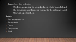
- 6. ŌĆó Otoscopy: may show perforation ŌĆó Cholesteatoma can be identified as a white mass behind the tympanic membrane or coming to the external canal through a perforation. Causes: ŌĆó Staphylococcus aureus ŌĆó Streptococcus ŌĆó Proteus ŌĆó Pseudomonas ŌĆó E.coli
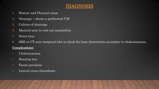
- 7. DIAGNOSIS 1. History and Physical exam 2. Otoscopy ŌĆō shows a perforated T.M 3. Culture of drainage 4. Mastoid xray to rule out mastoiditis 5. Sinus xray 6. MRI or CT scan temporal lobe to check for bone destruction secondary to cholesteatoma. Complications: ŌĆó Cholesteatoma ŌĆó Hearing loss ŌĆó Facial paralysis ŌĆó Lateral sinus thrombosis
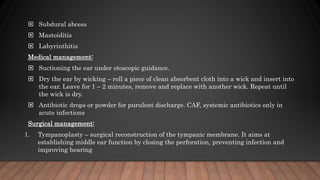
- 8. ’é© Subdural abcess ’é© Mastoiditis ’é© Labyrinthitis Medical management: ’é© Suctioning the ear under otoscopic guidance. ’é© Dry the ear by wicking ŌĆō roll a piece of clean absorbent cloth into a wick and insert into the ear. Leave for 1 ŌĆō 2 minutes, remove and replace with another wick. Repeat until the wick is dry. ’é© Antibiotic drops or powder for purulent discharge. CAF, systemic antibiotics only in acute infections Surgical management: 1. Tympanoplasty ŌĆō surgical reconstruction of the tympanic membrane. It aims at establishing middle ear function by closing the perforation, preventing infection and improving hearing
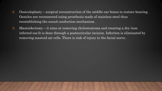
- 9. 2. Ossiculoplasty ŌĆō surgical reconstruction of the middle ear bones to restore hearing. Ossicles are reconnected using prosthesis made of stainless steel thus reestablishing the sound conduction mechanism. 3. Mastoidectomy ŌĆō it aims at removing cholesteatoma and creating a dry /non infected ear.It is done through a postauricular incision. Infection is eliminated by removing mastoid air cells. There is risk of injury to the facial nerve.