Acute appendicitis
Download as PPTX, PDF4 likes859 views

Acute appendicitis is the acute inflammation of the appendix, typically due to an obstruction of the appendiceal lumen. It is the most common cause of acute abdomen requiring emergency surgical intervention in both children and adults.


More Related Content
What's hot (20)
Similar to Acute appendicitis (20)


Recently uploaded (20)


Acute appendicitis
- 1. ACUTE APPENDICITIS DR. ASIF ALI
- 2. DEFINITIONS Appendicitis: ïĩ acute inflammation of the vermiform appendix Uncomplicated appendicitis: ïĩ appendicitis with no evidence of an appendiceal fecalith, an appendiceal tumor, or complications, such as perforation, gangrene, abscess, or mass Complicated appendicitis: ïĩ appendicitis associated with perforation, gangrene, abscess, an inflammatory mass, an appendiceal fecalith, or an appendiceal tumor
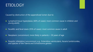
- 3. ETIOLOGY Caused by obstruction of the appendiceal lumen due to: ïĩ Lymphoid tissue hyperplasia; (60% of cases): most common cause in children and young adults ïĩ Fecalith; and fecal stasis (35% of cases): most common cause in adult ïĩ Neoplasm; (uncommon): more likely in patients > 50 years of age ïĩ Parasitic infestation (uncommon): e.g., Enterobius vermicularis, Ascaris lumbricoides, and species of the Taenia and Schistosoma genera
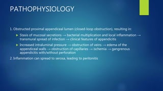
- 4. PATHOPHYSIOLOGY 1. Obstructed proximal appendiceal lumen (closed-loop obstruction), resulting in: ïĩ Stasis of mucosal secretions â bacterial multiplication and local inflammation â transmural spread of infection â clinical features of appendicitis ïĩ Increased intraluminal pressure â obstruction of veins â edema of the appendiceal walls â obstruction of capillaries â ischemia â gangrenous appendicitis with/without perforation 2. Inflammation can spread to serosa, leading to peritonitis
- 5. CLINICAL PRESENTATION Symptoms ïĩ Abdominal pain: initial periumbilical pain with migration to the right lower quadrant (RLQ) ïĩ Anorexia ïĩ Nausea ïĩ Vomiting ïĩ Diarrhea ïĩ Constipation ïĩ Indigestion
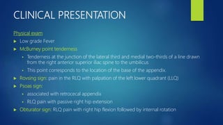
- 6. CLINICAL PRESENTATION Physical exam ïĩ Low grade Fever ïĩ McBurney point tenderness ï§ Tenderness at the junction of the lateral third and medial two-thirds of a line drawn from the right anterior superior iliac spine to the umbilicus ï§ This point corresponds to the location of the base of the appendix. ïĩ Rovsing sign: pain in the RLQ with palpation of the left lower quadrant (LLQ) ïĩ Psoas sign: ï§ associated with retrocecal appendix ï§ RLQ pain with passive right hip extension ïĩ Obturator sign: RLQ pain with right hip flexion followed by internal rotation
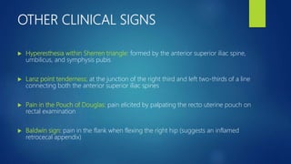
- 7. OTHER CLINICAL SIGNS ïĩ Hyperesthesia within Sherren triangle: formed by the anterior superior iliac spine, umbilicus, and symphysis pubis ïĩ Lanz point tenderness: at the junction of the right third and left two-thirds of a line connecting both the anterior superior iliac spines ïĩ Pain in the Pouch of Douglas: pain elicited by palpating the recto uterine pouch on rectal examination ïĩ Baldwin sign: pain in the flank when flexing the right hip (suggests an inflamed retrocecal appendix)
- 8. INVESTIGATIONS ïĩ CBC: mild leukocytosis with left shift; normal WBC count does not rule out acute appendicitis ïĩ CRP: elevated (> 10 mg/L) ïĩ Creatinine: maybe elevated ïĩ Serum electrolyte abnormalities may be present in patients with severe vomiting; and diarrhea Tests to rule out differential diagnoses ïĩ Urine/serum Îē-hCG test; : perform in all women of reproductive age to rule out pregnancy (including ectopic pregnancy) ACUTE APPENDICITIS IS CLINICAL DIAGNOSIS
- 9. Radiological Investigation ïĩ CT abdomen with IV contrast: preferred initial imaging modality in adults (except for pregnant women) ïĩ MRI abdomen without IV contrast: pregnant patients with inconclusive ultrasound findings ïĩ Abdominal ultrasound: ï§ Preferred initial imaging modality in children or pregnant patients ï§ As an alternative to CT scan if CT findings are inconclusive ï§ Abdominal ultrasound is more reliable for confirming acute appendicitis than ruling it out.
- 10. DIFFERENTIAL DIAGNOSIS ïĩ Ectopic pregnancy ïĩ Renal colic ïĩ Psoas abscess ïĩ Epiploid appendagitis ïĩ Constipation ïĩ Irritable Bowel syndrome
- 11. DIAGNOSTIC CRITERIA â ALVARDO SCORING
- 12. TREATMENT Supportive care ïĩ Bowel rest - Nil by mouth (NPO) ïĩ Intravenous fluids: Ringer Lactate and Normal saline ïĩ Electrolyte repletion as needed ïĩ IV analgesics: Ketorolac 30mg (Toradol) OR Tramadol 100mg (Tramol) ïĩ IV antiemetics as needed: Metoclopramide Or Dimenhydrinate ïĩ Antipyretic therapy: IV Paracetamol 1g/100ml x TDS/BD ïĩ IV antibiotic therapy: Ceftriaxone 1g x BD, + Metronidazole 500mg/100ml x TDS
- 13. SURGICAL TREATEMNT Non-perforated appendicitis ïĩ Appendectomy (laparoscopic or open) ï§ should be performed within 12 hours of diagnosis ï§ laparoscopic approach is more common and popular Perforated appendicitis with hemodynamic instability, sepsis, free perforation, or peritonitis ïĩ emergency appendectomy: irrigation and drainage of peritoneal cavity, colon resection if needed Stable perforated appendicitis ïĩ initial nonoperative management: IV antibiotics, percutaneous drainage of abscess if present ïĩ Rescue appendectomy for patients who do not respond to antibiotics
- 14. COMPLICATIONS ïĩ Appendiceal abscess ïĩ Perforation ïĩ Sepsis ïĩ Peritonitis ïĩ Hemodynamic instability ïĩ Death
- 15. THANK YOU