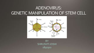
Adenovirus
- 3. The adenoid, also known as a pharyngeal tonsil or nasopharyngeal tonsil
- 14. Cell surface characterization and differentiation potential of MSCs. (A) Cell surface expression of various MSC markers were detected by staining with specific monoclonal antibodies and analysed by flow cytometry. MSCs are CD29, CD73, CD90 and CD44H positive, and CD45RA, CD71 low or negative. Shown are FACS histograms of CD MSCs stained with antibodies against surface markers as indicated or with appropriate isotype controls in triplicates. MSCs were tested for their potential to differentiate into (B) Aadipocytes and (C) Osteocytes by incubating the cells with specific formulations. Results shown are representative data of 3 separate isolations
- 15. Immunophenotyping and pro-inflammatory cytokine expression of untransduced and Ad-transduced MSCs. (A) Untransduced and Ad-transduced MSCs were stained for expression of the immunologically relevant markers MHC class I/II, CD80 and CD86 with specific antibodies and analysed by flow cytometry. (B) MSCs were transduced with Ad.GFP and after 24 h cells were collected and subjected to mRNA isolation and cDNA synthesis. RT-PCR analysis showing mRNA expression levels of IL-1β, IFN-γ and IL-6 from untransduced and Ad-transduced MSCs
- 16. T-cell proliferation in the presence or absence of untransduced and Ad.GFP transduced MSCs. (A) Histograms showing percent proliferation of CFSE-labelledT cells that were polyclonally stimulated with anti-CD3/anti- CD28 beads in the absence or presence of either untransduced or Ad.GFP transduced MSCs (ratio ofT cells to MSCs; 1‚à∂250). (B) Bar chart showing the percentage of cells divided greater than three generations following polyclonal stimulation in the absence or presence of either untransduced or Ad.GFP transduced MSCs. CFSE fluorescence was analysed on day 4 after co-culture.
- 18. Brokhman I, Pomp O, Fishman L,TennenbaumT, Amit M, Itzkovitz-Eldor J, Goldstein RS.


