ºÝºÝߣ

ºÝºÝߣShare a Scribd company logo
Bony Attachments
(LaPrade, JBJS, 2007)
Medial Epicondyle (ME)
Adductor Tubercle (AT)
Gastrocnemius Tubercle (GT)
Courtesy of Dr. LaPrade
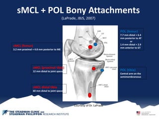
sMCL + POL Bony Attachments
(LaPrade, JBJS, 2007)
sMCL (femur)
3.2 mm proximal + 4.8 mm posterior to ME
sMCL (proximal tibia)
12 mm distal to joint space
sMCL distal tibia
60 mm distal to joint space
POL (femur)
7.7 mm distal + 6.4
mm posterior to AT
or
1.4 mm distal + 2.9
mm anterior to GT
POL (tibia)
Central arm on the
semimembranosus
Courtesy of Dr. LaPrade
PLC bony attachment
Fibular Collateral Ligament
FCL = 1o Varus stabilizer
Femoral attachment:
1.4 mm Proximal + 3.1 mm
posterior to lateral epicondyle
Fibula insertion:
Anterior lateral on the fibula
head
(8.2 mm post to the ant
margin of the fibular head
28.4 mm distal to the tip of
the fibular styloid)
Popliteus Tendon
PLT = 1o rotational stabilizer
Femoral attachment:
Average 18.5 mm anterior of the
FCL attachment at 70 degree flexion
Tibial attachment:
Posterior on the tibia
Courtesy of Dr. LaPrade
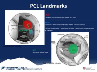
PCL Landmarks
ALB:
Between trochlear point and medial arch point
PCL:
Locate at bundle ridge
PMB:
Centered 8-9 mm posterior to edge of MFC articular cartilage
On wall anterior edge 5.8 mm from cartilage 2-4 mm bone-bridge between
ALB & PMB12.1 mm
Courtesy of Dr. LaPrade
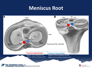
Meniscus Root
Posterior Lateral Root:
Approx. 9 mmm posterior to MTE
Approx. 8 mm anterior-lateral to PCL
Posterior Medial Root:
Approx. 4 mm lateral to LTE
Approx. 10 mm anterior to PCL
Courtesy of Dr. LaPrade

More Related Content

Anatomical Landmarks of the Knee

  • 1. Bony Attachments (LaPrade, JBJS, 2007) Medial Epicondyle (ME) Adductor Tubercle (AT) Gastrocnemius Tubercle (GT) Courtesy of Dr. LaPrade
  • 2. sMCL + POL Bony Attachments (LaPrade, JBJS, 2007) sMCL (femur) 3.2 mm proximal + 4.8 mm posterior to ME sMCL (proximal tibia) 12 mm distal to joint space sMCL distal tibia 60 mm distal to joint space POL (femur) 7.7 mm distal + 6.4 mm posterior to AT or 1.4 mm distal + 2.9 mm anterior to GT POL (tibia) Central arm on the semimembranosus Courtesy of Dr. LaPrade
  • 3. PLC bony attachment Fibular Collateral Ligament FCL = 1o Varus stabilizer Femoral attachment: 1.4 mm Proximal + 3.1 mm posterior to lateral epicondyle Fibula insertion: Anterior lateral on the fibula head (8.2 mm post to the ant margin of the fibular head 28.4 mm distal to the tip of the fibular styloid) Popliteus Tendon PLT = 1o rotational stabilizer Femoral attachment: Average 18.5 mm anterior of the FCL attachment at 70 degree flexion Tibial attachment: Posterior on the tibia Courtesy of Dr. LaPrade
  • 4. PCL Landmarks ALB: Between trochlear point and medial arch point PCL: Locate at bundle ridge PMB: Centered 8-9 mm posterior to edge of MFC articular cartilage On wall anterior edge 5.8 mm from cartilage 2-4 mm bone-bridge between ALB & PMB12.1 mm Courtesy of Dr. LaPrade Courtesy of Dr. LaPrade
  • 5. Meniscus Root Posterior Lateral Root: Approx. 9 mmm posterior to MTE Approx. 8 mm anterior-lateral to PCL Posterior Medial Root: Approx. 4 mm lateral to LTE Approx. 10 mm anterior to PCL Courtesy of Dr. LaPrade