1.Anatomy of the Medulla
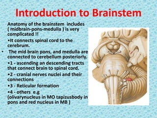
2. Introduction to Brainstem Anatomy of the brainstem includes ( midbrain-pons-medulla ) is very complicated !! ŌĆóIt connects spinal cord to the cerebrum. ŌĆó The mid brain pons, and medulla are connected to cerebellum posteriorly. ŌĆó1 - ascending an descending tracts that connect brain to spinal cord. ŌĆó2 - cranial nerves nuclei and their connections ŌĆó3 - Reticular formation ŌĆó4 - others e.g (olivarynucleus in MO tapizusbody in pons and red nucleus in MB )
3. Medulla oblongata ŌĆóThe medulla oblongata is the part of the brainstem between the pons and spinal cord ŌĆóIt extends through the foramen magnum to the level of the atlas. ŌĆóMedulla is vital for our function, without medulla we die. ŌĆóAbove the foramen magnum it is embraced dorsally by the cerebellar hemispheres. 1.The lower end which contains the upward continuation of the central canal of the spinal cord is the ŌĆśclosed part of the medullaŌĆÖ, 2.The upper end, where the canal comes to the surface as the lower part of the floor of the fourth ventricle, is the ŌĆśopen partŌĆÖ.
4. Medulla contdŌĆ”.. MO is lowest 3 cm of the brainstem ŌĆóit extend from the ponto- medullary junction until plane below foramina magnum for about 0.5 cm. ŌĆóMedulla spinalis have a central canal which prolonged into its lower half to open in the fourth ventricle at its upper half. ŌĆóCSF is encircle the MO from outside ( subarachnoid space ) and inside ( central canal ). ŌĆóMO is between the two lobes of cerebellum ( anterior cerebellar notch )
5. EXTERNAL FEATURES AND RELATIONS ŌĆó 3Cm long. ŌĆó Located at the caudal portion of brainstem ŌĆó Upper limit is cerebello-pontine angle ŌĆó Transverse plane that above C1 (suboccipital) intersects upper border of atlas dorsally and centre of dens ventrally marks lower limit
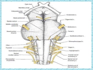
6. VENTRAL SURFACE ŌĆó Ventral median fissure extends from foramen coecum to caudal end of pyramid decussation ŌĆó Lateral to median fissure is pyramid ŌĆó Lat to pyramid is the ventrolateral sulcus (VLS) ŌĆó Hypoglossal nerve rootlets emerge from VLS ŌĆó Lat to VLS is olive which contains inf olivary nucleus ŌĆó Inferior cerebellar peduncle connects medulla with cerebellum and forms side wall of caudal half of fourth ventricle
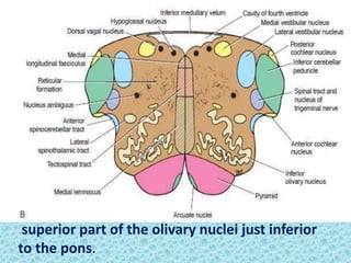
7. Ventral Surface Pyramid: Swelling on each side of anterior median fissure. ŌĆó Composed of bundles of nerve fibers, (corticospinal fibers) originate from the precentral gyrus of the cerebral cortex. ŌĆó The pyramids taper inferiorly and majority of the descending fibers decussate to the opposite side. Olive: ŌĆó Olives are the anterolateral oval elevations produced by the underlying inferior olivary nuclei. ŌĆó From the groove between the pyramid and the olive, the rootlets of the hypoglossal nerve emerge
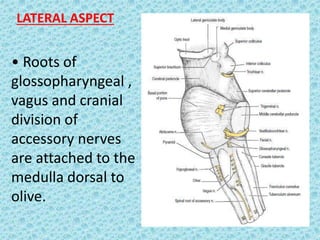
8. LATERAL ASPECT ŌĆó Roots of glossopharyngeal , vagus and cranial division of accessory nerves are attached to the medulla dorsal to olive.
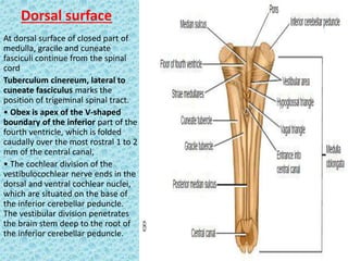
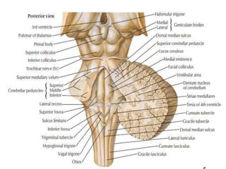
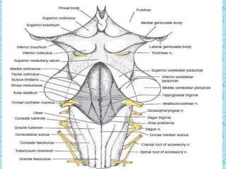
9. Dorsal surface At dorsal surface of closed part of medulla, gracile and cuneate fasciculi continue from the spinal
The document summarizes the development, anatomy, and histology of the pons and midbrain. It describes that the pons develops from the metencephalon and receives cells from the myelencephalon. The midbrain develops from the mesencephalon. The document then provides detailed descriptions of the structures, tracts, nuclei, and blood supply of both the pons and midbrain through multiple sections and diagrams.
The document describes the anatomy and functions of the medulla oblongata. It is the lowest part of the brainstem located in the posterior cranial fossa. It connects the spinal cord to the forebrain and contains nuclei of cranial nerves III-XII. Key structures in the medulla include the pyramids, olives, inferior cerebellar peduncles, and nuclei that control vital functions like respiration and cardiovascular regulation. The document discusses the medulla at different transverse section levels to describe its internal organization and pathways for motor and sensory signals.
The document describes the anatomy and vascular supply of the medulla oblongata. It discusses the dorsal nucleus of the vagus, nucleus ambiguus, hypoglossal nucleus, inferior olivary nucleus, and their locations. It also describes lateral and medial medullary syndromes which can result from vascular disorders affecting the posterior inferior cerebellar artery and vertebral artery respectively.
The cerebellum is located behind the brain stem and is divided into three lobes - anterior, posterior, and flocculonodular. It receives input from the spinal cord, vestibular system, and cerebral cortex. The cerebellar cortex consists of molecular, purkinje, and granular layers. Purkinje cells are the sole output, projecting to deep cerebellar nuclei which connect to motor and premotor areas. The cerebellum is involved in coordination, precision of movement, and maintaining balance and posture.
The fourth ventricle is located ventral to the cerebellum and dorsal to the pons and medulla. It is bounded laterally by the gracile and cuneate tubercles and inferior cerebellar peduncles, and superiorly by the superior cerebellar peduncle. Its roof is formed by the superior cerebellar peduncle and medullary velum. Its floor contains landmarks like the median sulcus, facial colliculus, and hypoglossal triangle. Cerebrospinal fluid circulates from the fourth ventricle through the median aperture and exits into the subarachnoid space through the foramina of Luschka and Magendi.
The thalamus is a paired symmetrical structure located in the center of the brain that relays sensory and motor signals between the brainstem and cerebral cortex. It is divided into several nuclei that have distinct connections and functions. The document provides detailed information on the anatomy, physiology, functional organization and clinical syndromes associated with lesions of different thalamic nuclei. Key points include a description of the gross anatomy and location of the thalamus, its blood supply, the nuclei and their connections, and syndromes associated with infarcts in the posterolateral and medial thalamic territories.
The cerebellum is located behind the brainstem and contains only 10% of the brain's volume. It receives input from muscles, joints, and the motor cortex, and provides corrective signals to the motor cortex to coordinate voluntary movement. The cerebellum evaluates and adjusts motor movements, integrating sensory information to ensure balance and motor learning. Damage to different parts of the cerebellum results in difficulties with coordination, posture, movement timing and sequencing.
The third ventricle is a midline cavity located between the two thalami and hypothalami. It communicates with the lateral ventricles via the foramen of Monroe and with the fourth ventricle via the cerebral aqueduct. The third ventricle's roof is formed by the fornix and tela choroidea, while its floor extends from the optic chiasm to the posterior perforated substance. The third ventricle can be accessed surgically through various anterior or posterior approaches between brain structures such as the fornix.
The document describes the anatomy of the cerebrum and base of the skull. It discusses the lobes and cortical regions of the cerebrum, including the frontal, parietal, occipital and temporal lobes. It also describes the structures and openings at the base of the skull, such as the foramen magnum, jugular foramen, optic canal and others.
The brainstem is located between the cerebrum and spinal cord. It consists of the midbrain, pons, and medulla oblongata. The midbrain connects the pons and cerebrum and contains the superior and inferior colliculi. The pons connects to the cerebellum via peduncles and contains pontine nuclei and cranial nerve nuclei. The medulla oblongata connects to the spinal cord and contains cranial nerve nuclei, the inferior olives, and tracts such as the gracile and cuneate fasciculi.
The cerebellum has three main parts - the vermis, two hemispheres, and four lobes. It receives sensory input from the spinal cord, brainstem, and cerebral cortex. There are three layers in the cerebellar cortex - molecular layer, purkinje cell layer, and granular layer. The cerebellum is connected to the brainstem via three cerebellar peduncles and plays a role in motor coordination and balance.
The document describes the four ventricles in the brain: the lateral ventricles located in the cerebral hemispheres, the third ventricle in the diencephalon, and the fourth ventricle between the pons, medulla, and cerebellum. Each ventricle is lined with ependyma and filled with cerebrospinal fluid. The lateral ventricles are C-shaped with three horns, the third ventricle connects to the lateral ventricles through the foramen of Monro, and the fourth ventricle roof projects into the cerebellum and is continuous with the cerebral aqueduct.
You can watch the video on my you tube channel: https://youtu.be/I0FaX-iQfa0
Medulla oblongata or more simply medulla is part of brain stem which forms base of the brain stem. It contains pyramid, olive and above pyramidal structure, there is decussation of pyramids which explains why each part of brain controls opposite part of body. Adding to that medulla also has several nuclei which controls activity of cardiovascular system and respiratory system. Medulla also has nuclei for controlling reflexes of vomiting, swallowing, hiccuping, coughing and sneezing. It has also nuclei for test, hearing and balance. Medulla also contains nuclei of cranial nerve number VIII, IX, X, XI and XII.
The document provides information on the gross anatomy and internal structure of the midbrain and cerebellum. It discusses:
- The midbrain connects the pons and cerebellum to the forebrain. It contains four colliculi, cranial nerve nuclei, motor and sensory tracts, and the cerebral aqueduct runs through it. Injuries can cause specific deficits depending on the structures involved.
- The cerebellum coordinates voluntary movement and balance. It has three lobes and is connected to the brainstem by three peduncles. It is divided into vermis, paravermis and hemispheres that serve different motor functions. Injuries can impact coordination and balance.
The cerebellum is located at the back of the brain and plays an important role in motor control. It develops from the metencephalon and can be divided into three lobes and three functional areas. The cerebellum coordinates precision and timing of movement as well as motor learning. Damage to different parts of the cerebellum can produce a variety of motor symptoms.
Thalamus, anatomy of thalamus, Thalamus PPTdrasarma1947
╠²
1. The document describes the anatomy and functions of the thalamus.
2. It is a midline paired structure located within the brain that relays sensory information (except smell) between the brainstem and cerebral cortex.
3. The thalamus has several nuclei that receive sensory information from different pathways and project to specific areas of the cerebral cortex, functioning as a relay for sensory integration.
The brainstem consists of the midbrain, pons, and medulla oblongata. It connects the forebrain to the spinal cord superiorly and inferiorly. The pons and medulla are separated posteriorly by the fourth ventricle. The brainstem contains nuclei that control vital functions like respiration, circulation, swallowing and eye movements. Damage to the ascending reticular activating system in the brainstem can disturb consciousness.
The brainstem consists of the medulla oblongata, pons, and midbrain. It connects the spinal cord to the forebrain and controls vital functions like breathing, heart rate, swallowing and sneezing. The medulla regulates breathing and circulation. The pons relays messages between the brain and cerebellum and is involved in sleep, hearing, taste and balance. The midbrain coordinates eye and body movements and processes vision and sound.
There are 12 pairs of cranial nerves in the brain with motor and/or sensory nuclei in the brain stem. Each cranial nerve has its own nucleus of origin or termination. These nuclei are arranged medio-laterally and include somatic, visceral, special, and general fibers. The medio-lateral arrangement includes sensory, special visceral efferent, general visceral efferent, general visceral afferent, special visceral afferent, general somatic afferent, and special somatic afferent. Cranial nerve nuclei are located in the midbrain, pons, and medulla.
The document provides information on the functional anatomy of the brainstem, including its three main parts - medulla, pons, and midbrain. It discusses the structures, functions, blood supply, and clinical correlates of each region. Key points include that the brainstem connects the spinal cord to the forebrain, contains important reflex centers, and cranial nerve nuclei. It describes nuclei and tracts at different levels, and clinical syndromes that can result from lesions in different areas, such as lateral medullary syndrome and Dejerine's anterior bulbar palsy.
The cerebellum is located in the posterior fossa of the skull below the brain. It has three main lobes - the anterior, middle, and flocculonodular lobes. The cerebellum coordinates voluntary movement through connections with other parts of the brain and spinal cord. It has an outer gray matter cortex containing Purkinje cells and an inner white matter. The cerebellum receives sensory information and regulates motor movements to produce smooth, coordinated activity.
This document provides an overview of the medulla oblongata. It begins with an introduction and outline. It then describes the gross appearance and internal structures of the medulla, including the pyramids, olives, and medial lemnisci. It discusses the blood supply, venous drainage, and functions of the medulla, which include respiration, cardiac and vasomotor centers, and reflex centers. The document concludes by covering diseases of the medulla such as genetic, developmental, vascular, degenerative, infectious, inflammatory, and neoplastic conditions.
The brainstem consists of three parts - the midbrain, pons, and medulla. It connects the spinal cord to the forebrain and contains nuclei that control vital functions like breathing and heart rate. It also contains tracts that relay signals between the spinal cord and higher brain centers. The reticular formation is a network of fibers and neurons throughout the brainstem that plays roles in motor control, sensory processing, autonomic functions, and maintaining alertness. Important structures in the brainstem include the cranial nerve nuclei, pyramidal tract, olives, and red nucleus.
The dural venous sinuses are lined with endothelium and lack muscles and valves. They collect blood from the brain, meninges, orbit, inner ear and diploe. The superior sagittal sinus begins at the crista galli and ends at the internal occipital protuberance, draining into the confluence of sinuses. Infection from the scalp, nasal cavity or diploic tissue can lead to septic thrombosis and obstruct CSF absorption, causing increased intracranial pressure. The paired transverse sinuses and sigmoid sinuses carry blood through the posterior compartment of the jugular foramen before joining the internal jugular vein.
The pons is located in the brainstem, between the midbrain and medulla oblongata. It contains fibers that connect the cerebellum and cerebrum, and nuclei for several cranial nerves including the trigeminal, facial, and abducent nerves. The pons receives its blood supply primarily from the basilar artery and its branches, as well as the anterior inferior cerebellar and superior cerebellar arteries. It plays an important role in motor functions and sensory processes.
The document provides an overview of cerebrum anatomy. It discusses that the cerebrum is the largest part of the brain and is divided into two hemispheres. It describes the lobes of the cerebrum including the frontal, parietal, temporal, and occipital lobes. It also discusses the internal structures of the cerebrum including the cerebral cortex, ventricles, basal ganglia, and white matter tracts.
The document describes the major structures and regions of the human brain. It begins by listing the main parts including the forebrain, cerebrum, diencephalon, pons, medulla oblongata, cerebellum, midbrain, and hindbrain. It then provides more detailed information about each region, their locations, functions, and relationships to other structures. Key details discussed include the lobes and areas of the cerebrum, structures within the diencephalon, cranial nerves originating from the pons and medulla, and connections of the cerebellum.
The brainstem consists of 3 parts - the midbrain, pons, and medulla. It connects the spinal cord to the forebrain and has important functions like transmitting signals between the two, controlling vital reflexes like respiration, and containing nuclei for cranial nerves III-XII. The reticular formation is a network of fibers and neurons that extends through the brainstem and is involved in functions like motor control, sensory processing, visceral control, and neuroendocrine regulation. Other structures in the brainstem include the cerebral peduncles, cerebral aqueduct, inferior and superior colliculi, cranial nerve nuclei, and the pontine nuclei.
The brainstem consists of 3 parts - midbrain, pons, and medulla. It connects the spinal cord to the forebrain and contains important centers that control respiration, cardiovascular function, and consciousness. It also contains nuclei for cranial nerves 3 through 12. The medulla contains pyramids, olives, and tracts. The pons connects the medulla to the midbrain. The midbrain connects the pons to the forebrain and contains the cerebral aqueduct and corpora quadrigemina. The reticular formation extends through the brainstem and is important for motor control, sensory processes, autonomic functions, and maintaining alertness.
The brainstem consists of three parts - the midbrain, pons, and medulla. It connects the spinal cord to the forebrain and serves functions like relaying signals between them and controlling vital processes. The midbrain contains structures like the cerebral peduncles and corpora quadrigemina. The pons connects the midbrain to the medulla and contains pontine nuclei. The medulla ends at the spinal cord and contains the pyramids, olives, and nuclei for cranial nerves III to XII.
OBJECTIVES
ŌĆó1.To understand anatomy cerebellum and brainstem
ŌĆó2.To explore the functional role of the cerebellum &brainstem and clinical relevances
ŌĆó3.Highlights the neural pathways involving the cerebellum and brainstem
ŌĆó4.Understand clinical implication of damaged to the cerebellum and brain stem
This document provides information about the rhombencephalon or hindbrain, which includes the medulla oblongata, pons, and cerebellum. It discusses the external and internal structures of the medulla oblongata and pons in detail. It describes the nuclei, tracts, and vascular supply found at different levels within these structures. Finally, it outlines some clinical correlates like lateral medullary syndrome, medial medullary syndrome, and how lesions in the hindbrain can cause specific neurological deficits or syndromes depending on their location.
The nervous system is divided into the central nervous system (CNS) and peripheral nervous system (PNS). The CNS comprises the brain and spinal cord and is responsible for sensory integration, coordination of motor actions. The PNS includes cranial and spinal nerves and connects to the CNS, carrying sensory information in and motor commands out. The brain is divided into the cerebrum, brainstem, and cerebellum. The cerebrum is the largest part and is divided into lobes. The brainstem connects the cerebrum to the spinal cord and contains nuclei that control vital functions. The cerebellum coordinates movement.
This document provides an overview of the brain stem, including its external features, internal structures, and clinical significance. It describes the medulla oblongata, pons, and midbrain in detail. Key points include:
- The brain stem connects the spinal cord to the forebrain and contains cranial nerve nuclei, reflex centers, and tracts connecting different brain regions.
- Structures in the medulla include the pyramids, olives, cranial nerve nuclei, and tracts like the medial lemniscus. The pons contains cranial nerve nuclei and connects the medulla to midbrain. The midbrain contains the cerebral aqueduct and superior and inferior colliculi.
- Trans
The brainstem is composed of the medulla, pons, and midbrain. It serves as a conduit between the spinal cord and forebrain, contains important reflex centers that control respiration, circulation, and consciousness, and contains the nuclei of cranial nerves III through XII. The medulla begins where the spinal cord meets the skull and houses centers that regulate vital functions like breathing, heart rate, and blood pressure. It contains the nuclei of cranial nerves IX through XII and conducts ascending and descending nerve tracts between the brain and spinal cord.
EXTERNAL FEATURES OF MIDBRAIN, ANATOMY OF INTERNAL FEATURES OF MIDBRAIN, CRUS CEREBRI, SUBSTANTIA NIGRA, CEREBRAL PEDUNCLE,INFERIOR COLLICULUS,LEMNISCI
This document provides an overview of the anatomy of the brain. It discusses the central nervous system and its major components including the brain, spinal cord, and meninges. It describes the protective coverings of the brain including the cranium, meninges, and cerebrospinal fluid. It then details the specific structures of the brain including the cerebrum, diencephalon, brainstem, cerebellum, and their lobes, sulci, gyri and surfaces. Key structures like the corpus callosum, ventricles, and basal ganglia are also summarized.
This document provides an overview of the anatomy of the brain. It describes the central nervous system including the brain and spinal cord. It then discusses the protective coverings of the brain including the cranium, meninges, and cerebrospinal fluid. The document proceeds to describe the various parts of the brain in detail, including the cerebrum, diencephalon, brainstem, cerebellum, and ventricles. It discusses the protective coverings, blood supply via the circle of Willis, and functions of different regions.
The brain stem consists of the medulla, pons, and midbrain. It is situated in the posterior cranial fossa. The medulla is the lowest part and connects with the spinal cord. It contains nuclei for cranial nerves and tracts for sensory and motor functions. The pons is in the middle and connects the midbrain with the medulla. It contains pontine nuclei and transverse fibers. The midbrain connects the hindbrain and forebrain. It contains the cerebral peduncles and tectum including the superior and inferior colliculi.
The document provides detailed information about the anatomy of the brainstem and related structures. It describes the three parts of the brainstem from superior to inferior as the midbrain, pons, and medulla. It then discusses the structures and features of each part in multiple paragraphs and levels of detail. It also covers the ventricles and cisterns of the brain as well as the cranial nerves and surrounding blood vessels.
The document provides information on the anatomy and structure of the spinal cord. It discusses the following key points:
- The spinal cord is surrounded by three meningeal coverings - dura mater, arachnoid mater, and pia mater. It has cervical and lumbar enlargements.
- Spinal nerves arise in pairs from the spinal cord. Each nerve has a dorsal root containing a ganglion and a ventral root.
- The spinal cord has gray matter on the outside containing nuclei and white matter on the inside. It also contains ascending and descending tracts that connect to the brain.
- Ascending tracts like the anterior and lateral spinothalamic tracts transmit sensory information from
Chair, Grzegorz (Greg) S. Nowakowski, MD, FASCO, discusses diffuse large B-cell lymphoma in this CME activity titled ŌĆ£Addressing Unmet Needs for Better Outcomes in DLBCL: Leveraging Prognostic Assessment and Off-the-Shelf Immunotherapy Strategies.ŌĆØ For the full presentation, downloadable Practice Aid, and complete CME information, and to apply for credit, please visit us at https://bit.ly/49JdxV4. CME credit will be available until February 27, 2026.
Chair, Joshua Sabari, MD, discusses NSCLC in this CME activity titled ŌĆ£Modern Practice Principles in Lung CancerŌĆöFirst Find the Targets, Then Treat With Precision: A Concise Guide for Biomarker Testing and EGFR-Targeted Therapy in NSCLC.ŌĆØ For the full presentation, downloadable Practice Aid, and complete CME information, and to apply for credit, please visit us at https://bit.ly/3VomnBV. CME credit will be available until February 26, 2026.
FAO's Support Rabies Control in Bali_Jul22.pptxWahid Husein
╠²
What is FAO doing to support rabies control programmes in Bali, Indonesia, using One Health approach with mass dog vaccination and integrated bite case management as main strategies
At Macafem, we provide 100% natural support for women navigating menopause. For over 20 years, we've helped women manage symptoms, and in 2024, we're proud to share their heartfelt experiences.
Dr. Jaymee ShellŌĆÖs Perspective on COVID-19Jaymee Shell
╠²
Dr. Jaymee Shell views the COVID-19 pandemic as both a crisis that exposed weaknesses and an opportunity to build stronger systems. She emphasizes that the pandemic revealed critical healthcare inequities while demonstrating the power of collaboration and adaptability.
Shell highlights that organizations with gender-diverse executive teams are 25% more likely to experience above-average profitability, positioning diversity as a business necessity rather than just a moral imperative. She notes that the pandemic disproportionately affected women of color, with one in three women considering leaving or downshifting their careers.
To combat inequality, Shell recommends implementing flexible work policies, establishing clear metrics for diversity in leadership, creating structured virtual collaboration spaces, and developing comprehensive wellness programs. For healthcare providers specifically, she advocates for multilingual communication systems, mobile health units, telehealth services with alternatives for those lacking internet access, and cultural competency training.
Shell emphasizes the importance of mental health support through culturally appropriate resources, employee assistance programs, and regular check-ins. She calls for diverse leadership teams that reflect the communities they serve and community-centered care models that address social determinants of health.
In her words: "The COVID-19 pandemic didn't create healthcare inequalities ŌĆō it illuminated them." She urges building systems that reach every community and provide dignified care to all.
Acute & Chronic Inflammation, Chemical mediators in Inflammation and Wound he...Ganapathi Vankudoth
╠²
A complete information of Inflammation, it includes types of Inflammation, purpose of Inflammation, pathogenesis of acute inflammation, chemical mediators in inflammation, types of chronic inflammation, wound healing and Inflammation in skin repair, phases of wound healing, factors influencing wound healing and types of wound healing.
Rabies Bali 2008-2020_WRD Webinar_WSAVA 2020_Final.pptxWahid Husein
╠²
A decade of rabies control programmes in Bali with support from FAO ECTAD Indonesia with Mass Dog Vaccination, Integrated Bite Case Management, Dog Population Management, and Risk Communication as the backbone of the programmes
Title: Regulation of Tubular Reabsorption ŌĆō A Comprehensive Overview
Description:
This lecture provides a detailed and structured explanation of the mechanisms regulating tubular reabsorption in the kidneys. It explores how different physiological and hormonal factors influence glomerular filtration and reabsorption rates, ensuring fluid and electrolyte balance in the body.
¤öŹ Who Should Read This?
This presentation is designed for:
Ō£ö’ĖÅ Medical Students (MBBS, BDS, Nursing, Allied Health Sciences) preparing for physiology exams.
Ō£ö’ĖÅ Medical Educators & Professors looking for structured teaching material.
Ō£ö’ĖÅ Healthcare Professionals (doctors, nephrologists, and physiologists) seeking a refresher on renal physiology.
Ō£ö’ĖÅ Postgraduate Students & Researchers in the field of medical sciences and physiology.
¤ōī What YouŌĆÖll Learn:
Ō£ģ Local Regulation of Tubular Reabsorption
Ō£ö’ĖÅ Glomerulo-Tubular Balance ŌĆō its mechanism and clinical significance
Ō£ö’ĖÅ Net reabsorptive forces affecting peritubular capillaries
Ō£ö’ĖÅ Role of peritubular hydrostatic and colloid osmotic pressures
Ō£ģ Hormonal Regulation of Tubular Reabsorption
Ō£ö’ĖÅ Effects of Aldosterone, Angiotensin II, ADH, and Natriuretic Peptides
Ō£ö’ĖÅ Clinical conditions like AddisonŌĆÖs disease & Conn Syndrome
Ō£ö’ĖÅ Mechanisms of pressure natriuresis and diuresis
Ō£ģ Nervous System Regulation
Ō£ö’ĖÅ Sympathetic Nervous System activation and its effects on sodium reabsorption
¤®║ Clinical Correlations & Case Discussions
Ō£ö’ĖÅ How renal regulation is altered in hypertension, hypotension, and proteinuria
Ō£ö’ĖÅ Comparison of Glomerulo-Tubular Balance vs. Tubulo-Glomerular Feedback
This presentation provides detailed diagrams, flowcharts, and calculations to enhance understanding and retention. Whether you are studying, teaching, or practicing medicine, this lecture will serve as a valuable resource for mastering renal physiology.
¤ōó Keywords for Easy Search:
#Physiology #RenalPhysiology #TubularReabsorption #GlomeruloTubularBalance #HormonalRegulation #MedicalEducation #Nephrology
2. Introduction to Brainstem
Anatomy of the brainstem includes
( midbrain-pons-medulla ) is very
complicated !!
ŌĆóIt connects spinal cord to the
cerebrum.
ŌĆó The mid brain pons, and medulla are
connected to cerebellum posteriorly.
ŌĆó1 - ascending an descending tracts
that connect brain to spinal cord.
ŌĆó2 - cranial nerves nuclei and their
connections
ŌĆó3 - Reticular formation
ŌĆó4 - others e.g
(olivarynucleus in MO tapizusbody in
pons and red nucleus in MB )
3. Medulla oblongata
ŌĆóThe medulla oblongata is the part of the
brainstem between the pons and spinal
cord
ŌĆóIt extends through the foramen magnum
to the level of the atlas.
ŌĆóMedulla is vital for our function, without
medulla we die.
ŌĆóAbove the foramen magnum it is
embraced dorsally by the cerebellar
hemispheres.
1.The lower end which contains the
upward continuation of the central canal
of the spinal cord is the ŌĆśclosed part of the
medullaŌĆÖ,
2.The upper end, where the canal comes to
the surface as the lower part of the floor
of the fourth ventricle, is the ŌĆśopen partŌĆÖ.
4. Medulla contdŌĆ”..
MO is lowest 3 cm of the
brainstem
ŌĆóit extend from the ponto-
medullary junction until plane
below foramina magnum for
about 0.5 cm.
ŌĆóMedulla spinalis have a central
canal which prolonged into its
lower half to open in the fourth
ventricle at its upper half.
ŌĆóCSF is encircle the MO from
outside ( subarachnoid space )
and inside ( central canal ).
ŌĆóMO is between the two lobes of
cerebellum ( anterior cerebellar
notch )
5. EXTERNAL FEATURES AND RELATIONS
ŌĆó 3Cm long.
ŌĆó Located at the caudal portion of brainstem
ŌĆó Upper limit is cerebello-pontine angle
ŌĆó Transverse plane that above C1 (suboccipital)
intersects upper border of atlas dorsally and
centre of dens ventrally marks lower limit
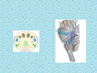
6. VENTRAL SURFACE
ŌĆó Ventral median fissure extends from foramen
coecum to caudal end of pyramid decussation
ŌĆó Lateral to median fissure is pyramid
ŌĆó Lat to pyramid is the ventrolateral sulcus (VLS)
ŌĆó Hypoglossal nerve rootlets emerge from VLS
ŌĆó Lat to VLS is olive which contains inf olivary
nucleus
ŌĆó Inferior cerebellar peduncle connects medulla
with cerebellum and forms side wall of caudal
half of fourth ventricle
8. Ventral Surface
Pyramid:
Swelling on each side of anterior
median fissure.
ŌĆó Composed of bundles of nerve fibers,
(corticospinal fibers) originate from
the precentral gyrus of the cerebral
cortex.
ŌĆó The pyramids taper inferiorly and
majority of the descending fibers
decussate to the opposite side.
Olive:
ŌĆó Olives are the anterolateral oval
elevations produced by the underlying
inferior olivary nuclei.
ŌĆó From the groove between the
pyramid and the olive, the rootlets of
the hypoglossal nerve emerge
11. LATERAL ASPECT
ŌĆó Roots of
glossopharyngeal ,
vagus and cranial
division of
accessory nerves
are attached to the
medulla dorsal to
olive.
12. Dorsal surface
At dorsal surface of closed part of
medulla, gracile and cuneate
fasciculi continue from the spinal
cord
Tuberculum cinereum, lateral to
cuneate fasciculus marks the
position of trigeminal spinal tract.
ŌĆó Obex is apex of the V-shaped
boundary of the inferior part of the
fourth ventricle, which is folded
caudally over the most rostral 1 to 2
mm of the central canal,
ŌĆó The cochlear division of the
vestibulocochlear nerve ends in the
dorsal and ventral cochlear nuclei,
which are situated on the base of
the inferior cerebellar peduncle.
The vestibular division penetrates
the brain stem deep to the root of
the inferior cerebellar peduncle.
17. Medial lemnisci
ŌĆó The great sensory decussation.
ŌĆó The lemnisci have been formed by the
internal arcuate fibers, which emerge
from the anterior aspects of the
nucleus gracilis and nucleus cuneatus.
ŌĆó The deussation takes place anterior to
the central grey mater.
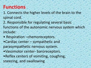
21. Functions
1. Connects the higher levels of the brain to the
spinal cord.
2. Responsible for regulating several basic
functions of the autonomic nervous system which
include:
ŌĆó Respiration ŌĆōchemoreceptors.
ŌĆóCardiac center ŌĆō sympathetic and
parasympathetic nervous system.
ŌĆóVasomotor center- baroreceptors.
ŌĆóReflex centers of vomiting, coughing,
sneezing, and swallowing
22. Applied Anatomy
ŌĆó The vital centres i.e respiratory and vasomotor
are situated in the lower part of floor of the
fourth ventricle formed by Medulla. An injury to
the medulla is therefore usually fatal.
ŌĆó Bulbar paralysis
ŌĆó Pseudobulbar palsy
ŌĆó Common vascular lesions involving the medulla
a) Thrombosis of Post-inf-Cerebellar artery
b) Thrombosis of vertibral artery