Annormalities of head size and shape
- 1. Abnormalities of Head Size and Shape Mr.Riyaz Khan Aug-2012 Batch VI th Semster Department of Pediatrics Nepalgunj Medical College-Kohalpur
- 2. MACROCEPHALY • Definition: Head circumference ( occipito frontal ) > 2 standard deviation(SD) above the mean for age and sex.
- 3. 1 SD = 1.25 CM Macrocephaly > 2 SD i.e. 2.5 cm Microcephaly < 3 SD i.e 3.75 cm Take 50 centile as base line
- 4. 1. Megalencephaly -Benign Familial -Neurocutaneous syndromes: Neurofibromatosis, tuberous sclerosis -Fragile X syndrome -Leucodystrophies & Lysosomal storage disease 2. Increased Cerebrospinal fluid -Hydrocephalous -Benign enlargement of subarachnoid space 3. Enlarged Vascular compartment -Arteriovenous malformation -Subdural, epidural, subarachnoid or intraventricular hemorrhage Causes of Macrocephaly
- 5. cont…. 4. Increase in bony compartment Bone disease: Achondroplasia, osteogenesis imperfecta, osteopetrosis Bone marrow expansion: Thalassemia major 5. Miscellaneous causes Intracranial mass lesions: Cyst, abscess or tumor Raised intracranial pressure: Idiopathic pseudotumor cerebri, lead poisoning, galactosemia
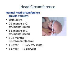
- 6. Head Circumference Normal head circumference growth velocity: • Birth-35cm • 0-3 months : +2 cm/month(41cm) • 3-6 months :+ 1 cm/month(44cm) • 6-12 months :+ 0.5cm/month(47cm) • 1-3 year : 0.25 cm/ mnth • 3-6 year : 1 cm/year
- 7. • History • Examination including auscultation of the skull for bruit • Developmental history • Rate of head growth – serial measurements Investigations : 1. Urea/electrolytes 2. Thyroid function test 3. Plasma amino acids 4. Urine amino acids and organic acids, glycosaminoglycans 5. CT head/MRI head preferably 6. Bone profile APPROACH
- 8. TREATMENT -generally require no tx - Infants with hydrocephalus may require neurological intervention( e.g. placement of a ventriculo-peritoneal shunt).
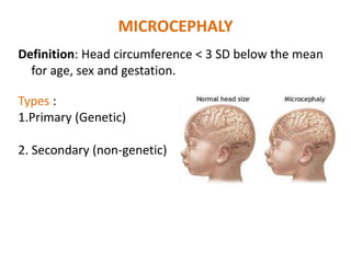
- 9. MICROCEPHALY Definition: Head circumference < 3 SD below the mean for age, sex and gestation. Types : 1.Primary (Genetic) 2. Secondary (non-genetic)
- 10. 1. PRIMARY ( Genetic ) MICROCEPHALY • Condition associated with reduced generation of neurons during neural development and migration. • Refers to group - associated with specific genetic syndromes. • Usually have slanting forehead. • Identified at birth itself
- 11. Causes for primary • Familial - AR • Autosomal dominant • Syndromes : 1. Down Syndrome 2. Cri du chat 3. Edward 4. Cornelia de Lange 5. Rubinstein Tyabi
- 12. • Results from noxious agents that may affect a fetus in utero or an infant during periods of rapid brain growth, particularly the first 2 years of life 2. Secondary ( non genetic) Microcephaly
- 13. 1. Radiation 2. Congenital infections – rubella, CMV, toxoplasmosis, HIV, Syphilis 3. Drugs – fetal alcohol, fetal hydantoin 4. Meningitis/encephalitis 5. Metabolic – maternal diabetes 6. Hypoxic ischemic encephalopathy 7. Malnutrition 8. Hyperthermia Causes for secondary microcepahaly
- 14. APPROACH • History (perinatal – family history) • Examination – dysmorphic features – malformations • Development • Growth – serial measurements of HC INVESTIGATIONS • Baseline biochemistry, metabolic screen • Genetic testing – karyotype, molecular genetics • TORCH screen • Ophthalmology • MRI brain
- 15. • No treatment for microcephaly • Baby’s head cannot be returned to a normal size & shape • According to the cause – Anticonvulsants – Physiotherapy – Hearing and speech therapy – Dietary management for failure to thrive – Genetic counseling Management
- 16. CRANIOSYNOSTOSIS Definition: premature fusion of one or more cranial sutures, either major(e.g metopic, coronal, sagittal, and lambdoid) or minor( frontnasal, temporosquamosal, and frontosphenoidal).
- 18. DEFORMITIES OF SKULL 1. Plagiocephaly 2. Scaphocephaly 3. Trigonocephaly 4. Turencephaly 5. Brachycephaly
- 19. -Fusion of either right or left side of the coronal suture -Causes the normal forehead and the brow to stop growing -Produces flattening of the forehead and the brow on the affected side, with the forehead tending to be excessively prominent on the opposite side PLAGIOCEPHALY
- 20. SCAPHOCEPHALY Early closure or fusion of the sagittal suture Fusion causes a long, narrow skull .Prominent occiput and forehead Usually only craniosynostosis which is relatively harmless
- 21. TRIGONOCEPHALY Fusion of the metopic (forehead) suture Fusion result in a prominent ridge running down the forehead -looks pointed, like a triangle, with closely placed eyes (hypotelorism).
- 22. • Turriencephaly – cone shaped head . Fusion of coronal and speno frontal or fronto ethmoid sutures. • Brachycephaly – premature closure of coronal suture expands skull parallel to coronal suture , thus broadening of forehead with short AP diameter. Eg – in many syndromes like Downs Syndrome
- 23. Diagnosis • Palpation of suture reveals prominent bony ridge. • Fusion may be confirmed by x-ray skull • Associated syndromes – Crouzon , Alperts, Carpenter
- 24. Management • Premature fusion of single suture rarely causes any neurological deficit . Thus, in this situation the only indication is cosmetics. • 2 or more suture fusion – more complications eg. ↑ ICT, hydrocephalus, optic atrophy, DNS, choanal atresia --- operative surgery essential – craniectomy with craniofacial correction. • Usually good prognosis with non syndromic infants……………
- 25. Thank You


