Applied Surgical Anatomy of the Brain and Spinal Cord
ŌĆóDownload as PPT, PDFŌĆó
5 likesŌĆó2,632 views

The document summarizes key anatomy related to the spinal cord and scalp. It describes the layers of the scalp from superficial to deep. It then discusses the skull vault and base, identifying several surface landmarks. It details the anatomy of the spinal cord, including its length, weight, segments, internal configuration of grey and white matter, and surrounding meninges. Finally, it lists the objectives which are to identify brain and spinal cord anatomy and relate it to surgical procedures and surface markings.


Applied Surgical Anatomy of the Brain and Spinal Cord
- 2. Objectives ŌĆó To identify key brain and spinal cord anatomy ŌĆó Be able to relate brain and spinal cord anatomy in relation to surface marking and surgical procedure
- 3. ŌĆó S: Skin (hair follicles and sweat glands) ŌĆó C: Connective tissue (Fibrous, vessels and nerves) ŌĆó A: Aponeurosis (Musculofibrous) ŌĆó L: Loose areolar tissue ŌĆó P: Pericranium The first three layers are united as a single unit and are difficult to separate. Epicranial muscles, where present, lie between the galea aponeurotica and the loose areolar tissue. The scalp
- 4. ŌĆó The galea aponeurotica is the most stable layer in the suturing of the scalp. ŌĆó After craniectomy, the galeal layer should be approximated to prevent dehiscence of the scalp incision due to swelling of the intracranial contents. The scalp (Galea aponeurotica)
- 5. ŌĆó Potential space (Free attachment) ŌĆó Emissary veins The scalp (Loose areolar tissue)
- 6. ŌĆó The fontanelles are located between the developing cranial bones and are covered by the pericranium (periosteum) externally and by the dura mater internally. ŌĆó Provides minimal blood supply to the underlying skull bone. Hence, detachment of the periosteum does not cause underlying bone necrosis but may produce some demineralization. The scalp (Pericranium)
- 7. Young Adult ŌĆó The periosteum is connected to the dura mater by connective tissue along these suture lines. ŌĆó The periosteum can be easily separated from the skull except at suture lines. Adult ŌĆó The suture lines are fused or obliterated. ŌĆó The periosteum does not have strong attachments within the suture lines. ŌĆó The periosteum has very low osteogenic activity in adults. The scalp (Pericranium)
- 8. The scalp: Blood supply
- 9. The scalp: Muscles and nerves
- 11. Anterior fontanelle: closes at about 18 months of age Posterior fontanelle: closes by the 3rd to 6th month . The skull (Vault)
- 13. 2 cm behind and 1.25 cm above the superior part of the posterior border of the mastoid process. 7 cm above the external occipitalprotuberance. 3.5 cm behind and 1.5 cm above the fronto-zygomatic suture. The skull (Vault): Surface Landmarks
- 14. Pterion
- 15. Asterion
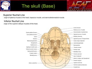
- 17. Superior Nuchal Line origin of splenius muscle of the head, trapezius muscle, and sternocleidomastoid muscle. Inferior Nuchal Line origin of the superior oblique muscles of the head. The skull (Base)
- 18. Petrous Sphenoid Anterior cranial fossa Middle cranial fossa Posterior cranial fossa The skull (Base)
- 19. The skull (Vault and skull base)
- 20. The skull (Vault): Diploic vein
- 21. ŌĆó Cranial Epidural Space ŌĆō Cranial venous plexuses ŌĆō Meningeal arteries ŌĆō Some cranial nerves, such as those to the orbit ŌĆó Dura Mater ŌĆó Subdural Space ŌĆó Arachnoid ŌĆó Subarachnoid space. ŌĆó Pia mater Leptomeningeal Dura and spaces
- 22. ŌĆó Falx Cerebri ŌĆō crista galli ŌĆō superior sagittal sinus ŌĆō inferior sagittal sinus ŌĆó Tentorium Cerebelli ŌĆō Posterior clinoid processes ŌĆō petrous ridge ŌĆō superior petrosal sinus ŌĆō straight sinus ŌĆó Small Falx Cerebelli ŌĆō occipitalis sinus. Dura Septa
- 23. Venous Sinuses and basilar plexus
- 24. Cavernous sinus
- 26. Cistern
- 27. Ventricles
- 28. Cerebrum
- 31. Central Sulcus of Rolando
- 32. Sylvian fissure / Lateral sulcus
- 35. Precentral and post central sulci
- 36. Cortical Area
- 37. Spinal Cord
- 38. CONTENT 1. Anatomy of spinal cord 2. Surface landmarks for pre-operative planning Objectives
- 39. 10 cm C4 to the T1 8 cm T9 to T12 45 cm males. 42 cm females 1 cm Weight of 30 grams Conus medullaris L1-L2 disk space embryonic spinal cord Filum terminale 2% 8 cervical 12 thoracic 5 lumbar 5 sacral 1 coccygeal Anatomy of the Spinal Cord
- 40. 31 pairs of spinal nerves Cervical vertebrae exit superior exit inferior C1-C7 C8 Other vertebras exit inferior C1 ventral roots (Branch of XI) ŌĆó The spinal cord has four surfaces: ŌĆō Ventral (Anterior) ŌĆō Dorsal (Posterior) ŌĆō 2 laterals Anatomy of the Spinal Cord
- 41. Ventral Surface
- 42. Dorsal Surface
- 43. Lateral Aspect
- 44. External Aspect
- 46. The neurons of the ventral horn are arranged in a somatotopic fashion Internal Configuration: Grey Matter of ventral horn
- 47. The dorsal horn ŌĆó Oblong ŌĆó Consists of cells arranged, according to Rexed, in laminae or layers, numbered from I to VI. Internal Configuration: Grey Matter of Dorsal horn
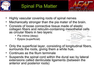
- 48. ŌĆó Highly vascular covering roots of spinal nerves ŌĆó Mechanically stronger than the pia mater of the brain. ŌĆó Consists of loose connective tissue made of elastic collagen fibers and reticulin-containing mesothelial cells as circular fibers in two layers: ŌĆó Pia intima (deep) ŌĆó Epipia (superficial) ŌĆó Only the superficial layer, consisting of longitudinal fibers, surrounds the roots, giving them a white hue. ŌĆó Continues as the filum terminale ŌĆó Suspends the spinal cord within the dural sac by lateral extensions called denticulate ligaments (between the anterior and posterior roots) Spinal Pia Matter
- 49. ŌĆó Composed of the pia medially and the dura laterally. ŌĆó 18 to 22 pairs ŌĆó Attach the lateral surface of the spinal cord to the dura transversely to support the spinal cord during flexion and extension. ŌĆó The medial border of each, thinner than the lateral border, is adherent to the lateral column Dentate Ligaments
- 50. ŌĆó The insertion of the dentate ligaments is between the DREZ and the anterior roots ŌĆō Landmark for the corticospinal tract posteriorly and the spinothalamic tract anteriorly ŌĆó At the cervical level, the ligament is located anterior to the spinal accessory nerve. ŌĆó Horizontal in cervical and Vertical in thoracic Dentate Ligaments
- 51. ŌĆó Not in the area anterior to the dentate ligaments ŌĆó Consisting of a dense, impermeable, avascular, fibroelastic membrane layer adherent to the dura, ŌĆó May contain calcifications. ŌĆó Laterally, the pia and arachnoid taper off at the point of contact between the spinal nerves and the dura, the latter becoming continuous with the epineurium. ŌĆō At this level, the arachnoid has intradural and transdural processes, as well as transvenous processes, similar to the pacchionian granulations found intracranially ŌĆō Involved in cerebrospinal fluid resorption. Spinal Arachnoid Matter
- 52. ŌĆó Encloses the caudal end of the medulla, the spinal roots of the spinal accessory nerves, the spinal cord, the filum terminale, and the cauda equina ŌĆó Extends from the foramen magnum to the sacrum. ŌĆó Forms a cylindrical sheath, is separated from the spinal canal by the epidural space (contains fat and the epidural venous plexuses). ŌĆó The thick rostral end of the dural sheath is adherent to the margin of the foramen magnum, to the dorsal aspect of the process of the axis, from which it is separated by the tectorial membrane (an extension of the common dorsal vertebral ligament) and to the dorsal craniospinal ligaments. (This adhesion disappears below the axis). Spinal Dura Matter
- 53. Vertebral Column
- 54. 1. Anatomy of spinal cord 2. Surface landmarks for pre-operative planning CONTENT
Editor's Notes
- #61: 3.1