Brachial Plexus Anatomy and Nerves .pptx
- 1. 1
- 2.  The brachial plexus (plexus brachialis) is a somatic nerve plexus formed by the ventral rami of C5-C8 and T1.  The brachial plexus provides the motor innervation and nearly all the sensory supply of the upper limb.  Based on its course brachial plexus can be divided into 5 parts. 2
- 3.  The brachial plexus (plexus brachialis) is a somatic nerve plexus formed by the ventral rami of C5-C8 and T1.  The brachial plexus provides the motor innervation and nearly all the sensory supply of the upper limb.  Based on its course brachial plexus can be divided into 5 parts. 3 Roots Trunks Divisions Cords Branches
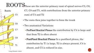
- 4.  These are the anterior primary rami of spinal nerves C5, C6, C7, C8 and T1, with contributions from the anterior primary rami of C4 and T2  The roots then joins together to form the trunk  Two anatomical Variations  PreFixed Brachial Plexus-the contribution by C4 is large and that from T2 is often absent  PostFixed Brachial Plexus-In a postfixed plexus, the contribution by T1 is large, T2 is always present, C4 is absent, and C5 is reduced in size. 4
- 5.  . As the roots of the brachial plexus emerge in the groove between the anterior and posterior tubercles of the transverse processes of the cervical vertebrae, they lie in a fibrofatty space between two sheaths of fibrous tissue.  The posterior part of the sheath - from the posterior tubercles and covers the front of scalenus medius;  the anterior part - from the anterior tubercles and covers the posterior aspect of scalenus anterior.  Laterally, the sheath extends as a covering around the brachial plexus as this emerges into the axilla 5
- 6.  Roots C5 and C6 join to form the upper trunk.  Root C7 forms the middle trunk.  Roots CB and T1 join to form the lower trunk.  The interscalene brachial plexus block technique therefore targets the trunks of the brachial plexus as they pass between the scalene muscles.  The three trunks emerge from between the scalene muscles and travels downwards and laterally. 6
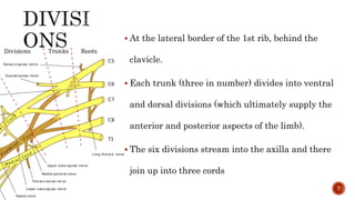
- 7.  At the lateral border of the 1st rib, behind the clavicle.  Each trunk (three in number) divides into ventral and dorsal divisions (which ultimately supply the anterior and posterior aspects of the limb).  The six divisions stream into the axilla and there join up into three cords 7
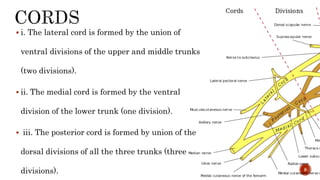
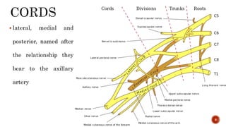
- 8.  i. The lateral cord is formed by the union of ventral divisions of the upper and middle trunks (two divisions).  ii. The medial cord is formed by the ventral division of the lower trunk (one division).  iii. The posterior cord is formed by union of the dorsal divisions of all the three trunks (three divisions). 8
- 9.  lateral, medial and posterior, named after the relationship they bear to the axillary artery 9
- 10.  Roots-Between the scalene muscles. The roots of the plexus lie above the second part of the subclavian artery  Trunks In the posterior triangle, the trunks of the plexus, invested in a sheath of prevertebral fascia, are superficially placed, being covered only by skin ,deep fascia and platysma. 10
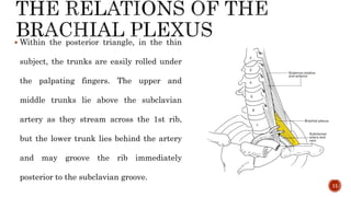
- 11.  Within the posterior triangle, in the thin subject, the trunks are easily rolled under the palpating fingers. The upper and middle trunks lie above the subclavian artery as they stream across the 1st rib, but the lower trunk lies behind the artery and may groove the rib immediately posterior to the subclavian groove. 11
- 12.  Divisions At the lateral border of the 1st rib, the trunks bifurcate into divisions that are situated behind the clavicle, the subclavius muscle and the suprascapular vessels (which lie immediately posterior to the clavicle) and then descend into the axilla. 12
- 13.  Cords The cords are formed at the apex of the axilla and become grouped around the axillary artery; at first, the medial cord lies behind the artery with the posterior and lateral cords lateral to this vessel, but behind pectoralis minor the cords take up their relations to the artery as signified by their 13
- 14.  Branches from Root Stage i. Dorsal scapular nerve or nerve to rhomboids (C5) ii. Long thoracic nerve or nerve of Bell (C5, C6, C7).  Branches of Upper Trunk i.Suprascapular nerve (C5, C6) ii. Nerve to subclavius (C5, C6) 14
- 15.  Branches from Lateral Cord Lateral root of median C5, C6, C7 Lateral pectoral - C5, C6, C7 Musculocutaneous C5, C6, C7 15
- 16.  Branches from Medial Cord • Medial root of median C8, T1 • Medial pectoral-C8, T1 • Medial cutaneous nerve of arm C8, T1 • Medial cutaneous nerve of forearm - C8, T1 • Ulnar nerve-C8, Т1 16
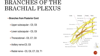
- 17.  Branches from Posterior Cord  Upper subscapular - C5, C6  Lower subscapular - C5, C6  Thoracodorsal - C6, C7, C8  Axillary nerve-C5, C6  Radial nerve - C5, C6, C7, C8, T1 17
- 18. 18
Editor's Notes
- #3: the area of the axilla (which is supplied by the supraclavicular nerve) and the dorsal scapula area, which is supplied by cutaneous branches of the dorsal rami.
- #4: the area of the axilla (which is supplied by the supraclavicular nerve) and the dorsal scapula area, which is supplied by cutaneous branches of the dorsal rami.
- #6: the area of the axilla (which is supplied by the supraclavicular nerve) and the dorsal scapula area, which is supplied by cutaneous branches of the dorsal rami.


