BRAIN TUMOR MRI IMAGE SEGMENTATION AND DETECTION IN IMAGE PROCESSING
•Download as PPTX, PDF•
18 likes•10,526 views

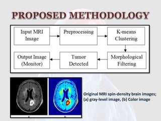
Image processing is an active research area in which medical image processing is a highly challenging field. Medical imaging techniques are used to image the inner portions of the human body for medical diagnosis. Brain tumor is a serious life altering disease condition. Image segmentation plays a significant role in image processing as it helps in the extraction of suspicious regions from the medical images. In this paper we have proposed segmentation of brain MRI image using K-means clustering algorithm followed by morphological filtering which avoids the misclustered regions that can inevitably be formed after segmentation of the brain MRI image for detection of tumor location.


![[1] R. P. Joseph, C. S. Singh, and M. Manikandan, “Brain Tumor
Mri Image Segmentation and Detection in Image Processing,” pp. 1–
5, 2014.
[2] M. Rakesh and T. Ravi, “Image Segmentation and Detection of
Tumor Objects in MR Brain Images Using FUZZY C-MEANS ( FCM )
Algorithm,” vol. 2, no. 3, pp. 2088–2094, 2012.
[3] H. P. S. P, G. K. Sundararaj, and A. Jayachandran, “Brain Tumor
Segmentation of Contras Material Applied MRI Using Enhanced Fuzzy
C-Means Clustering,” vol. 1, no. 2, pp. 161–166, 2012.
[4] B. Basavaprasad and M. Ravi, “A COMPARATIVE STUDY ON
CLASSIFICATION OF IMAGE SEGMENTATION METHODS WITH A FOCUS
ON GRAPH BASED TECHNIQUES,” pp. 310–315, 2014.
[5] K. I. Rahmani, “Clustering of Image Data Using K-Means and
Fuzzy,” vol. 5, no. 7, pp. 160–163, 2014.](https://image.slidesharecdn.com/imageprocessingbraintumordetection-160628111440/85/BRAIN-TUMOR-MRI-IMAGE-SEGMENTATION-AND-DETECTION-IN-IMAGE-PROCESSING-11-320.jpg)

BRAIN TUMOR MRI IMAGE SEGMENTATION AND DETECTION IN IMAGE PROCESSING
- 1. Miss.Dharshika Shreeganesh Reg No : 2012/SP/040 Index No : S 8288
- 2. Its me..! I am Sick..!
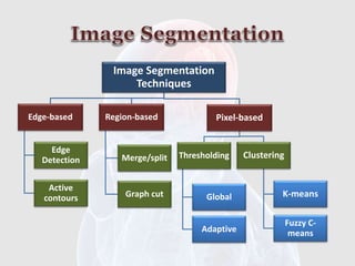
- 5. Image Segmentation Techniques Edge-based Edge Detection Active contours Region-based Merge/split Graph cut Pixel-based Clustering Fuzzy C- means K-means Thresholding Global Adaptive
- 6. Original MRI spin-density brain images; (a) gray-level image, (b) Color image
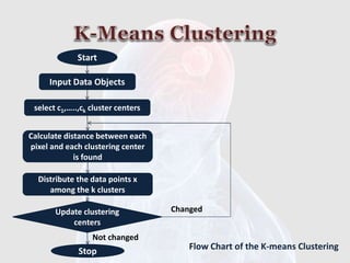
- 7. Start Input Data Objects select c1,…..,ck cluster centers Calculate distance between each pixel and each clustering center is found Distribute the data points x among the k clusters Update clustering centers Stop Changed Not changed Flow Chart of the K-means Clustering
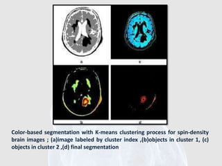
- 8. Color-based segmentation with K-means clustering process for spin-density brain images ; (a)image labeled by cluster index ,(b)objects in cluster 1, (c) objects in cluster 2 ,(d) final segmentation
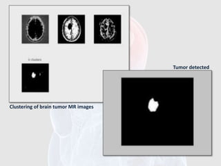
- 10. Clustering of brain tumor MR images Tumor detected
- 11. [1] R. P. Joseph, C. S. Singh, and M. Manikandan, “Brain Tumor Mri Image Segmentation and Detection in Image Processing,” pp. 1– 5, 2014. [2] M. Rakesh and T. Ravi, “Image Segmentation and Detection of Tumor Objects in MR Brain Images Using FUZZY C-MEANS ( FCM ) Algorithm,” vol. 2, no. 3, pp. 2088–2094, 2012. [3] H. P. S. P, G. K. Sundararaj, and A. Jayachandran, “Brain Tumor Segmentation of Contras Material Applied MRI Using Enhanced Fuzzy C-Means Clustering,” vol. 1, no. 2, pp. 161–166, 2012. [4] B. Basavaprasad and M. Ravi, “A COMPARATIVE STUDY ON CLASSIFICATION OF IMAGE SEGMENTATION METHODS WITH A FOCUS ON GRAPH BASED TECHNIQUES,” pp. 310–315, 2014. [5] K. I. Rahmani, “Clustering of Image Data Using K-Means and Fuzzy,” vol. 5, no. 7, pp. 160–163, 2014.
Editor's Notes
- #3: Brain, the important part of our body which is located on the most anterior to central nerves system. Brain tumor widely diagnosed and main problem among population of the world. Tumor the abnormal and uncontrolled growth and cell division in brain which like solid form.
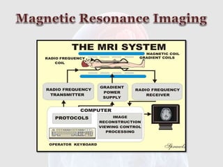
- #6: They are usually stored in the format called DICOM. The extension of the file is .dcm. DICOM stands for Digital Imaging and Communications in Medicine. The forum National Electrical Manufacturers Association, NEMAholds the copyright to this standard. As a further info, MATLAB contains dicomread as a function to read DICOM images.The data type is uint16,it needs to be converted to double for basic operations.