Bwat
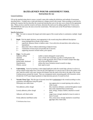
- 1. BATES-JENSEN WOUND ASSESSMENT TOOL Instructions for use General Guidelines: Fill out the attached rating sheet to assess a wound’s status after reading the definitions and methods of assessment described below. Evaluate once a week and whenever a change occurs in the wound. Rate according to each item by picking the response that best describes the wound and entering that score in the item score column for the appropriate date. When you have rated the wound on all items, determine the total score by adding together the 13-item scores. The HIGHER the total score, the more severe the wound status. Plot total score on the Wound Status Continuum to determine progress. Specific Instructions: 1. Size: Use ruler to measure the longest and widest aspect of the wound surface in centimeters; multiply length x width. 2. Depth: Pick the depth, thickness, most appropriate to the wound using these additional descriptions: 1= tissues damaged but no break in skin surface. 2= superficial, abrasion, blister or shallow crater. Even with, &/or elevated above skin surface (e.g., hyperplasia). 3= deep crater with or without undermining of adjacent tissue. 4= visualization of tissue layers not possible due to necrosis. 5= supporting structures include tendon, joint capsule. 3. Edges: Use this guide: Indistinct, diffuse = unable to clearly distinguish wound outline. Attached = even or flush with wound base, no sides or walls present; flat. Not attached = sides or walls are present; floor or base of wound is deeper than edge. Rolled under, thickened = soft to firm and flexible to touch. Hyperkeratosis = callous-like tissue formation around wound & at edges. Fibrotic, scarred = hard, rigid to touch. 4. Undermining: Assess by inserting a cotton tipped applicator under the wound edge; advance it as far as it will go without using undue force; raise the tip of the applicator so it may be seen or felt on the surface of the skin; mark the surface with a pen; measure the distance from the mark on the skin to the edge of the wound. Continue process around the wound. Then use a transparent metric measuring guide with concentric circles divided into 4 (25%) pie-shaped quadrants to help determine percent of wound involved. 5. Necrotic Tissue Type: Pick the type of necrotic tissue that is predominant in the wound according to color, consistency and adherence using this guide: White/gray non-viable tissue = may appear prior to wound opening; skin surface is white or gray. Non-adherent, yellow slough = thin, mucinous substance; scattered throughout wound bed; easily separated from wound tissue. Loosely adherent, yellow slough = thick, stringy, clumps of debris; attached to wound tissue. Adherent, soft, black eschar = soggy tissue; strongly attached to tissue in center or base of wound. Firmly adherent, hard/black eschar = firm, crusty tissue; strongly attached to wound base and edges (like a hard scab).  2001Barbara Bates-Jensen
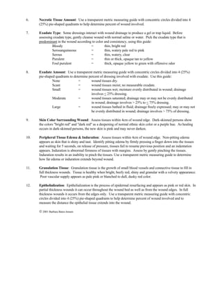
- 2. 6. Necrotic Tissue Amount: Use a transparent metric measuring guide with concentric circles divided into 4 (25%) pie-shaped quadrants to help determine percent of wound involved. 7. Exudate Type: Some dressings interact with wound drainage to produce a gel or trap liquid. Before assessing exudate type, gently cleanse wound with normal saline or water. Pick the exudate type that is predominant in the wound according to color and consistency, using this guide: Bloody = thin, bright red Serosanguineous = thin, watery pale red to pink Serous = thin, watery, clear Purulent = thin or thick, opaque tan to yellow Foul purulent = thick, opaque yellow to green with offensive odor 8. Exudate Amount: Use a transparent metric measuring guide with concentric circles divided into 4 (25%) pie-shaped quadrants to determine percent of dressing involved with exudate. Use this guide: None = wound tissues dry. Scant = wound tissues moist; no measurable exudate. Small = wound tissues wet; moisture evenly distributed in wound; drainage involves < 25% dressing. Moderate = wound tissues saturated; drainage may or may not be evenly distributed in wound; drainage involves > 25% to < 75% dressing. Large = wound tissues bathed in fluid; drainage freely expressed; may or may not be evenly distributed in wound; drainage involves > 75% of dressing. 9. Skin Color Surrounding Wound: Assess tissues within 4cm of wound edge. Dark-skinned persons show the colors "bright red" and "dark red" as a deepening of normal ethnic skin color or a purple hue. As healing occurs in dark-skinned persons, the new skin is pink and may never darken. 10. Peripheral Tissue Edema & Induration: Assess tissues within 4cm of wound edge. Non-pitting edema appears as skin that is shiny and taut. Identify pitting edema by firmly pressing a finger down into the tissues and waiting for 5 seconds, on release of pressure, tissues fail to resume previous position and an indentation appears. Induration is abnormal firmness of tissues with margins. Assess by gently pinching the tissues. Induration results in an inability to pinch the tissues. Use a transparent metric measuring guide to determine how far edema or induration extends beyond wound. 11. Granulation Tissue: Granulation tissue is the growth of small blood vessels and connective tissue to fill in full thickness wounds. Tissue is healthy when bright, beefy red, shiny and granular with a velvety appearance. Poor vascular supply appears as pale pink or blanched to dull, dusky red color. 12. Epithelialization: Epithelialization is the process of epidermal resurfacing and appears as pink or red skin. In partial thickness wounds it can occur throughout the wound bed as well as from the wound edges. In full thickness wounds it occurs from the edges only. Use a transparent metric measuring guide with concentric circles divided into 4 (25%) pie-shaped quadrants to help determine percent of wound involved and to measure the distance the epithelial tissue extends into the wound.  2001 Barbara Bates-Jensen
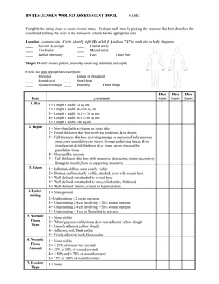
- 3. BATES-JENSEN WOUND ASSESSMENT TOOL NAME Complete the rating sheet to assess wound status. Evaluate each item by picking the response that best describes the wound and entering the score in the item score column for the appropriate date. Location: Anatomic site. Circle, identify right (R) or left (L) and use "X" to mark site on body diagrams: Sacrum & coccyx Lateral ankle Trochanter Medial ankle Ischial tuberosity Heel Other Site Shape: Overall wound pattern; assess by observing perimeter and depth. Circle and date appropriate description: Irregular Linear or elongated Round/oval Bowl/boat Square/rectangle Butterfly Other Shape Date Date Date Item Assessment Score Score Score 1. Size 1 = Length x width <4 sq cm 2 = Length x width 4--<16 sq cm 3 = Length x width 16.1--<36 sq cm 4 = Length x width 36.1--<80 sq cm 5 = Length x width >80 sq cm 2. Depth 1 = Non-blanchable erythema on intact skin 2 = Partial thickness skin loss involving epidermis &/or dermis 3 = Full thickness skin loss involving damage or necrosis of subcutaneous tissue; may extend down to but not through underlying fascia; &/or mixed partial & full thickness &/or tissue layers obscured by granulation tissue 4 = Obscured by necrosis 5 = Full thickness skin loss with extensive destruction, tissue necrosis or damage to muscle, bone or supporting structures 3. Edges 1 = Indistinct, diffuse, none clearly visible 2 = Distinct, outline clearly visible, attached, even with wound base 3 = Well-defined, not attached to wound base 4 = Well-defined, not attached to base, rolled under, thickened 5 = Well-defined, fibrotic, scarred or hyperkeratotic 4. Under- 1 = None present mining 2 =Undermining < 2 cm in any area 3 = Undermining 2-4 cm involving < 50% wound margins 4 = Undermining 2-4 cm involving > 50% wound margins 5 = Undermining > 4 cm or Tunneling in any area 5. Necrotic 1 = None visible Tissue 2 = White/grey non-viable tissue &/or non-adherent yellow slough Type 3 = Loosely adherent yellow slough 4 = Adherent, soft, black eschar 5 = Firmly adherent, hard, black eschar 6. Necrotic 1 = None visible Tissue 2 = < 25% of wound bed covered Amount 3 = 25% to 50% of wound covered 4 = > 50% and < 75% of wound covered 5 = 75% to 100% of wound covered 7. Exudate 1 = None Type
- 4. Date Date Date Item Assessment Score Score Score 2 = Bloody 3 = Serosanguineous: thin, watery, pale red/pink 4 = Serous: thin, watery, clear 5 = Purulent: thin or thick, opaque, tan/yellow, with or without odor 8. Exudate 1 = None, dry wound Amount 2 = Scant, wound moist but no observable exudate 3 = Small 4 = Moderate 5 = Large 9. Skin 1 = Pink or normal for ethnic group Color 2 = Bright red &/or blanches to touch Sur- 3 = White or grey pallor or hypopigmented rounding 4 = Dark red or purple &/or non-blanchable Wound 5 = Black or hyperpigmented 10. 1 = No swelling or edema Peripheral Tissue 2 = Non-pitting edema extends <4 cm around wound Edema 3 = Non-pitting edema extends >4 cm around wound 4 = Pitting edema extends < 4 cm around wound 5 = Crepitus and/or pitting edema extends >4 cm around wound 11. 1 = None present Peripheral Tissue 2 = Induration, < 2 cm around wound Induration 3 = Induration 2-4 cm extending < 50% around wound 4 = Induration 2-4 cm extending > 50% around wound 5 = Induration > 4 cm in any area around wound 12. Granu- 1 = Skin intact or partial thickness wound lation 2 = Bright, beefy red; 75% to 100% of wound filled &/or tissue Tissue overgrowth 3 = Bright, beefy red; < 75% & > 25% of wound filled 4 = Pink, &/or dull, dusky red &/or fills < 25% of wound 5 = No granulation tissue present 13. Epithe- 1 = 100% wound covered, surface intact lializa- 2 = 75% to <100% wound covered &/or epithelial tissue tion extends >0.5cm into wound bed 3 = 50% to <75% wound covered &/or epithelial tissue extends to <0.5cm into wound bed 4 = 25% to < 50% wound covered 5 = < 25% wound covered TOTAL SCORE SIGNATURE WOUND STATUS CONTINUUM 1 5 10 13 15 20 25 30 35 40 45 50 55 60 Tissue Wound Wound Health Regeneration Degeneration Plot the total score on the Wound Status Continuum by putting an "X" on the line and the date beneath the line. Plot multiple scores with their dates to see-at-a-glance regeneration or degeneration of the wound.  2001Barbara Bates-Jensen


