Case Study (2): CT-PA
•Download as PPTX, PDF•
1 like•157 views

Ms. K.M., a 39-year-old HIV-positive woman, presented with a week of hemoptysis and shortness of breath following a deep vein thrombosis in her right leg. Imaging investigations such as chest X-ray and venous duplex ultrasound can be normal or non-diagnostic in pulmonary embolism cases. CT pulmonary angiography has a high sensitivity of over 90% for detecting clots as small as 2mm, making it the gold standard for diagnosis. Indications for CT-PA include when the diagnosis needs to be confirmed to determine options for anticoagulation, IVC filter placement, or surgical embolectomy.


Case Study (2): CT-PA
- 2. Case Summary • Ms K.M, a 39 year old female, HIV +ve on ATRIPLA with a 1 week history of haemoptysis and SOB following a right leg DVT • Diagnosis: ? Recurrent Pulmonary Embolism
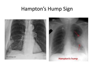
- 3. Imaging Investigations • CXR usually normal – Atelectasis or pleural effusion may be present – Classic signs such as hampton’s hump or westermark’s sign are very rare • Duplex u/s of the leg – Half of cases are negative
- 6. CT-PA • CT-Pulmonary Angiography has a sensetivity of > 90% • Visualise clots as small as 2mm • It is a Gold Standard
- 11. Indications For CT-Angiography • Pulmonary Embolism • Left ventricular stress/failure • Aortic Dissection • Teratology of Fallot • Aortic overloading
- 12. • Invasive and costs so.. • Usually reserved for patients in whom more – information or certainty of the diagnosis of PE are necessary.
- 13. Indications for CT-PA • Indications are – the need to confirm the diagnosis of PE in the presence of • contraindications to anticoagulation or • if IVC filter placement or • surgical embolectomy are contemplated
- 14. • In addition, patients with a – high index of clinical suspicion but non-diagnostic non-invasive studies and – patients with pulmonary hypertension of unknown cause
- 15. Contraindication for CT- Angio • Renal failure • Allergies to contrast • Pregnancy • Severe DM
- 16. • Life threatening complications are – typically secondary to acute cor pulmonale in patients with pre-existing severe pulmonary hypertension and – failing right ventricle.