Cell Signaling- Arijit.ppt
âĒDownload as PPT, PDFâĒ
0 likesâĒ28 views

The document discusses cell signaling and the JAK-STAT pathway. It notes that JAK-STAT signaling involves signal reception by a receptor, signal transduction through a series of protein phosphorylation steps, and a cellular response. STAT3 is constitutively activated in some cancers and promotes cell survival through upregulation of anti-apoptotic genes.


![CNS tumors
Retinoblastoma
[pRb]
Glioblastoma
[EGFR; PTEN; p53]
Medulloblastoma
De novo
[Primary]
Progressive
[Secondary]
P53 mutations and
accumulation
PTEN alteration and
EGFR amplification](https://image.slidesharecdn.com/cellsignaling-arijit-230329092409-b6bc57ce/85/Cell-Signaling-Arijit-ppt-36-320.jpg)


Cell Signaling- Arijit.ppt
- 2. Genetics: Studies of genes, heredity, and variations in organisms Gene: a unit of heredity Major Points âĒ Genes reside within chromosomes like beads on a string. âĒ Genes on the same chromosome are linked, but genes on different chromosomes assorted independently. âĒ New genes can be identified using linkage analysis based on recombination frequencies between known genes. âĒ Genes can become mutated to produce different phenotypes. âĒ Genes determine the activities of enzymes responsible for producing phenotypic traits (the âone gene-one enzymeâ hypothesis). âĒ The study of genes therefore is not just the study of inherited traits, but also the study of cellular functions.
- 3. The âOne gene-one enzymeâ hypothesis Nobel Prize, 1958 George Beadle Edward Tatum
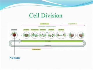
- 4. The Cell Nucleus Nuclear membrane Cytoplasm Cell membrane
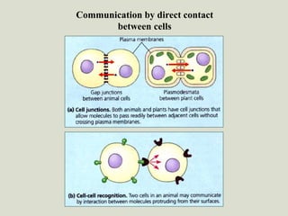
- 7. Communication by direct contact between cells
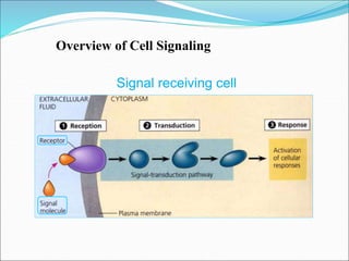
- 8. Signal receiving cell Overview of Cell Signaling
- 9. Transduction Signal transduction converts the change in the receptor to a form that can bring about a cellular response. This might involve a series of steps that alters and amplifies the change. Reception The cell targeted by a signal has a receptor molecule complementary to the signal molecule or ligand. The ligand fits like a key in a lock and triggers a change in the receptor molecule. Three stages of signaling process: âĒ Reception âĒ Transduction âĒ Response âĒ Activation of certain enzyme âĒ Rearrangement of the cytoskeleton âĒ Activation of specific genes Response This can be any of many cellular activities, such as:
- 12. âĒ When the signal molecule binds to the receptor, the receptor becomes activated.
- 15. Amplification of signals Protein kinases are important links in Many signal-transduction pathways. One kinase may activate many molecules of the next type of kinases in the chain, thus amplifying the signal, until the last kinase activates many protein molecules that carries out the final cellular response.
- 16. Non-protein molecules that act as intermediates e.g., cyclic AMP, calcium ions etc. Second messengers Calcium ion also acts as second messengers cAMP-mediated Signal Transduction
- 17. Cytoplasmic response to a signal Ultimate effect: Cellular response, such as- âĒ Alteration of metabolism âĒ Modulation of gene activity âĒ Rearrangement of cytoskeleton
- 18. âĒ Signaling pathways with a multiplicity of steps have two important benefits Protein kinase might activate a gene and trigger the synthesis of a new protein B. Specificity of response A. Signal amplification
- 21. Oligomerization Domain DNA-binding Domain SH2 Domain Activation Domain Dimerization Domain Y S N C N C JH1 JH2 JH3 JH4 JH5 JH6 JH7 Receptor-binding Region Pseudo-kinase Domain Kinase Domain Janus Kinase (JAK) Signal Transducer and Activator of Transcription (STAT)
- 23. âĒ Small protein molecules produced by mammalian cells âĒ Involved in cell-to-cell communication network âĒ Control division, differentiation and death of mammalian cells âĒ Essential for the development and function of the immune system âĒ Function in redundant and/or pleiotropic fashion Cytokines A B C D
- 24. Stat3 Signaling in Oncogenesis EGF, TGF-a, PDGF Src-family Kinase IL-6-family Cytokines Stat3 (Activated) BclXL Mcl1 Survival Bcl2 Pim Myc Cdc25A Division p16 p21 Differentiation
- 25. Stat Stat P PTK PTP Stat-Tyrosine Phosphorylation Is a Reversible Reaction
- 26. Question Why is Stat3 constitutively activated in certain human tumors?
- 27. Stat Recognition Sequences in Cytokine Responsive Genes N3-GAS AAG NNN CTT TTC NNN GAA AAN NNN NTT TTN NNN NAA N5-GAS Homodimers and Heterodimers of All Stats N4-GAS AAG NNNN CTT TTC NNNN GAA AAN NNNN NTT TTN NNNN NAA N6-GAS Homodimer of Stat6
- 28. ï· Receptor Inactivation - Receptor Antagonist - Decoy Receptor - Protein-tyrosine Phosphatase (PTP) - Suppressor of Cytokine Signaling (SOCS) - Proteolytic Degradation Negative Regulation of Jak-Stat Signaling ï· Jak Inactivation - Protein-tyrosine Phosphatase ï· Stat Inactivation - Protein Inhibitor of Activated Stat (PIAS) - Suppressor of Cytokine Signaling - Proteolytic Degradation - Protein-tyrosine Phosphatase - Proteolytic Degradation
- 29. Mission-critical Cellular and Molecular Events Underlying Cancer Progression âĒ Increased cell division âĒ Decreased cell death âĒ Gain-of-function status of Oncogenes/Oncoproteins âĒ Loss-of-function mutations of Tumor Suppressor Genes
- 30. Cellular and Molecular Biology of Gliomas Glioblastoma Multiforme Anaplastic Astrocytoma Astrocytoma Proliferation +/- ++ +++ Invasion ++ ++ +++ Angiogenesis - - +++ Rx Response - ++ - Survival 5-10 Years 2-3 Years 9-12 Months âĒ Mutation of p53 âĒ Over-expression of PDGF/PDGFR âĒ Mutation of Rb âĒ Amplification of CDK4 âĒ Loss of INK4A/ARF âĒ Loss of PTEN âĒ Amplification of EGFR âĒ Mutation of EGFR âĒ Loss of INK4A/ARF âĒ Loss of PTEN âĒ Mutation of Rb âĒ Amplification of IL-6
- 31. Oligomerization Domain DNA-binding Domain SH2 Domain Activation Domain Dimerization Domain F S Dominant Negative Mutant Stat3 N C
- 32. Common properties of most cancer cell lines for enhanced proliferation and survival âĒ Constitutive Stat3/other Stat activation. âĒ Constitutive Akt activation. âĒ Activation of Ras-MAPK pathway. âĒ Decreased p27 expression. âĒ Mutation in pRb family genes. âĒ Mutation /deletion of PTEN gene.
- 33. The Hallmarks of Cancer âĒ Self-sufficiency in growth signals Hanahan D and Weinberg RA Cell 100: 57-70, 2000 âĒ Insensitivity to anti-growth signals âĒ Evasion of programmed cell death âĒ Unlimited doubling potential âĒ Sustained angiogenesis âĒ Tissue invasion and metastasis
- 34. Mission-critical Cellular and Molecular Events Underlying Cancer Progression âĒ Increased cell division âĒ Gain-of-function status of Oncogenes/Oncoproteins âĒ Decreased cell death âĒ Loss-of-function mutations of Tumor Suppressor Genes
- 35. EGFR Signaling Pathways Stat3 PTEN MEK1 Erk1/Erk2 Proliferation PI3K AKT LY294002 Wortmannin U0126 mTOR S6 kinase Other S/T kinases Survival Transcription factors PD153035 AG1478
- 36. CNS tumors Retinoblastoma [pRb] Glioblastoma [EGFR; PTEN; p53] Medulloblastoma De novo [Primary] Progressive [Secondary] P53 mutations and accumulation PTEN alteration and EGFR amplification
- 37. âĒ Malignant gliomas are the most common subtype of primary brain tumors âĒ Glioma cells are migrating away from the main tumor mass through the brain parenchyma âĒ Clinically, gliomas are divided into four grades: Grade-I : Pilocytic astrocytoma Grade-II : Astrocytoma Grade-III : Anaplastic astrocytoma Grade-IV : Glioblastoma Multiforme (GBM) âĒ GBM(s) are multiforme in microscopically and genetically with various deletions, amplifications and point mutations âĒ GBM(s) are highly proliferative and resistant to apoptosis âĒ GBM (s) are resistant to radiation therapy and chemotherapy The Gliomas
- 38. Stat Family Members âĒ Stat1 : is crucial for interferon (IFN)-induced viral resistance âĒ Stat2 : is critical for IFN-a and IL-10 signaling âĒ Stat3 : deficiency results in very early embryonic lethality, for unknown reasons âĒ Stat4 : is critical for interleukin-12 signaling âĒ Stat5 : are activated in the response to a variety of (5A & 5B) cytokines including IL-13, EPO, OSM, GH, prolactin and IL-2 âĒ Stat6 : specifically mediates the effects of IL-4 and IL-13 on B or T cells
- 39. { Ras C-Myc Raf E2F Apoptosis and proliferation Survival and proliferation Net Loss of cells Net expansion of cells Survival signals Invasion Angiogenesis Metastasis Immune evasion