Chest and heart x ray
âĒDownload as PPTX, PDFâĒ
14 likesâĒ1,441 views

This document provides an overview of chest x-ray criteria, normal anatomy, and abnormalities that can be seen on chest x-rays. It discusses the proper technique for chest x-rays and includes sections on evaluating the heart, lungs, bones, diaphragm and mediastinum. Examples of common pathologies that can be identified on chest x-rays are given, such as pneumonia, lung cancer, tuberculosis, and metastases. Different abnormal heart shapes seen in conditions like cardiomegaly, transposition of the great vessels, and Fallot's tetralogy are also described.


Chest and heart x ray
- 1. CHEST AND HEART X-RAY BY DR. MOHAMED ABDEL-TAWAB LECTURER OF RADIOLOGY - ASSIUT UNIVERSITY
- 2. CRITERIA OF GOOD XR FILM âĒ ID âĒ Side RT, LT âĒ PA view âĒ Erect, Supine âĒ Penetration; contrast âĒ Field
- 4. CRITERIA OF GOOD XR FILM âĒ ID âĒ Side RT, LT âĒ PA view âĒ Erect, Supine âĒ Penetration; contrast âĒ Field
- 7. LOBES
- 8. REPORTING
- 9. 1- PLAIN X RAY Barium swallow: left atrial ++ indents esophagus
- 10. 2- VIEW OF X â RAY Lateral XR
- 11. 3- CENTRALIZATION IN THE X â RAY Medial clavicle at equidistance from spinous process
- 12. 4- BONY THORAX Fracture ribs ïĻ pneumothorax
- 13. 5- DIAPHRAGM Right copula > left copula
- 14. 6- MEDIASTIUM Bilateral hilar adenopathy Sarcoidosis
- 15. 7- HEART
- 16. 8- LUNG FIELDS APICAL, UPPER, MIDDLE, LOWER
- 17. REPORT ITEMS + SUMMARY OF LESIONS âĒ Clear both lung fields âĒ Clear costophrenic angles = No pleural effusion. âĒ Normal cardiac size and shape. âĒ No hilar adenopathy or mediastinal masses âĒ Intact bony thorax. No bony cervical ribs Impression NORMAL CHEST X-RAY
- 19. CARDIOMEGALY
- 20. CARDIOMEGALY
- 21. CARDIOMEGALY
- 22. CARDIOMEGALY Mitralization due to mitral stenosis
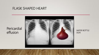
- 24. FLASK SHAPED HEART WATER BOTTLE SIGN Pericardial effusion
- 25. EGG ON SIDE = TRANSPOSITION OF GREAT VESSELS
- 26. BOOT SHAPED HEART (COEUR EN SABOT) = FALLOTâS TETRALOGY HEART IN FRENCH enlarged RV dilated aorta (RT) exaggerated waist
- 27. DUMBBELL SHAPED HEART âĒ Aneurysmal dilatation of both pa in primary pulmonary hypertension, cor pulmonale
- 28. COLLAPSE
- 29. COLLAPSE
- 30. COLLAPSE - CT
- 31. PNEUMONIA
- 32. PNEUMONIA
- 33. PNEUMONIA
- 36. LUNG ABSCESS
- 37. T.B. GHONS FOCUS
- 39. T.B. MILIARY TB
- 40. PLEURAL EFFUSION
- 42. PNEUMOTHORAX
- 43. PNEUMOTHORAX
- 44. BRONCHIECTASIS
- 45. EMPHYSEMA
- 46. EMPHYSEMA âĒ marked hyperinflation of the lungs âĒ flattened diaphragm, âĒ increased retrosternal air space âĒ hyperlucency of the lungs âĒ distorted parenchymal architecture. âĒ Ribbon shaped heart
- 48. ???
- 49. MEDIASTINAL MASS
- 50. LUNG CANCER
- 51. LUNG CANCER
- 52. LUNG CANCER
- 57. METASTASIS CANNON BALL âĒrenal cell carcinoma âĒchoriocarcinoma âĒprostate carcinoma âĒendometrial carcinoma