


Chronic inflammation
- 1. CHRONIC INFLAMMATION DR PREETI AGRAWAL
- 2. DEFINITION Chronic inflammation is inflammation of prolonged duration (weeks or months) in which inflammation, tissue injury, and attempts at repair coexist, in varying combinations.
- 3. CAUSES OF CHRONIC INFLAMMATION • Persistent infections by microorganisms that are difficult to eradicate, such as mycobacteria, and certain viruses, fungi, and parasites. • Immune-mediated inflammatory diseases - autoimmune diseases • Prolonged exposure to potentially toxic agents, either exogenous or endogenous.egs. silicosis , Atherosclerosis
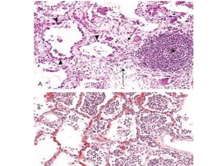
- 4. MORPHOLOGIC FEATURES chronic inflammation is characterized by: • Infiltration with mononuclear cells, which include macrophages, lymphocytes, and plasma cells • Tissue destruction, induced by the persistent offending agent or by the inflammatory cells • Attempts at healing by connective tissue replacement of damaged tissue, accomplished by proliferation of small blood vessels (angiogenesis) and, in particular, fibrosis
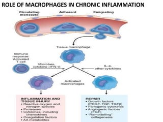
- 6. ROLE OF MACROPHAGES IN CHRONIC INFLAMMATION
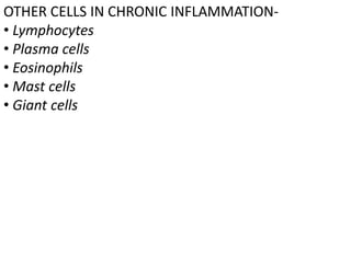
- 7. OTHER CELLS IN CHRONIC INFLAMMATION- • Lymphocytes • Plasma cells • Eosinophils • Mast cells • Giant cells
- 8. GRANULOMATOUS INFLAMMATION Granulomatous inflammation is a distinctive pattern of chronic inflammation that is encountered in a limited number of infectious and some noninfectious conditions.
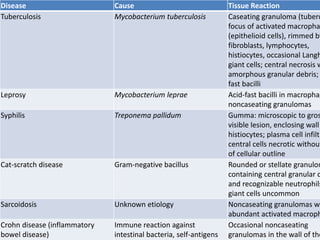
- 9. Disease Cause Tissue Reaction Tuberculosis Mycobacterium tuberculosis Caseating granuloma (tuberc focus of activated macropha (epithelioid cells), rimmed by fibroblasts, lymphocytes, histiocytes, occasional Langh giant cells; central necrosis w amorphous granular debris; fast bacilli Leprosy Mycobacterium leprae Acid-fast bacilli in macrophag noncaseating granulomas Syphilis Treponema pallidum Gumma: microscopic to gros visible lesion, enclosing wall histiocytes; plasma cell infiltr central cells necrotic without of cellular outline Cat-scratch disease Gram-negative bacillus Rounded or stellate granulom containing central granular d and recognizable neutrophils giant cells uncommon Sarcoidosis Unknown etiology Noncaseating granulomas wi abundant activated macroph Crohn disease (inflammatory bowel disease) Immune reaction against intestinal bacteria, self-antigens Occasional noncaseating granulomas in the wall of the
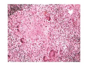
- 10. A granuloma is a focus of chronic inflammation consisting of a microscopic aggregation of macrophages that are transformed into epithelium- like cells, surrounded by a collar of mononuclear leukocytes, principally lymphocytes and occasionally plasma cells
- 11. Epithelioid cells fuse to form giant cells in the periphery or sometimes in the center of granulomas. These giant cells may attain diameters of 40 to 50 μm. They have a large mass of cytoplasm containing 20 or more small nuclei arranged either peripherally (Langhans-type giant cell) or haphazardly (foreign body–type giant cell). There is no known functional difference between these two types of giant cells