ºÝºÝߣ

ºÝºÝߣShare a Scribd company logo
The effects of bed height and time on the quality of chest compressions (and
                                                  resultant intrathoracic pressures) during Cardio Pulmonary Resuscitation (CPR).

                                                                     Dr Asher Lewinsohn, BSc, MB BS (Hons), Dr Peter Sherren MB BS, Dr Dhuleep Sanjay Wijayatilake, BSc, MB BS, FRCA

                                                                                               Department of Anaesthesia & Intensive Care Medicine, Queens Hospital, Essex, England.
                                                                                                                                                           Hospital,

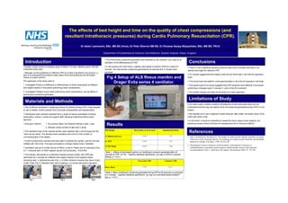
                                                                                                ß The intra thoracic pressures generated were detected by the ventilator, and used as an
  Introduction                                                                                  indication of the effectiveness of CPR.                                                          Conclusions
  Sudden cardiac arrest is a leading cause of death in Europe, affecting about 700,000          ß In the second part of the study, subjects were asked to perform CPR for a total of 2
                                                                                                                                                                                                ß There is now a significant growing evidence base which indicates that there is an
individuals every year1.                                                                        minutes. The intra thoracic pressures generated at 30 seconds and 2 minutes were
                                                                                                                                                                                                optimal bed height for effective CPR.
  Although current guidelines on effective CPR try to take most factors into account, a         recorded.
lack of a comprehensive evidence base means that most guidelines are still based                                                                                                                ß Our results suggest that this height is with the top of the bed in line with the operators
mainly on expert opinion2.                                                                        Fig 4 Setup of ALS Resus manikin and                                                          knee.
The objectives of this study were to:
                                                                                                      Drager Evita series 4 ventilator.                                                         ß This would have the patient s chest approximately in line with the operator s mid-thigh.
1. Investigate if there is a difference in effectiveness of chest compression at different                                                                                                      ß The second part of our study suggests that CPR would be more effective if the person
bed heights relative to the person performing chest compressions.                                                                                                                               performing it changed every 2 minutes (1 cycle of the ALS protocol) .
2. Investigate if fatigue occurs when performing chest compressions, and its effect on                                                                                                          ß This simple change can easily be achieved by a team approach.
intra thoracic pressures generated.

                                                                                                                                                                                                 Limitations of Study
  Materials and Methods
                                                                                                                                                                                                ß Our study used a manikin model to simulate the human chest wall cavity and we
ß Due to ethical constraints in obtaining consent for patients having CPR, it was decided                                                                                                       understand that this may not exactly mirror the pressures generated during CPR in a live
to use a manikin model (Laerdal ALS) to provide comparability and reproducibility.                                                                                                              subject.

ß Participants were randomly selected from a range of various specialities including                                                                                                            ß We decided not to use a cadaveric model because, after death, the elastic recoil of the
paramedics, doctors, nurses and support staff, following institutional ethics board                                                                                                             chest wall cavity is lost.
approval.
                                                                                                                                                                                                ß In the future, it would be interesting to repeat the study using human subjects, but
ß Exclusion criterion:     1. No previous Basic Life Support training in past 1 year.
                           2. Refused verbal consent to take part in study.
                                                                                                  Results                                                                                       obtaining consent (which will likely be retrospective) will of course be difficult.


ß The standard lungs of the Laerdal manikin were replaced with a set of lungs from the
resus annie series. This allowed direct intubation and a limit on the number of
                                                                                                Bed Height                        Mean Pmax at 30 seconds            Standard Deviation
                                                                                                                                                                                                  References
                                                                                                1) Xiphisternal area                        15.36 *                         2.52
connecting parts in the system.                                                                                                                                                                 1. Sans S, Kesteloot H, Kromhout D. The burden of cardiovascular diseases mortality in Europe.
                                                                                                                                                                                                   Task Force of the European Society of Cardiology on Cardiovascular Mortality and Morbidity
ß A size 9 portex endo tracheal tube was used to intubate the manikin, and the cuff was         2) ASIS                                     17.69 *                         2.91                   Statistics in Europe. Eur Heart J 1997;18:1231-48.
inflated with 10ml of air. This was connected to a Dragor series Evita 4 Ventilator.
                                                                                                                                                                                                2. International Liaison Committee on Resuscitation. International Consensus on
                                                                                                3) Mid Thigh                                18.89 *                         2.80
ß Ventilation was set to a tidal volume of 500ml, a rate of 10bpm and an inspiratory time                                                                                                          cardiopulmonary resuscitation and emergency cardiovascular science with treatment
of 1.7 seconds with no PEEP applied (as per ALS protocols). FiO2=30%.                                                                                                                              recommendations. Part 2. Adult basic life support. Resuscitation 2005; 67: 187-201
                                                                                                Table 1 : Effects of bed height position on intrathoracic pressure generated after 30
ß The manikin was placed on a standard hospital recovery trolley, and CPR was                   seconds of CPR. N=100. * signifies statistical significance. (by way of ANOVA variance
performed for 2 minutes at 3 different bed heights relative to the subjects before              testing, p < 0.01.)
recording data: 1) Xiphisternal area (Fig 1), 2) ASIS (Anterior Superior Iliac Spine (Fig2),                                           30 seconds CPR                 2 minutes CPR
3) Mid Thigh (Fig 3). Between each set of readings, a 2 minute rest period was given.

                                                                                                Pmax Mean                                   17.52*                         14.5 *


                                                                                                Table 2: Mean intrathoracic pressures generated during CPR at 30 seconds as compared
                                                                                                to 2 minutes. * signifies statistical significance. (by way of a one-tailed paired student t-
                                                                                                test, p < 0.01.)
       Fig 1.                               Fig 2.                                Fig3.
More Related Content

Cpr poster

  • 1. The effects of bed height and time on the quality of chest compressions (and resultant intrathoracic pressures) during Cardio Pulmonary Resuscitation (CPR). Dr Asher Lewinsohn, BSc, MB BS (Hons), Dr Peter Sherren MB BS, Dr Dhuleep Sanjay Wijayatilake, BSc, MB BS, FRCA Department of Anaesthesia & Intensive Care Medicine, Queens Hospital, Essex, England. Hospital, ß The intra thoracic pressures generated were detected by the ventilator, and used as an Introduction indication of the effectiveness of CPR. Conclusions Sudden cardiac arrest is a leading cause of death in Europe, affecting about 700,000 ß In the second part of the study, subjects were asked to perform CPR for a total of 2 ß There is now a significant growing evidence base which indicates that there is an individuals every year1. minutes. The intra thoracic pressures generated at 30 seconds and 2 minutes were optimal bed height for effective CPR. Although current guidelines on effective CPR try to take most factors into account, a recorded. lack of a comprehensive evidence base means that most guidelines are still based ß Our results suggest that this height is with the top of the bed in line with the operators mainly on expert opinion2. Fig 4 Setup of ALS Resus manikin and knee. The objectives of this study were to: Drager Evita series 4 ventilator. ß This would have the patient s chest approximately in line with the operator s mid-thigh. 1. Investigate if there is a difference in effectiveness of chest compression at different ß The second part of our study suggests that CPR would be more effective if the person bed heights relative to the person performing chest compressions. performing it changed every 2 minutes (1 cycle of the ALS protocol) . 2. Investigate if fatigue occurs when performing chest compressions, and its effect on ß This simple change can easily be achieved by a team approach. intra thoracic pressures generated. Limitations of Study Materials and Methods ß Our study used a manikin model to simulate the human chest wall cavity and we ß Due to ethical constraints in obtaining consent for patients having CPR, it was decided understand that this may not exactly mirror the pressures generated during CPR in a live to use a manikin model (Laerdal ALS) to provide comparability and reproducibility. subject. ß Participants were randomly selected from a range of various specialities including ß We decided not to use a cadaveric model because, after death, the elastic recoil of the paramedics, doctors, nurses and support staff, following institutional ethics board chest wall cavity is lost. approval. ß In the future, it would be interesting to repeat the study using human subjects, but ß Exclusion criterion: 1. No previous Basic Life Support training in past 1 year. 2. Refused verbal consent to take part in study. Results obtaining consent (which will likely be retrospective) will of course be difficult. ß The standard lungs of the Laerdal manikin were replaced with a set of lungs from the resus annie series. This allowed direct intubation and a limit on the number of Bed Height Mean Pmax at 30 seconds Standard Deviation References 1) Xiphisternal area 15.36 * 2.52 connecting parts in the system. 1. Sans S, Kesteloot H, Kromhout D. The burden of cardiovascular diseases mortality in Europe. Task Force of the European Society of Cardiology on Cardiovascular Mortality and Morbidity ß A size 9 portex endo tracheal tube was used to intubate the manikin, and the cuff was 2) ASIS 17.69 * 2.91 Statistics in Europe. Eur Heart J 1997;18:1231-48. inflated with 10ml of air. This was connected to a Dragor series Evita 4 Ventilator. 2. International Liaison Committee on Resuscitation. International Consensus on 3) Mid Thigh 18.89 * 2.80 ß Ventilation was set to a tidal volume of 500ml, a rate of 10bpm and an inspiratory time cardiopulmonary resuscitation and emergency cardiovascular science with treatment of 1.7 seconds with no PEEP applied (as per ALS protocols). FiO2=30%. recommendations. Part 2. Adult basic life support. Resuscitation 2005; 67: 187-201 Table 1 : Effects of bed height position on intrathoracic pressure generated after 30 ß The manikin was placed on a standard hospital recovery trolley, and CPR was seconds of CPR. N=100. * signifies statistical significance. (by way of ANOVA variance performed for 2 minutes at 3 different bed heights relative to the subjects before testing, p < 0.01.) recording data: 1) Xiphisternal area (Fig 1), 2) ASIS (Anterior Superior Iliac Spine (Fig2), 30 seconds CPR 2 minutes CPR 3) Mid Thigh (Fig 3). Between each set of readings, a 2 minute rest period was given. Pmax Mean 17.52* 14.5 * Table 2: Mean intrathoracic pressures generated during CPR at 30 seconds as compared to 2 minutes. * signifies statistical significance. (by way of a one-tailed paired student t- test, p < 0.01.) Fig 1. Fig 2. Fig3.