際際滷

際際滷Share a Scribd company logo
Cyclovertical deviation
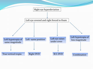
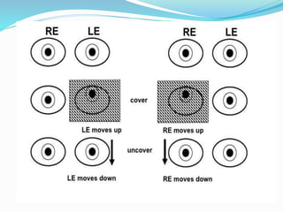
Right eye hyperdeviation
Left eye covered and right forced to fixate
Left hypotropia of
same magnitude
True vertical tropia
Left -same position
Right DVD
Left eye raises
under cover
B/L DVD
Left hypotropia of
less magnitude
Combination
Principles in cyclovertical
deviations
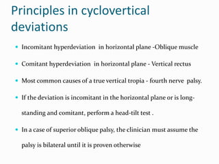
? Incomitant hyperdeviation in horizontal plane -Oblique muscle
? Comitant hyperdeviation in horizontal plane - Vertical rectus
? Most common causes of a true vertical tropia - fourth nerve palsy.
? If the deviation is incomitant in the horizontal plane or is long-
standing and comitant, perform a head-tilt test .
? In a case of superior oblique palsy, the clinician must assume the
palsy is bilateral until it is proven otherwise
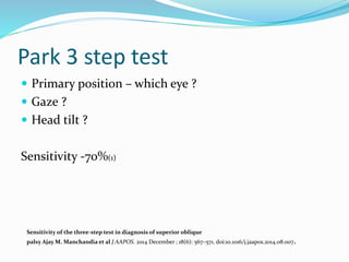
Park 3 step test
? Primary position C which eye ?
? Gaze ?
? Head tilt ?
Sensitivity -70%(1)
Sensitivity of the three-step test in diagnosis of superior oblique
palsy Ajay M. Manchandia et al J AAPOS. 2014 December ; 18(6): 567C571. doi:10.1016/j.jaapos.2014.08.007.
Cyclovertical deviation
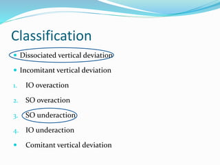
Classification
? Dissociated vertical deviation
? Incomitant vertical deviation
1. IO overaction
2. SO overaction
3. SO underaction
4. IO underaction
? Comitant vertical deviation
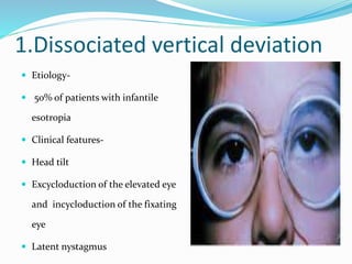
1.Dissociated vertical deviation
? Etiology-
? 50% of patients with infantile
esotropia
? Clinical features-
? Head tilt
? Excycloduction of the elevated eye
and incycloduction of the fixating
eye
? Latent nystagmus
Cyclovertical deviation
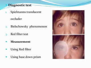
? Diagnostic test
1. Spielmanns translucent
occluder
2. Bielschowsky phenomenon
3. Red filter test
? Measurement
? Using Red filter
? Using base down prism
?Treatment
Medical
1. Refractive error
2. Occlusion
3. Prisms
Surgical
1. When ?
2. What ?
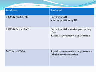
Condition Treatment
IOOA & mod. DVD Recession with
anterior positioning IO
IOOA & Severe DVD Recession with anterior positioning
IO +
Superior rectus-recession 7-10 mm
DVD & no IOOA: Superior rectus-recession 7-10 mm +
Inferior rectus resection
Inferior Oblique Muscle Over action
? Etiology
1. Primary
2. Secondary
? Clinical Features
? Management
? Weakening procedure on the
inferior oblique muscle
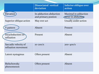
Dissociated vertical
deviation
Inferior oblique over
action
Elevation In adduction abduction
and primary postion
Maximal in adduction
never in abduction
Superior oblique action May over act Usually under action
V pattern Absent Present
Incycloduction on
refixation
Present Absent
Saccadic velocity of
refixation movement
10C200/s 200C400/s
Latent nystagmus Often present Absent
Bielschowsky
phenomenon
Often present Absent
Superior Oblique Muscle Over action
? Clinical Features
1. Overdepression in
adduction.
2. Associated with exo
? Management
? Superior oblique tendon
weakening procedure
4.Superior Oblique Muscle
Paralysis
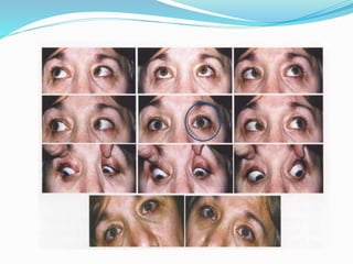
? Most common Single cyclovertical muscle paralysis.
? Etiology
? Congenital vs acquired
? Head trauma
? Vascular problems of the central nervous system,
? Diabetes mellitus, or a brain tumor
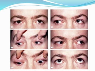
Clinical features
? Head tilt
? Hypertropia
? Excyclotorsion
? Esotropia
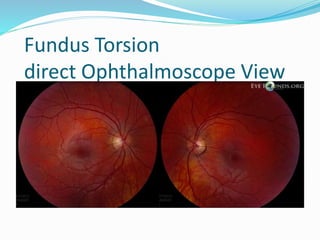
Fundus Torsion
direct Ophthalmoscope View
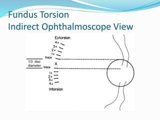
Fundus Torsion
Indirect Ophthalmoscope View
Cyclovertical deviation
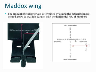
Maddox wing
? The amount of cyclophoria is determined by asking the patient to move
the red arrow so that it is parallel with the horizontal row of numbers
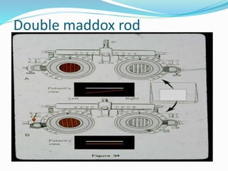
Double maddox rod
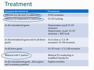
Treatment
Greatest deviation in Treatment
Affected eye elevated in adduction I/l IO recession
Affected eye depressed in adduction I/L SO tucking
In all contralateral gazes Hypertropia<25 pd I/L IO
recession
Hypertropia> 25 pd I/L IO
recession + RSO tuck
In all contralateral gazes and in all down
gazeS
As in class 3+ C/L IR
recession/ I/L SR recession
In all down gazes I/L SO tuck + C/L LIR recession
Bilateral with V pattern Bilateral IO weakening or
modified Harada Ito
In all contralatreral gazes , down gazes
and in primary position
Explore trochlea
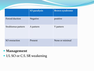
4.Inferior Oblique Muscle Paralysis
IO paralysis Brown syndrome
Forced duction Negative positive
Strabismus pattern A pattern V pattern
SO overaction Present None or minimal
? Management
? I/L SO or C/L SR weakening
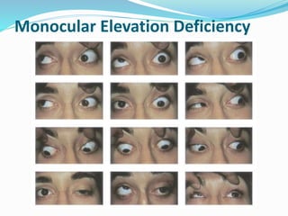
Monocular Elevation Deficiency
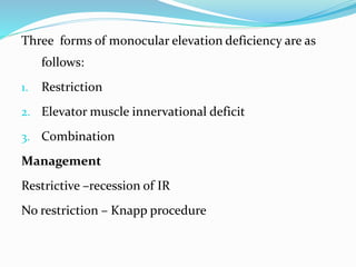
Three forms of monocular elevation deficiency are as
follows:
1. Restriction
2. Elevator muscle innervational deficit
3. Combination
Management
Restrictive Crecession of IR
No restriction C Knapp procedure
Refernces
? Binocular vision and occular motility - Von noorden
5th edition
? Handbook of Pediatric Strabismus and Amblyopia
Kenneth W. Wright, MD
? PEDIATRIC Ophthalmologyand Strabismus - Creig S
Hoyt -4thedition
? Pediatric Ophthalmology and Strabismus -Section
6 2015-2016- American academy of ophthalmology.

More Related Content

Cyclovertical deviation

  • 2. Right eye hyperdeviation Left eye covered and right forced to fixate Left hypotropia of same magnitude True vertical tropia Left -same position Right DVD Left eye raises under cover B/L DVD Left hypotropia of less magnitude Combination
  • 3. Principles in cyclovertical deviations ? Incomitant hyperdeviation in horizontal plane -Oblique muscle ? Comitant hyperdeviation in horizontal plane - Vertical rectus ? Most common causes of a true vertical tropia - fourth nerve palsy. ? If the deviation is incomitant in the horizontal plane or is long- standing and comitant, perform a head-tilt test . ? In a case of superior oblique palsy, the clinician must assume the palsy is bilateral until it is proven otherwise
  • 4. Park 3 step test ? Primary position C which eye ? ? Gaze ? ? Head tilt ? Sensitivity -70%(1) Sensitivity of the three-step test in diagnosis of superior oblique palsy Ajay M. Manchandia et al J AAPOS. 2014 December ; 18(6): 567C571. doi:10.1016/j.jaapos.2014.08.007.
  • 7. Classification ? Dissociated vertical deviation ? Incomitant vertical deviation 1. IO overaction 2. SO overaction 3. SO underaction 4. IO underaction ? Comitant vertical deviation
  • 8. 1.Dissociated vertical deviation ? Etiology- ? 50% of patients with infantile esotropia ? Clinical features- ? Head tilt ? Excycloduction of the elevated eye and incycloduction of the fixating eye ? Latent nystagmus
  • 10. ? Diagnostic test 1. Spielmanns translucent occluder 2. Bielschowsky phenomenon 3. Red filter test ? Measurement ? Using Red filter ? Using base down prism
  • 11. ?Treatment Medical 1. Refractive error 2. Occlusion 3. Prisms Surgical 1. When ? 2. What ?
  • 12. Condition Treatment IOOA & mod. DVD Recession with anterior positioning IO IOOA & Severe DVD Recession with anterior positioning IO + Superior rectus-recession 7-10 mm DVD & no IOOA: Superior rectus-recession 7-10 mm + Inferior rectus resection
  • 13. Inferior Oblique Muscle Over action ? Etiology 1. Primary 2. Secondary ? Clinical Features ? Management ? Weakening procedure on the inferior oblique muscle
  • 14. Dissociated vertical deviation Inferior oblique over action Elevation In adduction abduction and primary postion Maximal in adduction never in abduction Superior oblique action May over act Usually under action V pattern Absent Present Incycloduction on refixation Present Absent Saccadic velocity of refixation movement 10C200/s 200C400/s Latent nystagmus Often present Absent Bielschowsky phenomenon Often present Absent
  • 15. Superior Oblique Muscle Over action ? Clinical Features 1. Overdepression in adduction. 2. Associated with exo ? Management ? Superior oblique tendon weakening procedure
  • 16. 4.Superior Oblique Muscle Paralysis ? Most common Single cyclovertical muscle paralysis. ? Etiology ? Congenital vs acquired ? Head trauma ? Vascular problems of the central nervous system, ? Diabetes mellitus, or a brain tumor
  • 17. Clinical features ? Head tilt ? Hypertropia ? Excyclotorsion ? Esotropia
  • 21. Maddox wing ? The amount of cyclophoria is determined by asking the patient to move the red arrow so that it is parallel with the horizontal row of numbers
  • 23. Treatment Greatest deviation in Treatment Affected eye elevated in adduction I/l IO recession Affected eye depressed in adduction I/L SO tucking In all contralateral gazes Hypertropia<25 pd I/L IO recession Hypertropia> 25 pd I/L IO recession + RSO tuck In all contralateral gazes and in all down gazeS As in class 3+ C/L IR recession/ I/L SR recession In all down gazes I/L SO tuck + C/L LIR recession Bilateral with V pattern Bilateral IO weakening or modified Harada Ito In all contralatreral gazes , down gazes and in primary position Explore trochlea
  • 25. IO paralysis Brown syndrome Forced duction Negative positive Strabismus pattern A pattern V pattern SO overaction Present None or minimal ? Management ? I/L SO or C/L SR weakening
  • 27. Three forms of monocular elevation deficiency are as follows: 1. Restriction 2. Elevator muscle innervational deficit 3. Combination Management Restrictive Crecession of IR No restriction C Knapp procedure
  • 28. Refernces ? Binocular vision and occular motility - Von noorden 5th edition ? Handbook of Pediatric Strabismus and Amblyopia Kenneth W. Wright, MD ? PEDIATRIC Ophthalmologyand Strabismus - Creig S Hoyt -4thedition ? Pediatric Ophthalmology and Strabismus -Section 6 2015-2016- American academy of ophthalmology.