際際滷

際際滷Share a Scribd company logo
Surgical Technique
′ DCER stands for distraction, compression, extension, and reduction
′ reduce, realign and correct (even very severe) basilar invagination (BI),
atlantoaxial dislocation (AAD) with a posterior only, single staged approach
′ Involves motion in 2-axis using the lever principle
Sarat Chandra P, Bajaj J, Singh PK, Garg K, Agarwal D. Basilar Invagination and Atlantoaxial Dislocation: Reduction, Deformity Correction and Realignment Using the
DCER (Distraction, Compression, Extension, and Reduction) Technique With Customized Instrumentation and Implants. Neurospine. 2019 Jun;16(2):231-250. doi:
10.14245/ns.1938194.097
DCER (distraction, compression, extension, and reduction) technique
′ CVJ anomalies occur in roughly in 2 situations.
C1 arch is not fused with the occiput (reducible AAD): usually produces AAD.The
treatment of choice is C1 lateral mass and C2 pars screw fixation as described by
Goel and Shah
C1 arch is fused with occiput (previously called irreducible AAD): usually present
with moderate to severe BI.
DCER not only to reduce the AAD and BI but also to correct the hyper-lordosis of the
subaxial cervical spine
Sarat Chandra P, Bajaj J, Singh PK, Garg K, Agarwal D. Basilar Invagination and Atlantoaxial Dislocation: Reduction, Deformity Correction and Realignment Using the
DCER (Distraction, Compression, Extension, and Reduction) Technique With Customized Instrumentation and Implants. Neurospine. 2019 Jun;16(2):231-250. doi:
10.14245/ns.1938194.097
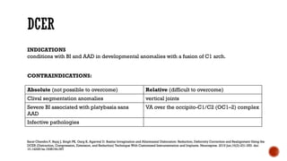
Absolute (not possible to overcome) Relative (difficult to overcome)
Clival segmentation anomalies vertical joints
Severe BI associated with platybasia sans
AAD
VA over the occipito-C1/C2 (OC1C2) complex
Infective pathologies
INDICATIONS
conditions with BI and AAD in developmental anomalies with a fusion of C1 arch.
CONTRAINDICATIONS:
Sarat Chandra P, Bajaj J, Singh PK, Garg K, Agarwal D. Basilar Invagination and Atlantoaxial Dislocation: Reduction, Deformity Correction and Realignment Using the
DCER (Distraction, Compression, Extension, and Reduction) Technique With Customized Instrumentation and Implants. Neurospine. 2019 Jun;16(2):231-250. doi:
10.14245/ns.1938194.097
′ Type I surgery
′ SI ranges from 90<C100<
′ DCER only would suffice
Sarat Chandra P, Bajaj J, Singh PK, Garg K, Agarwal D. Basilar Invagination and Atlantoaxial Dislocation: Reduction, Deformity Correction and Realignment Using the
DCER (Distraction, Compression, Extension, and Reduction) Technique With Customized Instrumentation and Implants. Neurospine. 2019 Jun;16(2):231-250. doi:
10.14245/ns.1938194.097
′ Type II surgery
′ SI ranges from 100<C160<
′ a joint remodeling should be performed, followed by DCER
′ Type III surgery
′ SI ranges between 160<C180<, i.e., the joint is almost vertical/ vertical
′ an extra-articular spacer is placed within the `pseudo-joint¨ to perform distraction
′ followed by technique of compression and extension (extra-articular distraction
with DCER)
Sarat Chandra P, Bajaj J, Singh PK, Garg K, Agarwal D. Basilar Invagination and Atlantoaxial Dislocation: Reduction, Deformity Correction and Realignment Using the
DCER (Distraction, Compression, Extension, and Reduction) Technique With Customized Instrumentation and Implants. Neurospine. 2019 Jun;16(2):231-250. doi:
10.14245/ns.1938194.097
DCER (distraction, compression, extension, and reduction) technique
′ Positioned prone on horseshoe with eye padding
′ No overnight skeletal traction
′ Intraoperative skeletal traction may be used with mild weight (around 2 kg)
′ Iliac crest bone graft is prepared.
Sarat Chandra P, Bajaj J, Singh PK, Garg K, Agarwal D. Basilar Invagination and Atlantoaxial Dislocation: Reduction, Deformity Correction and Realignment Using the
DCER (Distraction, Compression, Extension, and Reduction) Technique With Customized Instrumentation and Implants. Neurospine. 2019 Jun;16(2):231-250. doi:
10.14245/ns.1938194.097
′ Exposure is made from occiput till C4.
′ C1C2 joints exposed by following the superior border of the C2 lamina laterally
′ The C2 root ganglion seen over the joint may be saved by retracting it superiorly
′ If `hypertrophic¨ or is entrapped between the true and the pseudo-joint, will have to
be cut
Sarat Chandra P, Bajaj J, Singh PK, Garg K, Agarwal D. Basilar Invagination and Atlantoaxial Dislocation: Reduction, Deformity Correction and Realignment Using the
DCER (Distraction, Compression, Extension, and Reduction) Technique With Customized Instrumentation and Implants. Neurospine. 2019 Jun;16(2):231-250. doi:
10.14245/ns.1938194.097
′ Posterior rim of occiput should be drilled to just 2C3 cm
′ Raised as a small bone flap from the margin of the foramen magnum
′ Drill the lateral margin of foramen magnum to prevent any lateral compression
during compression and extension.
Sarat Chandra P, Bajaj J, Singh PK, Garg K, Agarwal D. Basilar Invagination and Atlantoaxial Dislocation: Reduction, Deformity Correction and Realignment Using the
DCER (Distraction, Compression, Extension, and Reduction) Technique With Customized Instrumentation and Implants. Neurospine. 2019 Jun;16(2):231-250. doi:
10.14245/ns.1938194.097
DCER (distraction, compression, extension, and reduction) technique
′ optimized for placement of spacer
′ denuding the cartilage to joint remodeling.
′ Type I surgery: the joint surfaces are drilled gently using a diamond drill
′ Type II surgery: posterior margins of the joints are drilled to change the SI to
around 90
′ Type III surgery: an extra-articular spacer is placed within the `pseudo-joint¨ to
perform distraction.This is then followed by technique of compression and
extension (extra-articular distraction with DCER)
Sarat Chandra P, Bajaj J, Singh PK, Garg K, Agarwal D. Basilar Invagination and Atlantoaxial Dislocation: Reduction, Deformity Correction and Realignment Using the
DCER (Distraction, Compression, Extension, and Reduction) Technique With Customized Instrumentation and Implants. Neurospine. 2019 Jun;16(2):231-250. doi:
10.14245/ns.1938194.097
DCER (distraction, compression, extension, and reduction) technique
′ The height of spacer is roughly 80%C90% the height of BI as calculated by
Chamberlain line
′ Initially trial implant (solid, smooth biconvex)followed by placement of the final
implant (biconvex `bullet shaped¨ hollow structure with serrated margins).
Sarat Chandra P, Bajaj J, Singh PK, Garg K, Agarwal D. Basilar Invagination and Atlantoaxial Dislocation: Reduction, Deformity Correction and Realignment Using the
DCER (Distraction, Compression, Extension, and Reduction) Technique With Customized Instrumentation and Implants. Neurospine. 2019 Jun;16(2):231-250. doi:
10.14245/ns.1938194.097
′ Braided no. 20 stainless-steel (SS) wire tied between the temporary occipital screw
and inferior border of the C2 spine
′ Gradual turning of the wire leads first to compression between OC1C2 tightly
holding the spacer in place followed by extension at the OC1C2 joints
′ CT using O-arm is taken to confirm complete reduction
′ Incase of incomplete reduction, higher height spacers considered
Sarat Chandra P, Bajaj J, Singh PK, Garg K, Agarwal D. Basilar Invagination and Atlantoaxial Dislocation: Reduction, Deformity Correction and Realignment Using the
DCER (Distraction, Compression, Extension, and Reduction) Technique With Customized Instrumentation and Implants. Neurospine. 2019 Jun;16(2):231-250. doi:
10.14245/ns.1938194.097
′ 4 occipital screws fixed to a contoured rod with 4 clamps (VERTEX, Medtronic
Sofamor Danek,Warsaw, IN, USA)
′ Long segment 3-point fixation performed (OC2C3 or OC3C4)
′ Occipital and C2 spine margins are roughened using a diamond drill and bone
graft is placed to enhance bony fusion
Sarat Chandra P, Bajaj J, Singh PK, Garg K, Agarwal D. Basilar Invagination and Atlantoaxial Dislocation: Reduction, Deformity Correction and Realignment Using the
DCER (Distraction, Compression, Extension, and Reduction) Technique With Customized Instrumentation and Implants. Neurospine. 2019 Jun;16(2):231-250. doi:
10.14245/ns.1938194.097
′ Titanium C1C2 spacers (PSC Spacers) (4 mm to 22 mm)
′ Universal craniovertebral junction reducer (UCVJR)
Sarat Chandra P, Bajaj J, Singh PK, Garg K, Agarwal D. Basilar Invagination and Atlantoaxial Dislocation: Reduction, Deformity Correction and Realignment Using the
DCER (Distraction, Compression, Extension, and Reduction) Technique With Customized Instrumentation and Implants. Neurospine. 2019 Jun;16(2):231-250. doi:
10.14245/ns.1938194.097
′ The ventral end is wedge shaped so that the spacer may introduced easily.
′ The superior and inferior surfaces are serrated so that the spacer can get a proper
grip over the C1C2 joint surface.
′ The central portion is a hollow core which may be filled with autologous bone or
bone like material.
′ The dorsal end is used to attach a screw driver.
′ The whole shape is like a `bullet shape¨ to convert the joint into a pivot joint to allow
the technique of DCER
Sarat Chandra P, Bajaj J, Singh PK, Garg K, Agarwal D. Basilar Invagination and Atlantoaxial Dislocation: Reduction, Deformity Correction and Realignment Using the
DCER (Distraction, Compression, Extension, and Reduction) Technique With Customized Instrumentation and Implants. Neurospine. 2019 Jun;16(2):231-250. doi:
10.14245/ns.1938194.097
DCER (distraction, compression, extension, and reduction) technique
Spacer Screw
′ The C2 dorsal listhesis cannot reduce unless an additional ventral `push¨ is
provided to C2
′ UCVJR/C2 translator has a fulcrum over the occipital plate
′ A screw is passed through the hole in the structure of the occipital plate.
′ Tightening this screw pushes the C2 anteriorly
Sarat Chandra P, Bajaj J, Singh PK, Garg K, Agarwal D. Basilar Invagination and Atlantoaxial Dislocation: Reduction, Deformity Correction and Realignment Using the
DCER (Distraction, Compression, Extension, and Reduction) Technique With Customized Instrumentation and Implants. Neurospine. 2019 Jun;16(2):231-250. doi:
10.14245/ns.1938194.097
DCER (distraction, compression, extension, and reduction) technique
′ Type I surgery: extubate
′ Type II and III surgeries: electively ventilate them overnight and extubated the
next day.
′ Philadelphia hard collar for a period of 3 months.
′ Isomeric neck exercises started within 1 week, as soon as the pain subsides to
prevent disuse atrophy
Sarat Chandra P, Bajaj J, Singh PK, Garg K, Agarwal D. Basilar Invagination and Atlantoaxial Dislocation: Reduction, Deformity Correction and Realignment Using the
DCER (Distraction, Compression, Extension, and Reduction) Technique With Customized Instrumentation and Implants. Neurospine. 2019 Jun;16(2):231-250. doi:
10.14245/ns.1938194.097
Complications (14%) Management
Vertebral Artery Injury (anomalous VA) 3% Primary repair; angiogram and stenting
CSF leak 4% Lumbar drain placement
Pneumonia (poor respiratory effort) 3% Tracheostomy
Deterioration of power 1% Conservative
Implant infection 1% Implant removal
Implant slippage 1% Repositioning
Extradural hematoma 1% Conservative/evacuation
Mortality (4%) due to VA injuries, acute quadriplegia, pneumonia ( all had severe spastic
quadriparesis, bedridden, malnourished and poor respiratory effort), and myocardial
infarction)
Sarat Chandra P, Bajaj J, Singh PK, Garg K, Agarwal D. Basilar Invagination and Atlantoaxial Dislocation: Reduction, Deformity Correction and Realignment Using the
DCER (Distraction, Compression, Extension, and Reduction) Technique With Customized Instrumentation and Implants. Neurospine. 2019 Jun;16(2):231-250. doi:
10.14245/ns.1938194.097
THANKYOU

More Related Content

DCER (distraction, compression, extension, and reduction) technique

  • 2. ′ DCER stands for distraction, compression, extension, and reduction ′ reduce, realign and correct (even very severe) basilar invagination (BI), atlantoaxial dislocation (AAD) with a posterior only, single staged approach ′ Involves motion in 2-axis using the lever principle Sarat Chandra P, Bajaj J, Singh PK, Garg K, Agarwal D. Basilar Invagination and Atlantoaxial Dislocation: Reduction, Deformity Correction and Realignment Using the DCER (Distraction, Compression, Extension, and Reduction) Technique With Customized Instrumentation and Implants. Neurospine. 2019 Jun;16(2):231-250. doi: 10.14245/ns.1938194.097
  • 6. ′ CVJ anomalies occur in roughly in 2 situations. C1 arch is not fused with the occiput (reducible AAD): usually produces AAD.The treatment of choice is C1 lateral mass and C2 pars screw fixation as described by Goel and Shah C1 arch is fused with occiput (previously called irreducible AAD): usually present with moderate to severe BI. DCER not only to reduce the AAD and BI but also to correct the hyper-lordosis of the subaxial cervical spine Sarat Chandra P, Bajaj J, Singh PK, Garg K, Agarwal D. Basilar Invagination and Atlantoaxial Dislocation: Reduction, Deformity Correction and Realignment Using the DCER (Distraction, Compression, Extension, and Reduction) Technique With Customized Instrumentation and Implants. Neurospine. 2019 Jun;16(2):231-250. doi: 10.14245/ns.1938194.097
  • 7. Absolute (not possible to overcome) Relative (difficult to overcome) Clival segmentation anomalies vertical joints Severe BI associated with platybasia sans AAD VA over the occipito-C1/C2 (OC1C2) complex Infective pathologies INDICATIONS conditions with BI and AAD in developmental anomalies with a fusion of C1 arch. CONTRAINDICATIONS: Sarat Chandra P, Bajaj J, Singh PK, Garg K, Agarwal D. Basilar Invagination and Atlantoaxial Dislocation: Reduction, Deformity Correction and Realignment Using the DCER (Distraction, Compression, Extension, and Reduction) Technique With Customized Instrumentation and Implants. Neurospine. 2019 Jun;16(2):231-250. doi: 10.14245/ns.1938194.097
  • 8. ′ Type I surgery ′ SI ranges from 90<C100< ′ DCER only would suffice Sarat Chandra P, Bajaj J, Singh PK, Garg K, Agarwal D. Basilar Invagination and Atlantoaxial Dislocation: Reduction, Deformity Correction and Realignment Using the DCER (Distraction, Compression, Extension, and Reduction) Technique With Customized Instrumentation and Implants. Neurospine. 2019 Jun;16(2):231-250. doi: 10.14245/ns.1938194.097
  • 9. ′ Type II surgery ′ SI ranges from 100<C160< ′ a joint remodeling should be performed, followed by DCER ′ Type III surgery ′ SI ranges between 160<C180<, i.e., the joint is almost vertical/ vertical ′ an extra-articular spacer is placed within the `pseudo-joint¨ to perform distraction ′ followed by technique of compression and extension (extra-articular distraction with DCER) Sarat Chandra P, Bajaj J, Singh PK, Garg K, Agarwal D. Basilar Invagination and Atlantoaxial Dislocation: Reduction, Deformity Correction and Realignment Using the DCER (Distraction, Compression, Extension, and Reduction) Technique With Customized Instrumentation and Implants. Neurospine. 2019 Jun;16(2):231-250. doi: 10.14245/ns.1938194.097
  • 11. ′ Positioned prone on horseshoe with eye padding ′ No overnight skeletal traction ′ Intraoperative skeletal traction may be used with mild weight (around 2 kg) ′ Iliac crest bone graft is prepared. Sarat Chandra P, Bajaj J, Singh PK, Garg K, Agarwal D. Basilar Invagination and Atlantoaxial Dislocation: Reduction, Deformity Correction and Realignment Using the DCER (Distraction, Compression, Extension, and Reduction) Technique With Customized Instrumentation and Implants. Neurospine. 2019 Jun;16(2):231-250. doi: 10.14245/ns.1938194.097
  • 12. ′ Exposure is made from occiput till C4. ′ C1C2 joints exposed by following the superior border of the C2 lamina laterally ′ The C2 root ganglion seen over the joint may be saved by retracting it superiorly ′ If `hypertrophic¨ or is entrapped between the true and the pseudo-joint, will have to be cut Sarat Chandra P, Bajaj J, Singh PK, Garg K, Agarwal D. Basilar Invagination and Atlantoaxial Dislocation: Reduction, Deformity Correction and Realignment Using the DCER (Distraction, Compression, Extension, and Reduction) Technique With Customized Instrumentation and Implants. Neurospine. 2019 Jun;16(2):231-250. doi: 10.14245/ns.1938194.097
  • 13. ′ Posterior rim of occiput should be drilled to just 2C3 cm ′ Raised as a small bone flap from the margin of the foramen magnum ′ Drill the lateral margin of foramen magnum to prevent any lateral compression during compression and extension. Sarat Chandra P, Bajaj J, Singh PK, Garg K, Agarwal D. Basilar Invagination and Atlantoaxial Dislocation: Reduction, Deformity Correction and Realignment Using the DCER (Distraction, Compression, Extension, and Reduction) Technique With Customized Instrumentation and Implants. Neurospine. 2019 Jun;16(2):231-250. doi: 10.14245/ns.1938194.097
  • 17. ′ optimized for placement of spacer ′ denuding the cartilage to joint remodeling. ′ Type I surgery: the joint surfaces are drilled gently using a diamond drill ′ Type II surgery: posterior margins of the joints are drilled to change the SI to around 90 ′ Type III surgery: an extra-articular spacer is placed within the `pseudo-joint¨ to perform distraction.This is then followed by technique of compression and extension (extra-articular distraction with DCER) Sarat Chandra P, Bajaj J, Singh PK, Garg K, Agarwal D. Basilar Invagination and Atlantoaxial Dislocation: Reduction, Deformity Correction and Realignment Using the DCER (Distraction, Compression, Extension, and Reduction) Technique With Customized Instrumentation and Implants. Neurospine. 2019 Jun;16(2):231-250. doi: 10.14245/ns.1938194.097
  • 19. ′ The height of spacer is roughly 80%C90% the height of BI as calculated by Chamberlain line ′ Initially trial implant (solid, smooth biconvex)followed by placement of the final implant (biconvex `bullet shaped¨ hollow structure with serrated margins). Sarat Chandra P, Bajaj J, Singh PK, Garg K, Agarwal D. Basilar Invagination and Atlantoaxial Dislocation: Reduction, Deformity Correction and Realignment Using the DCER (Distraction, Compression, Extension, and Reduction) Technique With Customized Instrumentation and Implants. Neurospine. 2019 Jun;16(2):231-250. doi: 10.14245/ns.1938194.097
  • 20. ′ Braided no. 20 stainless-steel (SS) wire tied between the temporary occipital screw and inferior border of the C2 spine ′ Gradual turning of the wire leads first to compression between OC1C2 tightly holding the spacer in place followed by extension at the OC1C2 joints ′ CT using O-arm is taken to confirm complete reduction ′ Incase of incomplete reduction, higher height spacers considered Sarat Chandra P, Bajaj J, Singh PK, Garg K, Agarwal D. Basilar Invagination and Atlantoaxial Dislocation: Reduction, Deformity Correction and Realignment Using the DCER (Distraction, Compression, Extension, and Reduction) Technique With Customized Instrumentation and Implants. Neurospine. 2019 Jun;16(2):231-250. doi: 10.14245/ns.1938194.097
  • 21. ′ 4 occipital screws fixed to a contoured rod with 4 clamps (VERTEX, Medtronic Sofamor Danek,Warsaw, IN, USA) ′ Long segment 3-point fixation performed (OC2C3 or OC3C4) ′ Occipital and C2 spine margins are roughened using a diamond drill and bone graft is placed to enhance bony fusion Sarat Chandra P, Bajaj J, Singh PK, Garg K, Agarwal D. Basilar Invagination and Atlantoaxial Dislocation: Reduction, Deformity Correction and Realignment Using the DCER (Distraction, Compression, Extension, and Reduction) Technique With Customized Instrumentation and Implants. Neurospine. 2019 Jun;16(2):231-250. doi: 10.14245/ns.1938194.097
  • 22. ′ Titanium C1C2 spacers (PSC Spacers) (4 mm to 22 mm) ′ Universal craniovertebral junction reducer (UCVJR) Sarat Chandra P, Bajaj J, Singh PK, Garg K, Agarwal D. Basilar Invagination and Atlantoaxial Dislocation: Reduction, Deformity Correction and Realignment Using the DCER (Distraction, Compression, Extension, and Reduction) Technique With Customized Instrumentation and Implants. Neurospine. 2019 Jun;16(2):231-250. doi: 10.14245/ns.1938194.097
  • 23. ′ The ventral end is wedge shaped so that the spacer may introduced easily. ′ The superior and inferior surfaces are serrated so that the spacer can get a proper grip over the C1C2 joint surface. ′ The central portion is a hollow core which may be filled with autologous bone or bone like material. ′ The dorsal end is used to attach a screw driver. ′ The whole shape is like a `bullet shape¨ to convert the joint into a pivot joint to allow the technique of DCER Sarat Chandra P, Bajaj J, Singh PK, Garg K, Agarwal D. Basilar Invagination and Atlantoaxial Dislocation: Reduction, Deformity Correction and Realignment Using the DCER (Distraction, Compression, Extension, and Reduction) Technique With Customized Instrumentation and Implants. Neurospine. 2019 Jun;16(2):231-250. doi: 10.14245/ns.1938194.097
  • 26. ′ The C2 dorsal listhesis cannot reduce unless an additional ventral `push¨ is provided to C2 ′ UCVJR/C2 translator has a fulcrum over the occipital plate ′ A screw is passed through the hole in the structure of the occipital plate. ′ Tightening this screw pushes the C2 anteriorly Sarat Chandra P, Bajaj J, Singh PK, Garg K, Agarwal D. Basilar Invagination and Atlantoaxial Dislocation: Reduction, Deformity Correction and Realignment Using the DCER (Distraction, Compression, Extension, and Reduction) Technique With Customized Instrumentation and Implants. Neurospine. 2019 Jun;16(2):231-250. doi: 10.14245/ns.1938194.097
  • 29. ′ Type I surgery: extubate ′ Type II and III surgeries: electively ventilate them overnight and extubated the next day. ′ Philadelphia hard collar for a period of 3 months. ′ Isomeric neck exercises started within 1 week, as soon as the pain subsides to prevent disuse atrophy Sarat Chandra P, Bajaj J, Singh PK, Garg K, Agarwal D. Basilar Invagination and Atlantoaxial Dislocation: Reduction, Deformity Correction and Realignment Using the DCER (Distraction, Compression, Extension, and Reduction) Technique With Customized Instrumentation and Implants. Neurospine. 2019 Jun;16(2):231-250. doi: 10.14245/ns.1938194.097
  • 30. Complications (14%) Management Vertebral Artery Injury (anomalous VA) 3% Primary repair; angiogram and stenting CSF leak 4% Lumbar drain placement Pneumonia (poor respiratory effort) 3% Tracheostomy Deterioration of power 1% Conservative Implant infection 1% Implant removal Implant slippage 1% Repositioning Extradural hematoma 1% Conservative/evacuation Mortality (4%) due to VA injuries, acute quadriplegia, pneumonia ( all had severe spastic quadriparesis, bedridden, malnourished and poor respiratory effort), and myocardial infarction) Sarat Chandra P, Bajaj J, Singh PK, Garg K, Agarwal D. Basilar Invagination and Atlantoaxial Dislocation: Reduction, Deformity Correction and Realignment Using the DCER (Distraction, Compression, Extension, and Reduction) Technique With Customized Instrumentation and Implants. Neurospine. 2019 Jun;16(2):231-250. doi: 10.14245/ns.1938194.097