Dermatology shuvo
- 1. Common Skin Diseases (Subcutaneous) of Large Animals in Bangladesh Md. Samyan Id. No. : 19VMed JJ 02M Reg. No. : 42082 Session : 2013-14 Bangladesh Agricultural University, Mymensingh-2202
- 2. Outline • Anasarca • Subcutaneous emphysema • Subcutaneous haemorrhage • Gangrene • Subcutaneous abscess
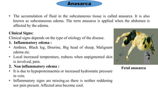
- 3. Anasarca • The accumulation of fluid in the subcutaneous tissue is called anasarca. It is also known as subcutaneous edema. The term anasarca is applied when the abdomen is affected by the edema. Clinical Signs: Clinical signs depends on the type of etiology of the disease. 1. Inflammatory edema : • Anthrax, Black leg, Dourine, Big head of sheep, Malignant edema etc. • Local increased temperature, redness when unpigmented skin is involved, pain. 2. Non inflammatory edema : • It is due to hypoproteinaemia or increased hydrostatic pressure in vein. • Inflammatory signs are missing,so there is neither reddening nor pain present. Affected area become cool. Fetal anasarca
- 4. Cont… Diagnosis : • History and characteristic clinical findings of visible swelling either local or diffuse. • Presence of swelling which pits on pressure when pressed firmly with the finger tips. • Inflammatory edema- Fever, anorexia, hot, hard and painful swelling on palpation. • Non inflammatory edema- Soft, cold, painless swelling on palpation. Treatment : • Use of diuretic drugs. Eg. Lasix@2ml/100lb body weight, IV once or twice daily for 3-5 days. • Edema producing cause should be corrected. • Supportive treatment including removal of the fluid by drainage – intubation or incisions.
- 5. Subcutaneous emphysema • Accumulation of gas in subcutaneous tissue space is called subcutaneous emphysema. Clinical Signs: • Visible swelling occurs over the body. • No pain or external lesions, except gas gangrene. • Swelling are soft, fluctuating and crepitating sound is present in palpation. • Stiffness in gait, interference with feeding and respiration. Subcutaneous emphysema
- 6. Diagnosis : • Tentative diagnosis can be made on history and characteristic clinical signs. • It is recognized by the presence of a soft, painless, yielding swelling that crepitates. • Bacteriological examination of the emphysematous fluid. Cont… Treatment : • Primary cause should be ascertained and treated. • Supportive treatment is necessary when the emphysema is extensive- skin incision. • Non infectious emphysema needs no treatment. • Gas gangrene needs immediate treatment with antibiotics. Eg. Penicillin 100000 IU, IM daily for 7 days.
- 7. Subcutaneous haemorrhage • Subcutaneous haemorrhage occurs as a result of extravasation of whole blood into the subcutaneous tissue. Clinical Signs: • Leakage of blood from the vascular system can cause local swelling with interference with normal body function. • Swelling is diffuse and soft with no visible effect on the skin surface. Subcutaneous haemorrhage
- 8. Cont… Diagnosis : • Tentative diagnosis can be made on history and characteristic clinical signs. • Diagnosis is confirmed by opening the swelling preferably by needle puncture. • Ascertaining the primary cause by platelet counts, histamine in blood, and prothrombin, clotting and bleeding times. Treatment : • Primary treatment includes removal of the causal agents. • Supportive treatment – haemorrhage should not be opened until clotting is completed. • Blood transfusion may be needed in heavy blood loss cases. • Parenteral administration of coagulants may be used.
- 9. Gangrene • Necrosis means death of the cells or a limited portion of the tissue. • Gangrene means death of a part of the body accompanied by putrefaction. Infection following necrosis may lead to gangrene after burns, scalds, frostbite, crush, wounds and puncture wounds etc. • When it occurs in skin it usually involves dermis, epidermis, and subcutaneous tissues. Dry gangrene Gangrenous mastitis
- 10. Clinical Signs: • Clinical signs depends on the types of gangrene. Cont… 1. Moist gangrene 2. Dry gangrene • The initial lesions are moist and the tissues are swollen with fluid, discoloured and cold. • Separation occurs at the margin and sloughing. • Underlying surface is raw and weeping. • Lesion is dry from the beginning. • Underlying surface usually consists of granulation tissues. Secondary bacterial infection • Putrefaction and absorption of toxins into the general circulation. • General signs of fever, rapid pulse and respiration.
- 11. Cont… Diagnosis : • Tentative diagnosis can be made on history and characteristic clinical signs. • Isolation and identification of the specific etiological agents. Treatment : • Primary treatment includes removal of the causal agents and surgical interventions. • Supportive treatment comprises the application of astringents and antibacterial ointments to facilitate separation of the gangrenous tissue and to prevent bacterial infection.
- 12. Subcutaneous abscess • Abscess is the localized collection of pus produced by an inflammatory reaction to pyogenic organisms in the subcutaneous tissues. • It is a circumscribed area of inflammation containing pus if matured. Clinical Signs : • The classic symptoms are redness, warmth, swelling, pain and tense. • Later the tip of the abscess become less tense, then breaks and the contents are discharged. Subcutaneous abscess
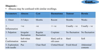
- 13. Cont… Diagnosis : • Abscess may be confused with similar swellings. Parameters Abscess Cyst Haematoma Tumour Hernia 1. Onset 3-5 days Months Recent Months Weeks 2. Inflammation +ve -ve +/- ve Usually – ve Usually – ve 3. Palpation Irregular fluctuation Regular fluctuation Crepitate No fluctuation No fluctuation 4. Conformation Soft when mature Hard Hard, soft in old cases Hard Soft 5. Exploration with needle Pus Clear fluid Clotted blood Fresh blood Abdominal contents
- 14. Treatment : • Abscess must be surgically drained and treated as open wound. • Gauze should be introduced into the wound after painted with antiseptic solution and should dressed in every alternative day for 10 days. • An antibiotic should be given for atleast 5 days. Cont…
- 15. Thank You…


