Digital Techniques For Myocardial Perfusion Spect
Download as pptx, pdf6 likes3,599 views

This document provides an overview of digital techniques for myocardial perfusion SPECT imaging. It describes SPECT equipment components like the gamma camera and workstation. It explains tomographic acquisition parameters and technical considerations. Image processing techniques are covered like filtered back projection, filtering, and iterative reconstruction. Quantitative analysis methods like bullseye displays and segmental analysis are also summarized.
![Filter Example: Butterworth
Butterworth
[Cutoff = 0.3 Order =5] Filtered Raw
image
Filter Reconstructed
slice
Too smooth!](https://image.slidesharecdn.com/digitaltechniquesformyocardialperfusionspect-13260389564903-phpapp02-120108101226-phpapp02/85/Digital-Techniques-For-Myocardial-Perfusion-Spect-28-320.jpg)
![Filter Example: Butterworth
Butterworth
[Cutoff = 0.5 Order =5]
Filter Filtered Raw Reconstructed
image slice
Good quality](https://image.slidesharecdn.com/digitaltechniquesformyocardialperfusionspect-13260389564903-phpapp02-120108101226-phpapp02/85/Digital-Techniques-For-Myocardial-Perfusion-Spect-29-320.jpg)

Digital Techniques For Myocardial Perfusion Spect
- 1. Digital Techniques for Myocardial Perfusion SPECT Dr. Muhammad Ayub Diplomate Certification Board of Nuclear Cardiology Diplomate Certification Board of Cardiovascular CT Punjab Institute of Cardiology, Lahore
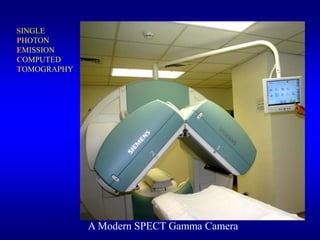
- 2. SINGLE PHOTON EMISSION COMPUTED TOMOGRAPHY A Modern SPECT Gamma Camera
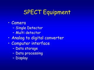
- 3. SPECT Equipment ? Camera ¿C Single Detector ¿C Multi detector ? Analog to digital converter ? Computer interface ¿C Data storage ¿C Data processing ¿C Display
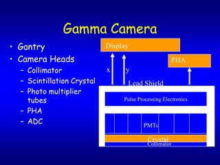
- 4. Gamma Camera ? Gantry Display ? Camera Heads PHA ¿C Collimator x y ¿C Scintillation Crystal Lead Shield ¿C Photo multiplier tubes Pulse Processing Electronics ¿C PHA ¿C ADC PMTs Crystal Collimator
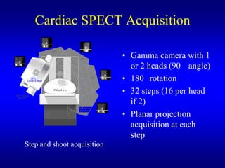
- 5. Cardiac SPECT Acquisition ? Gamma camera with 1 or 2 heads (90 angle) ? 180 rotation ? 32 steps (16 per head if 2) ? Planar projection acquisition at each step Step and shoot acquisition
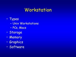
- 6. Workstation ? Types ¿C Unix Workstations ¿C PCs, Macs ? Storage ? Memory ? Graphics ? Software
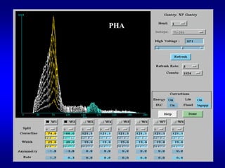
- 7. PHA
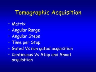
- 8. Tomographic Acquisition ? Matrix ? Angular Range ? Angular Steps ? Time per Step ? Gated Vs non gated acquisition ? Continuous Vs Step and Shoot acquisition
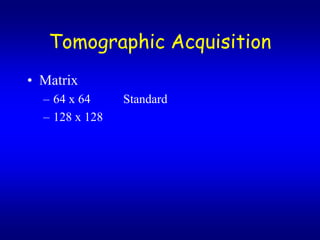
- 9. Tomographic Acquisition ? Matrix ¿C 64 x 64 Standard ¿C 128 x 128
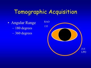
- 12. Tomographic Acquisition ? Angular Range RAO 135 ¿C 180 degrees ¿C 360 degrees 215 LPO
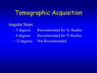
- 13. Tomographic Acquisition Angular Steps ¿C 3 degrees Recommended for Tc Studies ¿C 6 degrees Recommended for Tl Studies ¿C 12 degrees Not Recommended
- 14. Image Acquisition ? Orbit ¿C Circular Standard ¿C Non-circular Optional
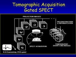
- 15. Tomographic Acquisition Gated SPECT
- 16. Technical Considerations ? QC ¿C Energy ¿C Uniformity ¿C Linearity ¿C COR
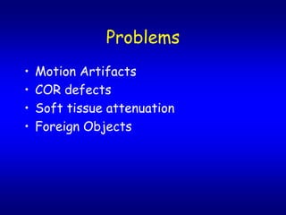
- 17. Problems ? Motion Artifacts ? COR defects ? Soft tissue attenuation ? Foreign Objects
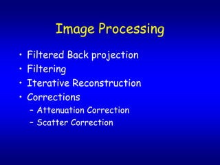
- 18. Image Processing ? Filtered Back projection ? Filtering ? Iterative Reconstruction ? Corrections ¿C Attenuation Correction ¿C Scatter Correction
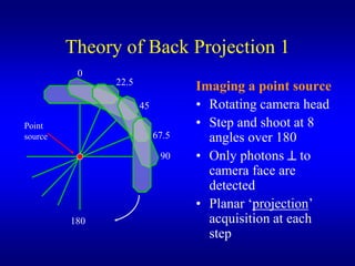
- 19. Theory of Back Projection 1 0 22.5 Imaging a point source 45 ? Rotating camera head Point ? Step and shoot at 8 source 67.5 angles over 180 90 ? Only photons ? to camera face are detected ? Planar í«projectioní» 180 acquisition at each step
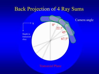
- 20. Back Projection of 4 Ray Sums Camera angle x 0íÒ 22.5íÒ Depth in 45íÒ transaxial slice 67.5íÒ Transaxial Plane
- 21. Filtering ? Pre filtering ? 3D Post Filtering ? Types ¿C Low pass ¿C Metz ¿C Wiener ¿C Band
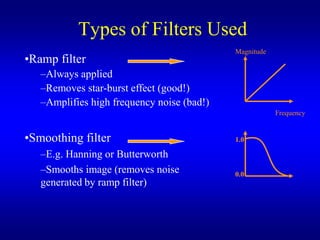
- 22. Types of Filters Used Magnitude ?Ramp filter ¿CAlways applied ¿CRemoves star-burst effect (good!) ¿CAmplifies high frequency noise (bad!) Frequency ?Smoothing filter 1.0 ¿CE.g. Hanning or Butterworth ¿CSmooths image (removes noise 0.0 generated by ramp filter)
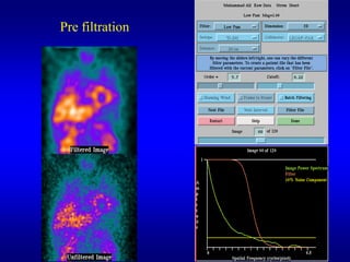
- 23. Pre filtration
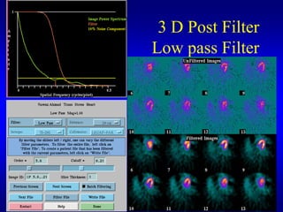
- 24. 3 D Post Filter Low pass Filter
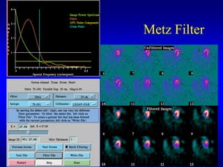
- 25. Metz Filter
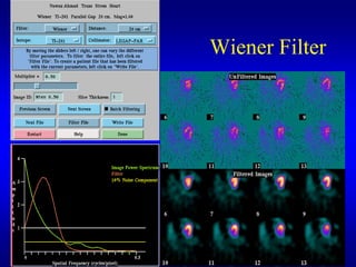
- 26. Wiener Filter
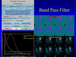
- 27. Band Pass Filter
- 28. Filter Example: Butterworth Butterworth [Cutoff = 0.3 Order =5] Filtered Raw image Filter Reconstructed slice Too smooth!
- 29. Filter Example: Butterworth Butterworth [Cutoff = 0.5 Order =5] Filter Filtered Raw Reconstructed image slice Good quality
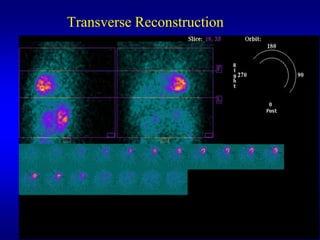
- 30. Image Reconstruction ? Short Axis ? Vertical Long Slices ? Horizontal Long Slices ? Slice Thickness
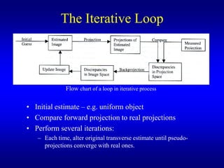
- 32. The Iterative Loop Flow chart of a loop in iterative process ? Initial estimate ¿C e.g. uniform object ? Compare forward projection to real projections ? Perform several iterations: ¿C Each time, alter original transverse estimate until pseudo- projections converge with real ones.
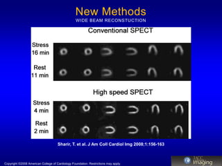
- 33. New Methods WIDE BEAM RECONSTUCTION Sharir, T. et al. J Am Coll Cardiol Img 2008;1:156-163 Copyright ?2008 American College of Cardiology Foundation. Restrictions may apply.
- 35. Image Display ? Cardiac Stress Rest Display Format ? Colored and Black & White Display ? 3D display ? Cine Display ? Animation
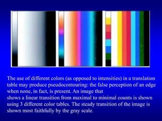
- 36. The use of different colors (as opposed to intensities) in a translation table may produce pseudocontouring: the false perception of an edge when none, in fact, is present. An image that shows a linear transition from maximal to minimal counts is shown using 3 different color tables. The steady transition of the image is shown most faithfully by the gray scale.
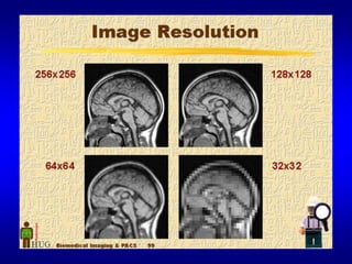
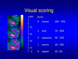
- 37. Visual scoring 2 1 100% Score 4 normal 100 - 70% 0 4 70 3 2 3 mild 70 - 50% 50 1 4 2 moderate 50 - 30% 4 30 4 1 severe 30 - 10% 2 10 4 0 0 absent 10 - 0%
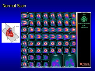
- 38. Normal Scan
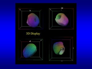
- 39. 3D Display
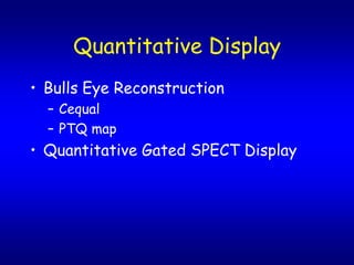
- 40. Quantitative Display ? Bulls Eye Reconstruction ¿C Cequal ¿C PTQ map ? Quantitative Gated SPECT Display
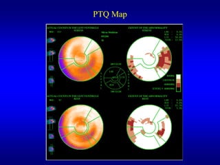
- 41. PTQ Map
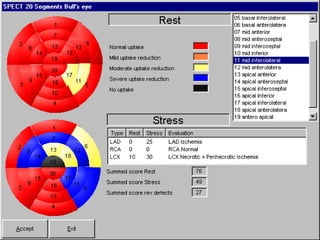
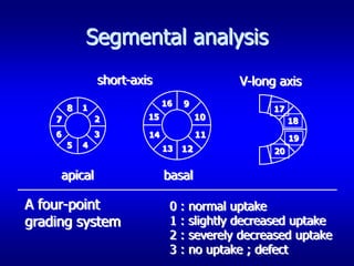
- 43. Segmental analysis short-axis V-long axis 16 9 8 1 17 7 2 15 10 18 6 3 14 11 19 5 4 13 12 20 apical basal A four-point 0 : normal uptake grading system 1 : slightly decreased uptake 2 : severely decreased uptake 3 : no uptake ; defect
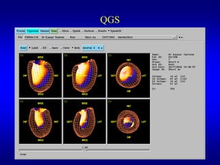
- 45. QGS
- 46. Thank You