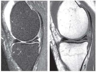
Findings of meniscal tears in mri knee
- 1. FINDINGS OF MENISCAL TEARS ON MRI KNEE Presented By Zubair Younis
- 2. Anatomy
- 6. BASIC MRI
- 7. Tools in Musculoskeletalimaging ’āś T1Weighted ’āśT2 Weighted ’āśFATSuppressed T2 ’āśSTIR ’āśGradientEchoImages. ’āśGadolinium studies ’āśMR Arthrography ’āśMR Angiography
- 8. MRIRules T1 T2 Fat Hyperintense Hyperintense Water Hypointense hyperintense Cortical bone Hypointense Hypointense Fibrous tissue Hypointense Hypointense Cartilage Isointense Isointense
- 9. T1-Weighted and T2-Weighted Images One should be able to determine whether an image is T1- weighted or T2-weighted using the following techniques: Recognition of an area within an image that is known to contain fluid, such as the knee or other joints (intraarticular fluid). If this fluid is noted to be bright or of high signal intensity, that image is likely T2-weighted. If the region of the fluid is noted to be dark, that image is likely T1-weighted.
- 11. Fat-suppressed T2-weighted images are acquired using techniques similar to those for conventional T2-weighted images and then various computer algorithms and processes are used to ŌĆ£suppressŌĆØ the signal that is coming from fat. This technique facilitates the evaluation of bone marrow edema and edema secondary to other pathologic processes.
- 12. Postgadolinium weighted images are typically obtained for the evaluation of infection, tumor, and postsurgical changes or scar.
- 13. MR Arthrography Images MR arthrography images are obtained after the joint of interest is injected with gadolinium or normal saline.They may be diffi cult to differentiate from T2-weighted images, which also contain bright fluid within the joint. MR Angiography Images MR angiography images highlight the blood vessels and allow for evaluation of the arterial and venous vascular structures.
- 16. Indications of MRI ’āśOccult fracture ’āśMarrow abnormality ’āśLigament pathology ’āśTendon pathology ’āśMuscular injury ’āśInfection ’āśBone and soft tissue tumour
- 17. SECTIONS- Coronal- Ant. To Post. Saggital- Lateral to Medial Axial- From abovedownward
- 18. Meniscal Tear ’āś Imaging Criteria 1. Presence of linear signal intensity whether reaching superior or inferior articular surface or not 2. Abnormal meniscal morphology
- 21. Grade I Grade II Grade III
- 29. Meniscal cyst Joint fluid isexpressed into adjacent soft tissue through the tear Mostly occur in medial compartment Most common associated tear is horizontal cleavage tear
- 30. Discoid Meniscus This entity occurs in approximately 5% of the population. The lateral meniscus is much more commonly involved than is the medial meniscus. Discoid menisci were first classified by Wantanabe et al as follows: Grade I (complete) ŌĆó Grade II (incomplete) ŌĆó Grade III (Wrisberg or lateral meniscal variant, i.e., a meniscus that is not secured to the posterior tibia by coronary ligaments) Monllau et al described a fourth, ring-shaped type.
- 33. Meniscocapsularseparation Fluid signal between posterior portion of medial meniscus and joint capsule
- 34. THANK YOU


