


![Approach to Anemic Child [Autosaved].pptx](https://cdn.slidesharecdn.com/ss_thumbnails/approachtoanemicchildautosaved-240427131658-64d6d32d-thumbnail.jpg?width=560&fit=bounds)


More Related Content
Similar to FOLIC ACID DEFICIENCY I N CLINICAL PRACTICEpptx (20)
More from DR Venkata Ramana (20)


Recently uploaded (20)


FOLIC ACID DEFICIENCY I N CLINICAL PRACTICEpptx
- 1. FOLIC ACID DEFICIENCY Dr G VENKATA RAMANA MBBS DNB FAMILY MEDICINE
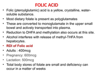
- 2. FOLIC ACID ŌĆó Folic (pteroylglutamic) acid is a yellow, crystalline, water- soluble substance. ŌĆó Most dietary folate is present as polyglutamates ŌĆó These are converted to monoglutamate in the upper small bowel and actively transported into plasma. ŌĆó Reduction to DHFA and methylation also occurs at this site. ŌĆó Alcohol interferes with release of methyl-THFA from hepatocytes. ŌĆó RDI of Folic acid ŌĆó Adults : 400mcg ŌĆó Pregnancy: 600mcg ŌĆó Lactation: 500mcg ŌĆó Total body stores of folate are small and deficiency can occur in a matter of weeks
- 5. Physiologic roles of folic acid ŌĆó DNA synthesis, RNA synthesis, DNA methylation ŌĆó Folic acid play a critical role in DNA and RNA synthesis ŌĆó Folic acid deficiency can therefore impair DNA synthesis, which in turn can cause a cell to arrest in the DNA synthesis (S) phase of the cell cycle, make DNA replication errors, and/or undergo apoptotic death ŌĆó Hematopoiesis ŌĆó Hematopoietic precursor cells are among the most rapidly dividing cells in the body and hence are one of the cell types most sensitive to abnormal DNA synthesis
- 6. ŌĆó Two major effects of the deficiency on hematopoiesis ŌĆó Megaloblastic changes ŌĆó caused by slowing of the nuclear division cycle relative to the cytoplasmic maturation cycle (ie, nuclear- cytoplasmic dyssynchrony). ŌĆó Ineffective erythropoiesis ŌĆó occurs when there is premature death (eg, phagocytosis or apoptosis) of the developing erythropoietic precursor cells in the bone marrow . ŌĆó There may be hypercellularity of the bone marrow ŌĆó laboratory findings of hemolysis, including elevated serum iron, indirect bilirubin, and lactate dehydrogenase (LDH), and low haptoglobin. ŌĆó The reticulocyte count is typically low
- 11. Clinical presentation ŌĆó Macrocytic anemia ŌĆó Symptoms of anemia-fatigue, irritability, cognitive decline,chest pain, shortness of breath,palpitations,light- headedness ŌĆó Yellowed skin ŌĆó Gastrointestinal symptoms ŌĆó Oral ulcers ŌĆó Glossitis ŌĆó Neuropsychiatric changes ŌĆó Depression, irritability, or forgetfulness ŌĆó Neural tube defects ŌĆó Spina bifida
- 12. lnvestigations ŌĆó CBC and blood smear ŌĆó Anemia ŌĆó Macrocytic red blood cells (MCV >100 fL) or macro- ovalocytosis ŌĆó An MCV value >115 fL is more specific to vitamin B12 or folate deficiency ŌĆó Mild leukopenia and/or thrombocytopenia ŌĆó Low reticulocyte count ŌĆó Hypersegmented neutrophils on the peripheral blood smear (ie, >5 percent of neutrophils with Ōēź5 lobes or Ōēź1 percent of neutrophils with Ōēź6 lobes) ŌĆó Increased lactate dehydrogenase ŌĆó Increased bilirubin
- 13. Peripheral smear and Bone marrow Peripheral blood smear showing a hypersegmented neutrophil (seven lobes) and macroovalocytes, a pattern that can be seen with vitamin B12 (cobalamin) or folate deficiency Erythroid precursors in the bone marrow (Left panel) Normal erythropoiesis. (Right panel) Megaloblastic erythropoiesis
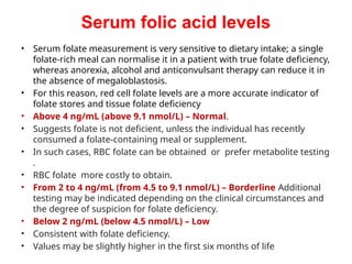
- 14. Serum folic acid levels ŌĆó Serum folate measurement is very sensitive to dietary intake; a single folate-rich meal can normalise it in a patient with true folate deficiency, whereas anorexia, alcohol and anticonvulsant therapy can reduce it in the absence of megaloblastosis. ŌĆó For this reason, red cell folate levels are a more accurate indicator of folate stores and tissue folate deficiency ŌĆó Above 4 ng/mL (above 9.1 nmol/L) ŌĆō Normal. ŌĆó Suggests folate is not deficient, unless the individual has recently consumed a folate-containing meal or supplement. ŌĆó In such cases, RBC folate can be obtained or prefer metabolite testing . ŌĆó RBC folate more costly to obtain. ŌĆó From 2 to 4 ng/mL (from 4.5 to 9.1 nmol/L) ŌĆō Borderline Additional testing may be indicated depending on the clinical circumstances and the degree of suspicion for folate deficiency. ŌĆó Below 2 ng/mL (below 4.5 nmol/L) ŌĆō Low ŌĆó Consistent with folate deficiency. ŌĆó Values may be slightly higher in the first six months of life
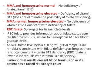
- 15. ŌĆó MMA and homocysteine normal ŌĆō No deficiency of folate,vitamin B12. ŌĆó MMA and homocysteine elevated ŌĆō Deficiency of vitamin B12 (does not eliminate the possibility of folate deficiency). ŌĆó MMA normal, homocysteine elevated ŌĆō No deficiency of vitamin B12. Consistent with deficiency of folate. ŌĆó RBC folate Surrogate for tissue folate levels. ŌĆó RBC folate provides information about folate status over the lifetime of RBCs, similar to hemoglobin A1C for blood glucose levels. ŌĆó An RBC folate level below 150 ng/mL (<150 mcg/L; <340 nmol/L) is consistent with folate deficiency as long as there is not concomitant vitamin B12 deficiency (RBC folate is lower in individuals with vitamin B12 deficiency). ŌĆó False-normal results -Recent blood transfusion or if a patient has a raised reticulocyte count
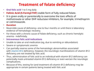
- 16. Treatment of folate deficiency ŌĆó Oral folic acid 1 to 5 mg daily ŌĆó Folinic Acid (5-Formyl-THF) stable form of fully reduced folate. ŌĆó It is given orally or parenterally to overcome the toxic effects of methotrexate or other DHF reductase inhibitors, for example, trimethoprim or cotrimoxazole. ŌĆó Duration ŌĆó Reversible cause of deficiency- one to four months or until there is laboratory evidence of hematologic recovery. ŌĆó For those with a chronic cause of folate deficiency, such as chronic hemolytic anemia - indefinitely. ŌĆó Intravenous folic acid indications ŌĆó Unable to take an oral medication (eg, due to vomiting or obtundation) ŌĆó Severe or symptomatic anemia ŌĆó Can partially reverse some of the hematologic abnormalities associated with vitamin B12 deficiency .however, the neurologic manifestations of vitamin B12 deficiency are not treated by folic acid. ŌĆó Thus, administration of folic acid to an individual with vitamin B12 deficiency can potentially mask untreated vitamin B12 deficiency or even worsen the neurologic complications. ŌĆó Because of this, testing for (and treatment of) vitamin B12 deficiency may be appropriate in certain patients being treated with folic acid
- 17. ŌĆó Adverse effects ŌĆó Oral folic acid is entirely nontoxic ŌĆó Injections rarely cause sensitivity reactions ŌĆó Prevention of folate deficiency ŌĆó Enrich cereals and grain products with folic acid to reduce the risk of neural tube defects ŌĆó Folic acid prophylaxis ŌĆó Typical dose, 1 mg orally per day ŌĆó All women,from the moment they begin trying to conceive until 12 weeks of gestation to prevent neural tube defects ŌĆó Hemolytic anemias/hyperproliferative hematologic states ŌĆó Patients with rheumatoid arthritis or psoriasis on methotrexate ŌĆó Patients on antiepileptic drugs ŌĆó Patients with ulcerative colitis