General Examination of CVS physical examination.pptx
- 1. General Examination in Cardiovascular System PREPARED BY DR. RUQAYA AL-KATHIRY HEAD OF THE MEDICAL DEPARTMENT OF UST ŌĆ£Always listen to the patient they might be telling you the
- 2. PHYSICAL EXAMINATION: GENERAL EXAMINATION: I-General Assessment: The sequence & extent of examination depends on the pt's condition: ’āśPts. with C/P arrest requiring resuscitation: Rx 1st & examine later ’āśPts. requiring ER care: rapidly assess & Rx 1st, leave more detailed exam. for later ’āśStable pts.: examine thoroughly 1st. Look at the pt's general appearance, noting whether he or she: ŌĆólooks unwell ŌĆóis frightened or distressed. ŌĆóPhysique, form & general nourishment: BMI (??) ŌĆóobesity: C.A.D, HTN. ŌĆóthin: ŌĆócachexic: chr. HF. ŌĆóBreathlessness at minimal exertion (dressing) or rest (semi-sitting, on 1 side). ŌĆóVital signs II-Hands: 1-Cyanosis: Q.DEFINE?TYPES?SITES?CAUSES? 2-Pallor: Q.DEFINE?WHAT ARE THE CARDIAC CAUSES? 3-Jaundice: Q.DEFINE?WHAT ARE THE CARDIAC CAUSES? 4-Tar Stain: Q.WHAT IS ITS SIGNIFICANCE IN THE CARDIAC DISEASES?
- 3. 5-Clubbing: of Fingers & Toes: Q.DEFINE?GRADES? ASSIGNMENT:Q.WHAT ARE THE CARDIAC CAUSES OF CLUBBING? 6-Flapping Tremor (Asterixis): Q.DEFINE?TECHNIQUE?CAUSES? III-Eyes: IV-Neck: 1-JugularVenous Pulse (JVP):. ASSIGNMENT: Q.WHAT ARE THE CARDIAC CAUSES OF RAISED JVP? 2-Lymph Nodes: should be examined. V-Skin: Erythema Nodosum=Rheumatic Fever Rash of Systemic Lupus Erythematosus (SLE) or MS Signs of Hyperlipidaemia: ŌĆóCorneal arcus: precipitation of cholesterol crystals=creamy yellow discoloration at the boundary of the iris & cornea. This can occur in those >50 ys with no hyperlipidaemia. ŌĆóXanthelasma: yellowish cholesterol plaques around the eyelids and periorbital area. ŌĆóXanthomata: lipid deposits=yellow nodules in skin/tendons (patella/Achilles tendon).
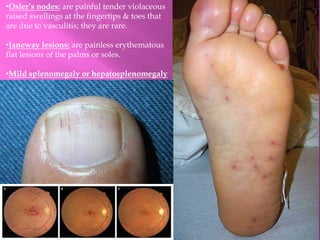
- 5. Signs of Infective Endocarditis: ŌĆóMultiple capillary haemorrhages (petechiae): In the skin, mostly on the legs & conjunctivae caused by vasculitis. ŌĆóD/D: rash of meningococcal disease ŌĆóRoth spots: Flame-shaped retinal haemorrhages with a 'cotton-wool' centre seen on ophthalmoscopy. They are due to circulating immune complexes. D/D: anaemia or leukaemia ŌĆóSplinter haemorrhages: Multiple, linear, reddish-brown marks along the axis of the fingernails and toenails. They are due to circulating immune complexes. D/D: 1-2 isolated 'splinters' are common in healthy individuals from trauma. ŌĆóFinger clubbing: A rare & late sign feature in chronic bacterial endocarditis. Microscopic haematuria.
- 6. ŌĆóOslerŌĆÖs nodes: are painful tender violaceous raised swellings at the fingertips & toes that are due to vasculitis; they are rare. ŌĆóJaneway lesions: are painless erythematous flat lesions of the palms or soles. ŌĆóMild splenomegaly or hepatosplenomegaly
- 7. WHAT IS THE DIAGNOSIS?
- 8. Q.WHAT ARE THE CAUSES OF IT UNILATERAL AND BILATERAL?
- 9. ŌĆ£Medicine is learned at the bedside and not in the classroomŌĆØ Sir William Osler (1849-1919) THANK YOU FOR YOUR ATTENDANCE


