This document discusses scanning electron microscopy (SEM) and the principles behind how it works. It provides details on:
- How an SEM uses an electron beam to scan sample surfaces and signals emitted are used to construct images.
- The interaction volume where primary electrons interact with the sample and the different types of signals (secondary electrons, backscattered electrons, X-rays) that can be detected from this volume.
- How the signals are amplified and synchronized with the beam position to create high-resolution digital images of the sample's surface structure and composition.
This document provides an overview of cell biology, including:
1. The origin of cells from spontaneous polymerization of organic molecules around 3.8 billion years ago. RNA was later found to be able to self-replicate.
2. The classification of prokaryotic and eukaryotic cells, and the endosymbiotic origin of mitochondria and chloroplasts in eukaryotic cells.
3. Experimental cell models used in research including E. coli, yeast, C. elegans, fruit flies, plants like Arabidopsis, and vertebrate models like zebrafish, frogs, and mice.
This document provides an overview of cell biology methods and techniques. It discusses the history of cell discovery using microscopes from Hooke to Leeuwenhoek. It then summarizes various microscopy methods like brightfield, phase contrast, fluorescence, confocal, and electron microscopy. Methods of subcellular fractionation like differential centrifugation and density gradient centrifugation are also outlined. The document briefly discusses cell culture techniques and growth factors as well as the use of viruses to study cell functions.
The document discusses different types of microscopes used in biology and medicine, including microscopes invented by Antonie van Leeuwenhoek and Robert Hooke that used simple lenses to magnify objects, as well as compound microscopes, transmission electron microscopes, and scanning electron microscopes, which use electrons rather than light and allow observation of cell structures at much higher magnifications. Key images show early microscope designs as well as examples of cells and structures observed using different microscope technologies.
This document discusses various indirect methods for measuring microbial growth, including turbidimetry, measuring metabolic activity, and dry weight determination. Turbidimetry uses a spectrophotometer to measure light passing through a cell suspension, where more cells cause more turbidity. Metabolic activity measurement involves assessing products like acid or oxygen levels proportional to population size. Dry weight determines the mass of filtered and dried cells, but cannot distinguish live from dead. Scintillation counters are also discussed for measuring radioisotopes through light excitation detection.
Recombinant DNA involves creating DNA molecules from different sources. Researchers improved fluorescent probes used to detect reactive oxygen species by replacing hydrogen with deuterium in the dyes. This increased the dyes' stability, shelf life, and ability to detect smaller concentrations of reactive oxygen species. Fluoromodules are fluorescent probes that monitor biological activities of individual proteins in real time. Researchers enhanced fluoromodule technology, creating probes that glow brighter than typical fluorescent proteins and allow proteins to be followed in real time. Fluorescent probes are essential for studying diseases like cancer and atherosclerosis as they detect and measure reactive oxygen species, which play a role in disease processes.
This document discusses various types of cell inclusions found in prokaryotic cells, including gas vesicles, carboxysomes, polyhydroxybutyrate (PHB) granules, metachromatic granules, glycogen bodies, starch granules, magnetosomes, sulfur granules, and chlorosomes. It describes the structure, function, and organisms where each type of inclusion is found. For example, it notes that gas vesicles allow planktonic bacteria to float and position themselves, while carboxysomes concentrate carbon dioxide for photosynthesis and PHB granules store carbon and energy.
The document discusses the dark field microscope, which was invented in 1830 by J.J. Lister. It describes how a dark field microscope works by placing an opaque disc under the condenser lens so that only light scattered by objects on the slide reaches the eye. This causes pigmented objects to appear in false colors. Some uses of the dark field microscope include examining unstained cells and samples in pond water or prepared slides, as well as determining mobility in cultures. While it produces beautiful images, it also has some disadvantages.
Bacterial cytology ribosomes, chromosomal and extrachromosomal materialVishrut Ghare
油
This document discusses bacterial cytology, focusing on ribosomes, chromosomal material, and extrachromosomal plasmids. It describes the structure and function of ribosomes, which are composed of RNA and proteins and are the site of protein synthesis. It also discusses the nucleoid, which contains the bacterial chromosome and proteins. The chromosome carries all the genetic information and can be circular or linear. Some bacteria contain extrachromosomal DNA molecules called plasmids, which are small, circular pieces of DNA that are not essential but can provide advantages like antibiotic resistance.
Bacteria can grow and divide very rapidly, every 20 minutes for some species under ideal conditions, or as slowly as every 100 years for bacteria in deep underground environments. The generation time, or doubling time, is the amount of time it takes for the number of bacterial cells to double in a culture. Optical density measurements using spectrophotometry is a common way to indirectly measure bacterial growth and calculate doubling times by tracking increases in turbidity over time. Direct microscopic counting and viability assays that measure colony forming units are other methods to directly measure bacterial cell numbers.
This document discusses various methods for measuring microbial growth, including direct cell counting, viable cell counting, and measurement of cell mass and constituents.
Direct cell counting can be done using a counting chamber under a microscope or with an electronic particle counter. Viable cell counts are determined using plate counting methods which allow colonies to form. Measurement of cell mass can be done by dry weight or turbidimetrically, while cell constituents like protein and ATP can also indicate growth. Overall, the document provides an overview of key techniques for quantifying and analyzing microbial cultures.
This document discusses a technique for preserving food using low-frequency magnetic fields. The technique takes advantage of the fact that magnetic fields disrupt ion transport across bacterial cell membranes, interfering with their ability to regulate pH and perform cellular reactions. The document reports on an experiment exposing Escherichia coli bacteria to low-frequency magnetic fields, which negatively impacted their growth, ability to form colonies, and oxidoreductive activity. Unlike other preservation methods, low wattage is required to kill bacteria using low-frequency magnetic fields.
Global warming is causing sea levels to rise through ocean expansion due to higher temperatures, melting glaciers and ice sheets, and changes to terrestrial water storage. Vietnam is highly vulnerable to sea level rise due to its long coastline and location of populated river deltas. A 1 meter rise in sea levels could impact over half of Vietnam's population and cause land loss and population displacement. Low-lying areas like the Xuan Thuy National Park are also at high risk of complete inundation from rising oceans. Human activities that produce greenhouse gases and destroy carbon-absorbing forests are accelerating global warming and sea level rise, threatening coastal communities and ecosystems.
This document provides an introduction to flow cytometry. It defines flow cytometry as a method for sensing individual cells in a fluid stream as they pass through a laser beam, measuring light scattering and fluorescence. Key aspects of flow cytometry systems and methodology are described, including hydrodynamic focusing of cells, light scattering measurements, use of fluorescent markers, optical and electronic components, data acquisition and analysis techniques like gating and compensation. The history of technological developments in flow cytometry is also summarized.
1) The document discusses various factors that affect radiation response, including linear energy transfer (LET), relative biological effectiveness (RBE), oxygen effect, and cell cycle phase.
2) It provides details on oxygen enhancement ratio (OER), describing how it ranges from 2.5-3.0 for x-rays and varies according to cell cycle phase and radiation type (higher for x-rays and neutrons vs. alpha particles).
3) LET is discussed in relation to radiation type (low for x-rays, medium for neutrons, high for alpha particles) and how it impacts ionization density and biological damage. An optimal LET of around 100 keV/亮m is described.
Flow cytometry is a technique that allows for the characterization of individual cells as they flow in a fluid stream through a laser beam. Parameters like cell size, granularity, and fluorescence from markers bound to cells can be measured. Key components include hydrodynamic focusing to align cells, lasers to excite fluorochromes, and detectors like photomultiplier tubes to quantify light scatter and emission. Developments in monoclonal antibodies, fluorochromes, and electronics have advanced the field since its origins in the 1930s measuring basic cell properties. Modern flow cytometers precisely route light through filters to characterize thousands of individual cells per second.
This document discusses several key concepts in radiation oncology:
1. The Law of Bergonie and Tribondeau states that radiosensitivity varies with cell maturation and metabolism, with stem cells being most radiosensitive. Younger and more metabolically active tissues are also more radiosensitive.
2. Factors like linear energy transfer (LET), relative biological effectiveness (RBE), fractionation, and oxygenation level impact radiation response. High LET radiation like alpha particles is more effective due to dense ionization.
3. The oxygen enhancement ratio (OER) compares radiation doses needed under hypoxic vs aerated conditions. It is typically 2.5-3.0 for X-rays but 1 for high
its about the microscopes types and there significance in the world for diagnostic purposes .advantages and disadvantages of the types of different microscopes
The document discusses luminescence and phosphorescence spectroscopy. It defines luminescence as light emission from a substance when an electron returns to the ground state from an excited state. Phosphorescence is luminescence from a triplet excited state with a longer lifetime than fluorescence which occurs from a singlet state. The document describes various types of luminescence and provides details on instrumentation, sample preparation, and applications of phosphorescence spectroscopy in different fields such as pharmaceutical, clinical, environmental, and forensic analyses.
1. Radiobiology is the study of the effects of ionizing radiation on living things. When radiation passes through living matter, it loses energy by interacting with atoms and molecules, causing ionization and excitation that can alter living cells.
2. The biological effects of radiation occur on different time scales - physical interactions are instantaneous, chemical changes occur within milliseconds, and biological effects can take hours to years to present.
3. Radiation can directly damage DNA through ionization, but the probability is low since DNA is a small target within cells. More likely, radiation interacts with water in cells, producing free radicals that can diffuse and indirectly damage critical targets like DNA through chemical reactions.
1. Lasers emit coherent, directional, monochromatic light that can be precisely focused. Lasers work by stimulating the emission of photons from excited atoms or molecules in a process called stimulated emission.
2. Ophthalmic lasers operate across a wide range of wavelengths from deep ultraviolet to infrared. Absorption of different wavelengths by chromophores like water, hemoglobin, and melanin leads to different tissue interactions including photochemical reactions or heating and thermal damage.
3. Laser light can interact with tissue through photochemical reactions at low intensities, through heating and thermal damage at higher intensities, or through rapid vaporization at very high intensities achieved with pulsed lasers. These interactions make lasers
This document provides information about electrophoresis. It discusses different types of electrophoretic techniques including slab electrophoresis, capillary electrophoresis, capillary zone electrophoresis, capillary gel electrophoresis, capillary isotachophoresis, and micellar electrokinetic chromatography. It also covers principles, instrumentation, applications in areas like DNA analysis and vaccine analysis.
An introduction to flow cytometry- Ashwini.RAshwini R
油
The document provides an introduction to flow cytometry. It describes flow cytometry as a technique that allows simultaneous multiparametric analysis of physical and chemical characteristics of single cells suspended in a fluid stream. Key components of a flow cytometer include fluidics, optics, detectors, and electronics. Cells are hydrodynamically focused into a single file stream and pass through a laser beam, where light scattering and fluorescence emissions provide information about cellular properties. Photodetectors convert light signals into electrical pulses that are analyzed. Flow cytometry has various applications including immunophenotyping, cell sorting, DNA content analysis, and cell cycle/proliferation analysis.
This document discusses x-ray crystallography techniques including Bragg's law and applications such as determining molecular arrangements of atoms. It explains key concepts like diffraction, constructive and destructive interference patterns, and how a crystal's perfection impacts the clarity of patterns. Bragg's law is derived and its importance explained. Techniques like isomorphous replacement that use heavy atoms to solve phase problems are covered. Considerations for selecting gas chromatography/mass spectrometry or liquid chromatography/mass spectrometry are presented. The importance of animals in biomedical research and various animal models are discussed. Alternatives to animal testing including cell culture and computer models are also summarized.
FACS and MACS with their applications in biological research.Deepak Agarwal
油
Flow cytometry (FACS) and magnetic activated cell sorting (MACS) are techniques used to analyze and separate cells based on their physical and chemical characteristics. FACS uses lasers to detect cell properties and sort cells into containers one by one, while MACS uses magnetic microbeads attached to cells to separate them in high gradient magnetic fields. These techniques have various applications in research including identifying stem cells, characterizing cancer cells, studying cell cycles, and isolating cell populations for further analysis.
Fluorescence and electron Microscope.pptxsaraso888
油
The fluorescence microscope uses fluorescence and phosphorescence instead of reflection and absorption to study organic and inorganic substances. It has a UV light source and filter to protect the viewer. Some organisms and substances naturally fluoresce, while others can be stained with fluorescent dyes. The fluorescence microscope is used widely in diagnostic microbiology to detect antigens, antibodies, and nucleic acids. Electron microscopes have much higher resolving power than light microscopes due to the small wavelength of electrons. Transmission electron microscopes allow viewing of inner structures while scanning electron microscopes image surfaces. Both have various applications in biology and medicine.
The document discusses electrophoresis, which is the movement of charged particles in an electric field. It migrates towards the oppositely charged electrode. Electrophoresis is used to separate biological molecules like proteins. It explains the principles and types of electrophoresis like paper, gel, and isolectric focusing electrophoresis. It provides details about agarose gel electrophoresis including properties, buffers used, procedure, and applications. It also briefly discusses polyacrylamide gel electrophoresis.
Introduction, the principle of immunofluorescence, Technique, Fluorescent microscope and its components, Application and types of immunofluorescence, Direct and indirect immunofluorescence, FACS (Fluorescence-activated cell sorting), Uses and limitations of Immunofluorescence
This document summarizes recent findings that some bacterial and viral DNA sequences can induce low frequency electromagnetic waves in high aqueous dilutions. Specifically:
1) Electromagnetic waves between 500-3000 Hz have been detected in certain high dilutions of filtrates containing DNA from various microorganisms.
2) The waves are produced from both whole genomes and isolated DNA fragments as short as 104 base pairs.
3) The waves appear to be triggered by excitation from the ambient extremely low frequency electromagnetic background.
4) This phenomenon could allow for highly sensitive detection of chronic bacterial and viral infections.
Andrew Goldsworthy to British Society for Ecological Medicine at the Royal Co...Martin Sharp
油
The document discusses how exposure to electromagnetic radiation from modern devices has increased and is linked to health issues like cancer and fertility problems. It explains that electromagnetic radiation can cause calcium ions to leak from cell membranes, weakening them. This membrane leakage can explain effects like cancer, DNA damage, neurological symptoms, and symptoms of electrosensitivity. Pulsed and low frequency radiation is most effective at causing this calcium leakage and biological effects.
Mate, a short story by Kate Grenvile.pptxLiny Jenifer
油
A powerpoint presentation on the short story Mate by Kate Greenville. This presentation provides information on Kate Greenville, a character list, plot summary and critical analysis of the short story.
Prelims of Kaun TALHA : a Travel, Architecture, Lifestyle, Heritage and Activism quiz, organized by Conquiztadors, the Quiz society of Sri Venkateswara College under their annual quizzing fest El Dorado 2025.
How to Manage Putaway Rule in Odoo 17 InventoryCeline George
油
Inventory management is a critical aspect of any business involved in manufacturing or selling products.
Odoo 17 offers a robust inventory management system that can handle complex operations and optimize warehouse efficiency.
QuickBooks Desktop to QuickBooks Online How to Make the MoveTechSoup
油
If you use QuickBooks Desktop and are stressing about moving to QuickBooks Online, in this webinar, get your questions answered and learn tips and tricks to make the process easier for you.
Key Questions:
* When is the best time to make the shift to QuickBooks Online?
* Will my current version of QuickBooks Desktop stop working?
* I have a really old version of QuickBooks. What should I do?
* I run my payroll in QuickBooks Desktop now. How is that affected?
*Does it bring over all my historical data? Are there things that don't come over?
* What are the main differences between QuickBooks Desktop and QuickBooks Online?
* And more
Database population in Odoo 18 - Odoo slidesCeline George
油
In this slide, well discuss the database population in Odoo 18. In Odoo, performance analysis of the source code is more important. Database population is one of the methods used to analyze the performance of our code.
Prelims of Rass MELAI : a Music, Entertainment, Literature, Arts and Internet Culture Quiz organized by Conquiztadors, the Quiz society of Sri Venkateswara College under their annual quizzing fest El Dorado 2025.
Reordering Rules in Odoo 17 Inventory - Odoo 際際滷sCeline George
油
In Odoo 17, the Inventory module allows us to set up reordering rules to ensure that our stock levels are maintained, preventing stockouts. Let's explore how this feature works.
APM People Interest Network Conference 2025
- Autonomy, Teams and Tension
- Oliver Randall & David Bovis
- Own Your Autonomy
Oliver Randall
Consultant, Tribe365
Oliver is a career project professional since 2011 and started volunteering with APM in 2016 and has since chaired the People Interest Network and the North East Regional Network. Oliver has been consulting in culture, leadership and behaviours since 2019 and co-developed HPTM速an off the shelf high performance framework for teams and organisations and is currently working with SAS (Stellenbosch Academy for Sport) developing the culture, leadership and behaviours framework for future elite sportspeople whilst also holding down work as a project manager in the NHS at North Tees and Hartlepool Foundation Trust.
David Bovis
Consultant, Duxinaroe
A Leadership and Culture Change expert, David is the originator of BTFA and The Dux Model.
With a Masters in Applied Neuroscience from the Institute of Organisational Neuroscience, he is widely regarded as the Go-To expert in the field, recognised as an inspiring keynote speaker and change strategist.
He has an industrial engineering background, majoring in TPS / Lean. David worked his way up from his apprenticeship to earn his seat at the C-suite table. His career spans several industries, including Automotive, Aerospace, Defence, Space, Heavy Industries and Elec-Mech / polymer contract manufacture.
Published in Londons Evening Standard quarterly business supplement, James Caans Your business Magazine, Quality World, the Lean Management Journal and Cambridge Universities PMA, he works as comfortably with leaders from FTSE and Fortune 100 companies as he does owner-managers in SMEs. He is passionate about helping leaders understand the neurological root cause of a high-performance culture and sustainable change, in business.
Session | Own Your Autonomy The Importance of Autonomy in Project Management
#OwnYourAutonomy is aiming to be a global APM initiative to position everyone to take a more conscious role in their decision making process leading to increased outcomes for everyone and contribute to a world in which all projects succeed.
We want everyone to join the journey.
#OwnYourAutonomy is the culmination of 3 years of collaborative exploration within the Leadership Focus Group which is part of the APM People Interest Network. The work has been pulled together using the 5 HPTM速 Systems and the BTFA neuroscience leadership programme.
https://www.linkedin.com/showcase/apm-people-network/about/
Finals of Kaun TALHA : a Travel, Architecture, Lifestyle, Heritage and Activism quiz, organized by Conquiztadors, the Quiz society of Sri Venkateswara College under their annual quizzing fest El Dorado 2025.
APM event hosted by the South Wales and West of England Network (SWWE Network)
Speaker: Aalok Sonawala
The SWWE Regional Network were very pleased to welcome Aalok Sonawala, Head of PMO, National Programmes, Rider Levett Bucknall on 26 February, to BAWA for our first face to face event of 2025. Aalok is a member of APMs Thames Valley Regional Network and also speaks to members of APMs PMO Interest Network, which aims to facilitate collaboration and learning, offer unbiased advice and guidance.
Tonight, Aalok planned to discuss the importance of a PMO within project-based organisations, the different types of PMO and their key elements, PMO governance and centres of excellence.
PMOs within an organisation can be centralised, hub and spoke with a central PMO with satellite PMOs globally, or embedded within projects. The appropriate structure will be determined by the specific business needs of the organisation. The PMO sits above PM delivery and the supply chain delivery teams.
For further information about the event please click here.
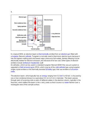
1. 1.
In a typical SEM, an electron beam is thermionically emitted from an electron gun fitted with
a tungsten filament cathode. Tungsten is normally used in thermionic electron guns because it
has the highest melting point and lowest vapor pressure of all metals, thereby allowing it to be
electrically heated for electron emission, and because of its low cost. Other types of electron
emitters include lanthanum hexaboride (LaB
6) cathodes, which can be used in a standard tungsten filament SEM if the vacuum system is
upgraded or field emission guns (FEG), which may be of the cold-cathode type using tungsten
single crystal emitters or the thermally assisted Schottky type, that use emitters of zirconium
oxide.
The electron beam, which typically has an energy ranging from 0.2 keV to 40 keV, is focused by
one or two condenser lenses to a spot about 0.4 nm to 5 nm in diameter. The beam passes
through pairs of scanning coils or pairs of deflector plates in the electron column, typically in the
final lens, which deflect the beam in the x and y axes so that it scans in a raster fashion over a
rectangular area of the sample surface.
2. Signals emitted from different parts of the interaction volume
Mechanisms of emission of secondary electrons, backscattered electrons, and characteristic X-
rays from atoms of the sample
When the primary electron beam interacts with the sample, the electrons lose energy by
repeated random scattering and absorption within a teardrop-shaped volume of the specimen
3. known as the interaction volume, which extends from less than 100 nm to approximately 5 袖m
into the surface. The size of the interaction volume depends on the electron's landing energy,
the atomic number of the specimen and the specimen's density.
The energy exchange between the electron beam and the sample results in the reflection of
high-energy electrons by elastic scattering, emission of secondary electrons by inelastic
scattering and the emission of electromagnetic radiation, each of which can be detected by
specialized detectors. The beam current absorbed by the specimen can also be detected and
used to create images of the distribution of specimen current.
Electronic amplifiers of various types are used to amplify the signals, which are displayed as
variations in brightness on a computer monitor (or, for vintage models, on a cathode ray tube).
Each pixel of computer video memory is synchronized with the position of the beam on the
specimen in the microscope, and the resulting image is therefore a distribution map of the
intensity of the signal being emitted from the scanned area of the specimen. In older
microscopes images may be captured by photography from a high-resolution cathode ray tube,
but in modern machines they are digitised and saved as digital images.
PRINCIPLE OF SEM
The types of signals produced by an SEM include secondary electrons (SE), reflected or back-
scattered electrons (BSE), photons of characteristic X-rays and light (cathodoluminescence)
(CL), absorbed current (specimen current) and transmitted electrons. Secondary electron
detectors are standard equipment in all SEMs, but it is rare that a single machine would have
detectors for all other possible signals.
The signals result from interactions of the electron beam with atoms at various depths within the
sample. In the most common or standard detection mode, secondary electron imaging or SEI,
the secondary electrons are emitted from very close to the specimen surface. Consequently,
SEM can produce very high-resolution images of a sample surface, revealing details less than
1 nm in size. Back-scattered electrons (BSE) are beam electrons that are reflected from the
sample by elastic scattering. They emerge from deeper locations within the specimen and
consequently the resolution of BSE images is generally poorer than SE images. However, BSE
are often used in analytical SEM along with the spectra made from the characteristic X-rays,
because the intensity of the BSE signal is strongly related to the atomic number (Z) of the
specimen. BSE images can provide information about the distribution of different elements in
the sample. For the same reason, BSE imaging can image colloidal gold immuno-labels of 5 or
10 nm diameter, which would otherwise be difficult or impossible to detect in secondary electron
images in biological specimens. Characteristic X-rays are emitted when the electron beam
4. removes an inner shell electron from the sample, causing a higher-energy electron to fill the
shell and release energy. These characteristic X-rays are used to identify the composition and
measure the abundance of elements in the sample.
Due to the very narrow electron beam, SEM micrographs have a large depth of field yielding a
characteristic three-dimensional appearance useful for understanding the surface structure of a
sample. This is exemplified by the micrograph of pollen shown above. A wide range of
magnifications is possible, from about 10 times (about equivalent to that of a powerful hand-
lens) to more than 500,000 times, about 250 times the magnification limit of the best light
microscopes.
2.
Xerophiles:-
are extremophilic organisms that can grow and reproduce in conditions with a low availability of
water, also known as water activity. Water activity (aw) is a measure of the amount of water
within a substrate that an organism can use to support sexual growth. Xerophiles are often said
to be "xerotolerant", meaning tolerant of dry conditions. They can survive in environments with
water activity below 0.8. Endoliths and halophiles are often xerotolerant.
The common food preservation method of reducing water activities may not prevent the growth
of xerophilic organisms, often resulting in food spoilage. Many mold and yeast species are
xerophilic. Mold growth on bread is an example of food spoilage by xerophilic organisms. This
naming comes from the Greek xeros meaning dry, and philos meaning "loving."
Halophiles are organisms that thrive in high salt concentrations. They are a type of
extremophile organisms. The name comes from the Greek word for "salt-loving". While most
halophiles are classified into the Archaea domain, there are also bacterial halophiles and some
eukaryota, such as the alga Dunaliella salina or fungus Wallemia ichthyophaga.
A basophileisatype of white bloodcell.Basophilsare the least commonof the granulocytes,
representingabout0.5 to 1% of circulatingwhite bloodcells.[1] However,theyare the largesttype of
granulocyte.Theyare responsible forinflammatoryreactionsduringimmune response,aswell asinthe
formationof acute and chronicallergicdiseases,includinganaphylaxis,asthma,atopicdermatitisand
5. hay fever.[2] Theycanperformphagocytosis(cell eating),produce histamineandserotoninthatinduce
inflammation,andheparinthatpreventsbloodclotting.[3]
Extremophilia, extremophile:Preference of livingextremalconditionsforsome microorganisms.
Geophilia, geophilic,geophileReferringtoorganismsthatpreferthe soil.
Halophilia,halophile:Attractiontosaltor salt-water.
Heliophilia,heliophile:Attractiontosunlight.
Hydrophilia:Attractiontowater.
Hyperthermophilia, hyperthermophile,hyperthermophilic:Organismsthatthrive inextremely
hot environments.
Acidophilia, acidophile: Preference of acidic conditions. Antonym: Acidophobia, acidophobe
Alkaliphilia, alkaliphile: Preference of alkaline environments.
Hydrostatic pressure is an important parameter influencing the distribution of microbial life in
the ocean. In this study, the response of marine bacterial populations from surface waters to
pressures representative of those under deep-sea conditions was examined.
3. Short notes
Bacterial Transformation:-
Transformation is one of three processes by which exogenous genetic material may be
introduced into a bacterial cell; the other two being conjugation (transfer of genetic material
between two bacterial cells in direct contact), and transduction (injection of foreign DNA by a
bacteriophage virus into the host bacterium).
Horizontal Genetic Transfer:-
Horizontal gene transfer (HGT) is the movement of genetic material between unicellular and/or
multicellular organisms other than via vertical transmission (the transmission of DNA from
parent to offspring.) HGT is synonymous with lateral gene transfer (LGT) and the terms are
interchangeable.
Vertical Genetic Transfer:-
vertical transfer, the transmission of genes from the parental generation to offspring via sexual
or asexual reproduction.
the insertion of copies of a gene into living cells in order to induce synthesis of the gene's
product: the desired gene may be microinjected directly into the cell or it may be inserted into
6. the core of a virus by gene splicing and the virus allowed to infect the cell for replication of the
gene in the cell's DNA.
Parthenogenesis:-
Parthenogenesis is most simply defined as reproduction without fertilization. More specifically, it
occurs when a female gamete develops a new individual without being fertilized by a male
gamete. It is often called a form of "asexual reproduction," but it is more accuratley defined as
an incomplete form of sexual reproduction. This is because it involves the production, activation,
and development of a female egg which is a specialized reproductive cell (Kaufman, 1983).
Parthenogenesis is not to be confused with hermaphroditic species which can also reproduce
by themselves. Hermaphroditic species reproduce by themselves because an organism can
produce both the male and female gamete.
4.Biochemical test for identification of bacteria, namely:-
Urease Test
Urease broth is a differential medium that tests the ability of an organism to produce an exoenzyme,
called urease, that hydrolyzes urea to ammonia and carbon dioxide. The broth contains two pH buffers,
urea, a very small amount of nutrients for the bacteria, and the pH indicator phenol red.
Catalase Test
The catalase test is used to differentiate staphylococci (catalase-positive) from streptococci (catalase-
negative). The enzyme, catalase, is produced by bacteria that respire using oxygen, and protects them
from the toxic by-products of oxygen metabolism.
7. Catalase is a common enzyme found in nearly all living organisms exposed to oxygen (such as bacteria,
plants, and animals). It catalyzes the decomposition of hydrogen peroxide to water and oxygen. It is a
very important enzyme in protecting the cell from oxidative damage by reactive oxygen species (ROS).
Starch Hydrolysis
The purpose is to see if the microbe can use starch, a complex carbohydrate made from glucose, as a
source of carbon and energy for growth. Use of starch is accomplished by an enzyme called alpha-
amylase.
Alpha-amylase: A medium containing starch is used. After inoculation and overnight incubation, iodine
reagent is added to detect the presence of starch. Iodine reagent complexes with starch to form a blue-
black color in the culture medium. Clear halos surrounding colonies is indicative of their ability to digest
the starch in the medium due to the presence of alpha-amylase.
Carbohydrate Utilization/Test
To test cells for the ability of an organism to utilize and digest several sugars or small carbohydrates. It is
useful in identifying Gram negative bacilli, especially Enterobacteriaceae, though it may aid in identifying
many other species as well.
The Carbohydrate Utilization test uses Phenol Red Broth (or Purple Broth) to test for the utilization of
different sugars. Phenol Red Broth is a differential media that includes the pH indicator Phenol Red,
peptones, and a series of tubes each with a different sugar. Different sugars and polyhydric alcohols may
be used, but glucose, sucrose, lactose, and mannitol are often studied among others. One inoculates
bacteria into each tube, if the strain of bacteria utilizes that sugar, an acid will form changing the color of
Phenol Red from reddish-orange to yellow.
Gelatin Utilization /Test
Nutrient gelatin is a differential medium that tests the ability of an organism to produce an exoenzyme,
called gelatinase, that hydrolyzes gelatin. Gelatin is commonly known as a component of gelled salads
and some desserts, but it's actually a protein derived from connective tissue.
At temperatures above 32属C, it is a viscous liquid. Gelatinase allows the organisms that produce it to
break down gelatin into smaller polypeptides, peptides, and amino acids that can cross the cell
membrane and be utilized by the organism.
5. Endotoxin and exotoxin properties and differences.
Exotoxins:
Definition: Toxins that are released extracellularly as the organism grows are called exotoxins. Exotoxins
may travel from a focus of infection to distant part of the body and cause damage. E.g. Neurotoxin
(botulinum toxin, tetanus toxin), Enterotoxin (cholera toxin), Cytotoxin
8. Endotoxins:
Definition: Endotoxins are lipopolysaccharides toxin produced by Gram negative bacteria. The name
endotoxin is derived from the fact that these toxins are generally cell bound and released only when the
cell lyses.
Basic properties and differences between Exotoxins and Endotoxins
Property Exotoxins Endotoxins
Biomolecule/Chemistry Proteins
Lipopolysaccharide-Lipoprotein
complex
Location of genes Plasmid or bacteriophage Bacterial chromosome
Source
Excreted by certain gram positive or gram
negative bacteria
Cell wall of Gram Negative bacteria,
released only after lysis of cells
Heat Stability
Destroyed rapidly at 60oC (except
staphylococcal enterotoxin) Stable at 100oC for one hour
Mode of Action
(Symptoms)
Specific. Either cytotoxin, enterotoxin or
neurotoxin with defined action on cells or
tissues General. fever, diarrhea, vomiting
Toxicity
Highly toxic, often fatal (fatal dose on the
order of 1 袖g)
Weakly toxic, rarely fatal (fatal dose
on the order of hundreds of
micrograms)
Immunogenicity
Highly immunogenic, stimulate the
production of neutralizing antibody
(antitoxins) Relatively poor immunogenicity
Toxoid
potential/Vaccines
Treatment of toxin with formaldehyde will
destroy toxicity, but treated toxins remain
immunogenic.
Toxids used as vaccines
No toxoid formed and no vaccine
available
Typical disease Tetanus, diphtheria, botulism
Meningococcemia, sepsis by gram
negative rods
9. S.N. Exotoxins Endotoxins
1 Excreted by organisms, living cell Integral part of cell wall
2
Found in both Gram positive and Gram
Negative bacteria
Found mostly in Gram Negative Bacteria
3 It is polypeptide It is lipopolysaccharide complex.
4 Relatively unstable, heat labile (60属C) Relatively stable, heat tolerant
5 Highly antigenic Weakly immunogenic
6 Toxoids can be madeby treating with
formalin
Toxoids cannot be made
7 Highly toxic, fatal in 袖g quantities Moderately toxic
8 Usually binds to specific receptors Specific receptors not found
9 Not pyrogenic usually, Toxin Specific
Fever by induction of interleukin 1 (IL-1)
production, Shock
10 Located on extrachromosomal genes
(e.g. plasmids)
Located on chromosomal genes
11 Filterable Not so
12 It has no enzymatic activity It has mostly enzymatic activity
13 Its molecular weight is 10KDa Its molecular weight is 50-1000KDa
14 On boiling it get denatured. On boiling it cannot be denatured.
15 Detected by many tests (neutralization,
precipitation, etc)
Detected by Limulus lysate assay
16
Examples: Toxins produced
by Staphylococcus aureus, Bacillus
cereus, Streptococcus pyogenes,
Bacillus anthrcis(Alpha-toxin, also
known as alpha-hemolysin (Hla))
Examples: Toxins produced by E.coli,
Salmonella Typhi, Shigella, Vibrio
cholera(Cholera toxin- also known as
choleragen)
17 Diseases: Tetanus, diphtheria, botulism
Diseases: Meningococcemia, sepsis by
gram negative rods