際際滷

際際滷Share a Scribd company logo
VIVEKANANDHA ARTS AND SCIENCE COLLEGE FOR WOMEN
VEERACHIPALAYAM - 6366007
SANKAGIRI, TAMIL NADU.
DEPARTMENT OF MICROBIOLOGY
SUBJECT INCHARGE
Dr.V.BHARATHI,M.Sc.,M.Phil.,Ph.D.
ASSISTANT PROFESSOR
DEPARTMENT OF MICROBIOLOGY
VIAAS, SANKAGIRI.
PRESENTED BY
DIVYAPRABHA K
I M.Sc MICROBIOLOGY
DEPARTMENT OF MICROBIOLOGY
VIAAS, SANKAGIRI
TOPIC : SCANNING ELECTRON MICROSCOPE
SUBJECT : GENERAL MICROBIOLOGY AND MICROBIAL DIVERSITY
INTRODUCTION
 The first Scanning Electron Microscope was initially made by Mafred
von Ardenne in 1937 with an aim to surpass the transmission electron
Microscope.
 He used high-resolution power to scan a small raster using a beam of
electrons that were focused on the raster.
 He also aimed at reducing the problems of chromatic aberrations
images produced by the Transmission electron Microscopes.
DEFINITION
 Scanning Electron Microscope (SEM) is a type of
electron microscope that scans surfaces of
microorganisms that uses a beam of electrons
moving at low energy to focus and scan specimens.
 The development of electron microscopes was due
to the inefficiency of the wavelength of light
microscopes. electron microscopes have very short
wavelengths in comparison to the light microscope
which enables better resolution power.
PRINCIPLES
 The scanning electron Microscope uses emitted electrons.
 The Scanning electron microscope works on the principle of applying
kinetic energy to produce signals on the interaction of the
electrons.
 The secondary electrons are emitted from the specimen play the
primary role of detecting the morphology and topography of the
specimen while the backscattered electrons show contrast in the
composition of the elements of the specimen.
Parts of a Scanning Electron Microscope (SEM)
The major components of the Scanning Electron Microscope include;
 Electron Source  This is where electrons are produced under thermal heat at a
voltage of 1-40kV.
 the electrons condense into a beam that is used for the creation of an image and
analysis.
 There are three types of electron sources that can be used i. e Tungsten filament,
Lanthanum hexaboride, and Field emission gun (FEG)
 Lenses  it has several condenser lenses that focus the beam of electrons from
the source through the column forming a narrow beam of electrons that form a spot
called a spot size.
 Scanning Coil  they are used to deflect the beam over the specimen surface.
 Detector  Its made up of several detectors that are able to differentiate the
secondary electrons, backscattered electrons, and diffracted backscattered
electrons.
 The functioning of the detectors highly depends on the voltage speed, the density
of the specimen.
 The display device (data output devices)
 Power supply
 Vacuum system
PARTS OF SCANNING ELECTRON MICROSCOPE
Scanning Electron Microscope (SEM) Images
Figure: SEM image of Tradescantia pollen and stamens. Figure: Low-temperature scanning electron
micrograph of soybean cyst nematode and its
egg. Magnified 1,000 times.
Figure: Photoresist SEM micrograph (1995) SEM= DSM
982 Gemini from Zeiss.
APPLICATIONS OF SCANNING ELECTRON MICROSCOPE (SEM)
 It is used in a variety of fields including industrial uses,nanoscience studies,
biomedical studies,microbiology.
 Used for spot chemical analysis in energy - Dispersive x-ray spectroscopy.
 Used in the analysis cosmetics components which are very tiny in size.
 Used to study the filament structures of microorganisms.
 Used to study the topography of elements used in industries.
Advantages of the Scanning Electron Microscope (SEM)
 They are easy to operate and have user-friendly interfaces.
 They are used in a variety of industrial applications to analyze surfaces of solid
objects.
 Some modern SEMs are able to generate digital data that can be portable.
 It is easy to acquire data from the SEM, within a short period of time of about 5
minutes.
Disadvantages of Scanning Electron Microscope
 Very cost effective.
 Samples must be slid and vacuum-compatible,so live specimens cant be studied.
 SEMs are large and bulky,and require a lot of space in lab.
 Special training is required to operate an SEM and prepare samples.
REFERENCES
 https://microbenotes.com/scanning-electron-microscope-sem/
 https://www.britannica.com/technology/scanning-electron-microscope
 https://blog.phenom-world.com/what-is-sem
Ad

Recommended

PPTX
Scanning electron microscope
SuganyaPaulraj
PPTX
Electron microscope
microbiology Notes
PPTX
Scanning Electron Microscope
Ketan Patil
PPTX
Scanning Electron Microscope
Marian L
PPTX
Stem
akshay567
PPTX
SEM & FESEM.pptx
THE CREATORS ACADEMY
PPTX
Scanning Electron Microscope (SEM).pptx
navaneethankv23mech
PPTX
electronmicroscope principle construction working & types
chaitanyamahesh06
PPTX
Electron microscope
Rasika Deshmukh
PPTX
scanning electron microscope
DrAkhilaCNV
PPTX
Scanning electron microscopy (SEM) .pptx
hirazamurd002
PPTX
Electron Microscopy - Scanning electron microscope, Transmission Electron Mic...
Sumer Pankaj
PPTX
scanning electron microscope for analysis
M Ali Mohsin
PPTX
Sem (2)
kanwalkayani1
PPTX
scanning electron microscope (SEM)
Himanshu Dixit
PPTX
Scanning electron microscope - SEM , Its applicability in rasashastra
Dr. Madduru Muni Haritha
PPTX
Scanning electron microscope
Senthil Arasan
PPTX
Scanning and Transmission Electron Microscope
JSPM Charak College of Pharmacy and Research
PPTX
Presentation forensic micoscopy SEM microscope.pptx
HarishKumar330845
PPTX
Chaitrali jadhav:- scanning electron microscope
Chaitrali Jadhav
PPTX
Confocal and electron microscopy.pptx
Sathish SV
PPTX
Presentation of Electron Microscopy.pptx
heminrostam169
PPTX
Presenatation topic SEM, M.Rashad.pptx
Chasim6
PPTX
Scanning electron microscopy (sem)
Sivasangari Shanmugam
PPTX
Scanning electron microscopy (SEM) Likhith K
LIKHITHK1
DOCX
CNM.docx
Thahsinabarveen
PPTX
Scanning electron microscope ppt.pptx
kavinkumars23mech
PPTX
Scanning electron microscope
Akhtar Kamal
PPTX
Code Profiling in Odoo 18 - Odoo 18 際際滷s
Celine George
PDF
University of Ghana Cracks Down on Misconduct: Over 100 Students Sanctioned
Kweku Zurek

More Related Content

Similar to General microbiology and microbial diversity - scanning electron microscope (20)

PPTX
Electron microscope
Rasika Deshmukh
PPTX
scanning electron microscope
DrAkhilaCNV
PPTX
Scanning electron microscopy (SEM) .pptx
hirazamurd002
PPTX
Electron Microscopy - Scanning electron microscope, Transmission Electron Mic...
Sumer Pankaj
PPTX
scanning electron microscope for analysis
M Ali Mohsin
PPTX
Sem (2)
kanwalkayani1
PPTX
scanning electron microscope (SEM)
Himanshu Dixit
PPTX
Scanning electron microscope - SEM , Its applicability in rasashastra
Dr. Madduru Muni Haritha
PPTX
Scanning electron microscope
Senthil Arasan
PPTX
Scanning and Transmission Electron Microscope
JSPM Charak College of Pharmacy and Research
PPTX
Presentation forensic micoscopy SEM microscope.pptx
HarishKumar330845
PPTX
Chaitrali jadhav:- scanning electron microscope
Chaitrali Jadhav
PPTX
Confocal and electron microscopy.pptx
Sathish SV
PPTX
Presentation of Electron Microscopy.pptx
heminrostam169
PPTX
Presenatation topic SEM, M.Rashad.pptx
Chasim6
PPTX
Scanning electron microscopy (sem)
Sivasangari Shanmugam
PPTX
Scanning electron microscopy (SEM) Likhith K
LIKHITHK1
DOCX
CNM.docx
Thahsinabarveen
PPTX
Scanning electron microscope ppt.pptx
kavinkumars23mech
PPTX
Scanning electron microscope
Akhtar Kamal
Electron microscope
Rasika Deshmukh
scanning electron microscope
DrAkhilaCNV
Scanning electron microscopy (SEM) .pptx
hirazamurd002
Electron Microscopy - Scanning electron microscope, Transmission Electron Mic...
Sumer Pankaj
scanning electron microscope for analysis
M Ali Mohsin
Sem (2)
kanwalkayani1
scanning electron microscope (SEM)
Himanshu Dixit
Scanning electron microscope - SEM , Its applicability in rasashastra
Dr. Madduru Muni Haritha
Scanning electron microscope
Senthil Arasan
Scanning and Transmission Electron Microscope
JSPM Charak College of Pharmacy and Research
Presentation forensic micoscopy SEM microscope.pptx
HarishKumar330845
Chaitrali jadhav:- scanning electron microscope
Chaitrali Jadhav
Confocal and electron microscopy.pptx
Sathish SV
Presentation of Electron Microscopy.pptx
heminrostam169
Presenatation topic SEM, M.Rashad.pptx
Chasim6
Scanning electron microscopy (sem)
Sivasangari Shanmugam
Scanning electron microscopy (SEM) Likhith K
LIKHITHK1
CNM.docx
Thahsinabarveen
Scanning electron microscope ppt.pptx
kavinkumars23mech
Scanning electron microscope
Akhtar Kamal

Recently uploaded (20)

PPTX
Code Profiling in Odoo 18 - Odoo 18 際際滷s
Celine George
PDF
University of Ghana Cracks Down on Misconduct: Over 100 Students Sanctioned
Kweku Zurek
PPTX
Tanja Vujicic - PISA for Schools contact Info
EduSkills OECD
PPTX
Values Education 10 Quarter 1 Module .pptx
JBPafin
PDF
Romanticism in Love and Sacrifice An Analysis of Oscar Wildes The Nightingal...
KaryanaTantri21
PDF
VCE Literature Section A Exam Response Guide
jpinnuck
PDF
K12 Tableau User Group virtual event June 18, 2025
dogden2
PPTX
OBSESSIVE COMPULSIVE DISORDER.pptx IN 5TH SEMESTER B.SC NURSING, 2ND YEAR GNM...
parmarjuli1412
PPTX
Wage and Salary Computation.ppt.......,x
JosalitoPalacio
PPTX
2025 Completing the Pre-SET Plan Form.pptx
mansk2
PPTX
How payment terms are configured in Odoo 18
Celine George
PPTX
June 2025 Progress Update With Board Call_In process.pptx
International Society of Service Innovation Professionals
PPTX
How to Manage Different Customer Addresses in Odoo 18 Accounting
Celine George
PPTX
List View Components in Odoo 18 - Odoo 際際滷s
Celine George
PPTX
NSUMD_M1 Library Orientation_June 11, 2025.pptx
Julie Sarpy
PPTX
LAZY SUNDAY QUIZ "A GENERAL QUIZ" JUNE 2025 SMC QUIZ CLUB, SILCHAR MEDICAL CO...
Ultimatewinner0342
PPTX
How to Add New Item in CogMenu in Odoo 18
Celine George
PDF
Learning Styles Inventory for Senior High School Students
Thelma Villaflores
PDF
LDMMIA Yoga S10 Free Workshop Grad Level
LDM & Mia eStudios
PPTX
Peer Teaching Observations During School Internship
AjayaMohanty7
Code Profiling in Odoo 18 - Odoo 18 際際滷s
Celine George
University of Ghana Cracks Down on Misconduct: Over 100 Students Sanctioned
Kweku Zurek
Tanja Vujicic - PISA for Schools contact Info
EduSkills OECD
Values Education 10 Quarter 1 Module .pptx
JBPafin
Romanticism in Love and Sacrifice An Analysis of Oscar Wildes The Nightingal...
KaryanaTantri21
VCE Literature Section A Exam Response Guide
jpinnuck
K12 Tableau User Group virtual event June 18, 2025
dogden2
OBSESSIVE COMPULSIVE DISORDER.pptx IN 5TH SEMESTER B.SC NURSING, 2ND YEAR GNM...
parmarjuli1412
Wage and Salary Computation.ppt.......,x
JosalitoPalacio
2025 Completing the Pre-SET Plan Form.pptx
mansk2
How payment terms are configured in Odoo 18
Celine George
June 2025 Progress Update With Board Call_In process.pptx
International Society of Service Innovation Professionals
How to Manage Different Customer Addresses in Odoo 18 Accounting
Celine George
List View Components in Odoo 18 - Odoo 際際滷s
Celine George
NSUMD_M1 Library Orientation_June 11, 2025.pptx
Julie Sarpy
LAZY SUNDAY QUIZ "A GENERAL QUIZ" JUNE 2025 SMC QUIZ CLUB, SILCHAR MEDICAL CO...
Ultimatewinner0342
How to Add New Item in CogMenu in Odoo 18
Celine George
Learning Styles Inventory for Senior High School Students
Thelma Villaflores
LDMMIA Yoga S10 Free Workshop Grad Level
LDM & Mia eStudios
Peer Teaching Observations During School Internship
AjayaMohanty7
Ad

General microbiology and microbial diversity - scanning electron microscope

  • 1. VIVEKANANDHA ARTS AND SCIENCE COLLEGE FOR WOMEN VEERACHIPALAYAM - 6366007 SANKAGIRI, TAMIL NADU. DEPARTMENT OF MICROBIOLOGY SUBJECT INCHARGE Dr.V.BHARATHI,M.Sc.,M.Phil.,Ph.D. ASSISTANT PROFESSOR DEPARTMENT OF MICROBIOLOGY VIAAS, SANKAGIRI. PRESENTED BY DIVYAPRABHA K I M.Sc MICROBIOLOGY DEPARTMENT OF MICROBIOLOGY VIAAS, SANKAGIRI TOPIC : SCANNING ELECTRON MICROSCOPE SUBJECT : GENERAL MICROBIOLOGY AND MICROBIAL DIVERSITY
  • 2. INTRODUCTION The first Scanning Electron Microscope was initially made by Mafred von Ardenne in 1937 with an aim to surpass the transmission electron Microscope. He used high-resolution power to scan a small raster using a beam of electrons that were focused on the raster. He also aimed at reducing the problems of chromatic aberrations images produced by the Transmission electron Microscopes.
  • 3. DEFINITION Scanning Electron Microscope (SEM) is a type of electron microscope that scans surfaces of microorganisms that uses a beam of electrons moving at low energy to focus and scan specimens. The development of electron microscopes was due to the inefficiency of the wavelength of light microscopes. electron microscopes have very short wavelengths in comparison to the light microscope which enables better resolution power.
  • 4. PRINCIPLES The scanning electron Microscope uses emitted electrons. The Scanning electron microscope works on the principle of applying kinetic energy to produce signals on the interaction of the electrons. The secondary electrons are emitted from the specimen play the primary role of detecting the morphology and topography of the specimen while the backscattered electrons show contrast in the composition of the elements of the specimen.
  • 5. Parts of a Scanning Electron Microscope (SEM) The major components of the Scanning Electron Microscope include; Electron Source This is where electrons are produced under thermal heat at a voltage of 1-40kV. the electrons condense into a beam that is used for the creation of an image and analysis. There are three types of electron sources that can be used i. e Tungsten filament, Lanthanum hexaboride, and Field emission gun (FEG) Lenses it has several condenser lenses that focus the beam of electrons from the source through the column forming a narrow beam of electrons that form a spot called a spot size.
  • 6. Scanning Coil they are used to deflect the beam over the specimen surface. Detector Its made up of several detectors that are able to differentiate the secondary electrons, backscattered electrons, and diffracted backscattered electrons. The functioning of the detectors highly depends on the voltage speed, the density of the specimen. The display device (data output devices) Power supply Vacuum system
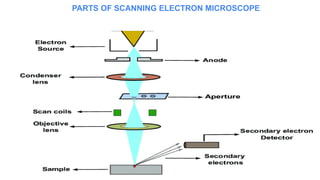
  • 7. PARTS OF SCANNING ELECTRON MICROSCOPE
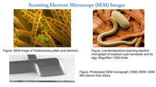
  • 8. Scanning Electron Microscope (SEM) Images Figure: SEM image of Tradescantia pollen and stamens. Figure: Low-temperature scanning electron micrograph of soybean cyst nematode and its egg. Magnified 1,000 times. Figure: Photoresist SEM micrograph (1995) SEM= DSM 982 Gemini from Zeiss.
  • 9. APPLICATIONS OF SCANNING ELECTRON MICROSCOPE (SEM) It is used in a variety of fields including industrial uses,nanoscience studies, biomedical studies,microbiology. Used for spot chemical analysis in energy - Dispersive x-ray spectroscopy. Used in the analysis cosmetics components which are very tiny in size. Used to study the filament structures of microorganisms. Used to study the topography of elements used in industries.
  • 10. Advantages of the Scanning Electron Microscope (SEM) They are easy to operate and have user-friendly interfaces. They are used in a variety of industrial applications to analyze surfaces of solid objects. Some modern SEMs are able to generate digital data that can be portable. It is easy to acquire data from the SEM, within a short period of time of about 5 minutes. Disadvantages of Scanning Electron Microscope Very cost effective. Samples must be slid and vacuum-compatible,so live specimens cant be studied. SEMs are large and bulky,and require a lot of space in lab. Special training is required to operate an SEM and prepare samples.