Hearing
•Download as PPT, PDF•
7 likes•2,795 views

The document describes the anatomy and physiology of the human ear and vestibular system. It explains that sound waves are channeled through the outer ear to the tympanic membrane, causing the ossicles to vibrate and transmit the vibrations through the oval window to the cochlea. Within the cochlea, vibrations stimulate hair cells which transduce the mechanical signal into neural impulses that travel to the brain. The vestibular system contains semicircular canals and otolith organs that provide the senses of balance and spatial orientation through the detection of head motion.

Hearing
- 1. Ears & Hearing 10-34
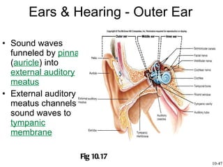
- 2. Sound waves funneled by pinna ( auricle ) into external auditory meatus External auditory meatus channels sound waves to tympanic membrane Ears & Hearing - Outer Ear Fig 10.17 10-47
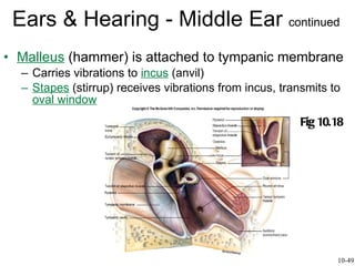
- 3. Malleus (hammer) is attached to tympanic membrane Carries vibrations to incus (anvil) Stapes (stirrup) receives vibrations from incus, transmits to oval window Ears & Hearing - Middle Ear continued Fig 10.18 10-49
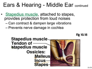
- 4. Stapedius muscle , attached to stapes, provides protection from loud noises Can contract & dampen large vibrations Prevents nerve damage in cochlea Ears & Hearing - Middle Ear continued 10-50 Fig 10.18
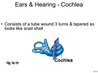
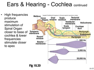
- 5. Ears & Hearing - Cochlea Consists of a tube wound 3 turns & tapered so looks like snail shell Fig 10.19 10-51
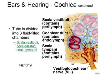
- 6. Ears & Hearing - Cochlea continued Tube is divided into 3 fluid-filled chambers Scala vestibuli , cochlear duct , scala tympani Fig 10.19 10-52
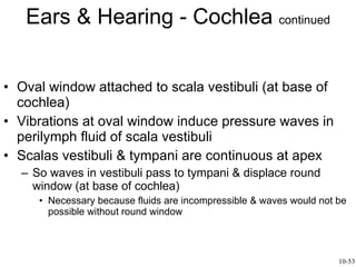
- 7. Ears & Hearing - Cochlea continued Oval window attached to scala vestibuli (at base of cochlea) Vibrations at oval window induce pressure waves in perilymph fluid of scala vestibuli Scalas vestibuli & tympani are continuous at apex So waves in vestibuli pass to tympani & displace round window (at base of cochlea) Necessary because fluids are incompressible & waves would not be possible without round window 10-53
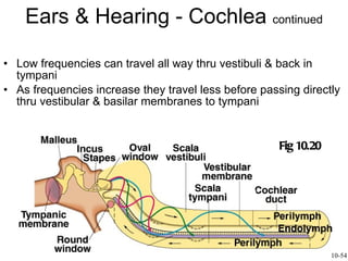
- 8. Ears & Hearing - Cochlea continued Low frequencies can travel all way thru vestibuli & back in tympani As frequencies increase they travel less before passing directly thru vestibular & basilar membranes to tympani Fig 10.20 10-54
- 9. Ears & Hearing - Cochlea continued High frequencies produce maximum stimulation of Spiral Organ closer to base of cochlea & lower frequencies stimulate closer to apex Fig 10.20 10-55
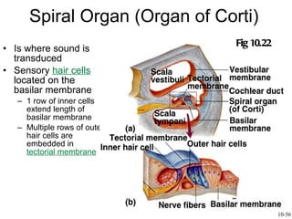
- 10. Spiral Organ (Organ of Corti) Is where sound is transduced Sensory hair cells located on the basilar membrane 1 row of inner cells extend length of basilar membrane Multiple rows of outer hair cells are embedded in tectorial membrane Fig 10.22 10-56
- 11. Spiral Organ (Organ of Corti) Pressure waves moving thru cochlear duct create shearing forces between basilar & tectorial membranes, moving & bending stereocilia Causing ion channels to open, depolarizing hair cells The greater the displacement, the greater the amount of NT released & APs produced 10-57
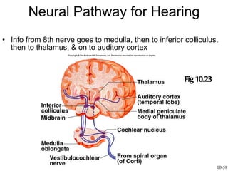
- 12. Neural Pathway for Hearing Info from 8th nerve goes to medulla, then to inferior colliculus, then to thalamus, & on to auditory cortex Fig 10.23 10-58
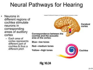
- 13. Neural Pathways for Hearing Neurons in different regions of cochlea stimulate neurons in corresponding areas of auditory cortex Each area of cortex represents different part of cochlea & thus a different pitch Fig 10.24 10-59
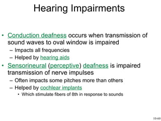
- 14. Hearing Impairments Conduction deafness occurs when transmission of sound waves to oval window is impaired Impacts all frequencies Helped by hearing aids Sensorineural ( perceptive ) deafness is impaired transmission of nerve impulses Often impacts some pitches more than others Helped by cochlear implants Which stimulate fibers of 8th in response to sounds 10-60
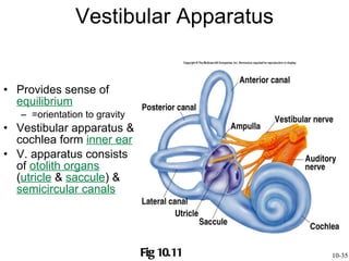
- 15. Vestibular Apparatus Provides sense of equilibrium =orientation to gravity Vestibular apparatus & cochlea form inner ear V. apparatus consists of otolith organs ( utricle & saccule ) & semicircular canals Fig 10.11 10-35
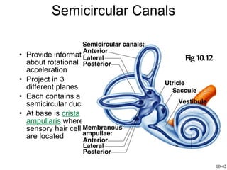
- 16. Semicircular Canals Provide information about rotational acceleration Project in 3 different planes Each contains a semicircular duct At base is crista ampullaris where sensory hair cells are located Fig 10.12 10-42
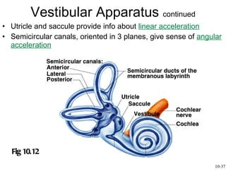
- 17. Utricle and saccule provide info about linear acceleration Semicircular canals, oriented in 3 planes, give sense of angular acceleration Vestibular Apparatus continued Fig 10.12 10-37
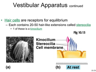
- 18. Hair cells are receptors for equilibrium Each contains 20-50 hair-like extensions called stereocilia 1 of these is a kinocilium Vestibular Apparatus continued Fig 10.13 10-38
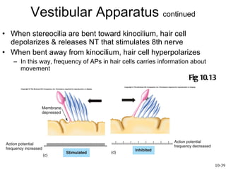
- 19. When stereocilia are bent toward kinocilium, hair cell depolarizes & releases NT that stimulates 8th nerve When bent away from kinocilium, hair cell hyperpolarizes In this way, frequency of APs in hair cells carries information about movement Vestibular Apparatus continued Fig 10.13 10-39
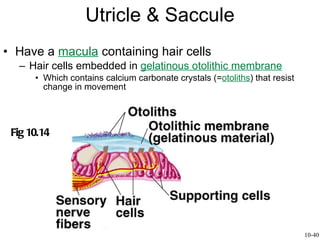
- 20. Utricle & Saccule Have a macula containing hair cells Hair cells embedded in gelatinous otolithic membrane Which contains calcium carbonate crystals (= otoliths ) that resist change in movement Fig 10.14 10-40
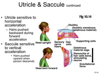
- 21. Utricle & Saccule continued Utricle sensitive to horizontal acceleration Hairs pushed backward during forward acceleration Saccule sensitive to vertical acceleration Hairs pushed upward when person descends Fig 10.14 10-41
- 22. Semicircular Canals Provide information about rotational acceleration Project in 3 different planes Each contains a semicircular duct At base is crista ampullaris where sensory hair cells are located Fig 10.12 10-42
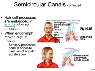
- 23. Semicircular Canals continued Hair cell processes are embedded in cupula of crista ampullaris When endolymph moves cupula moves Sensory processes bend in opposite direction of angular acceleration Fig 10.15 10-43
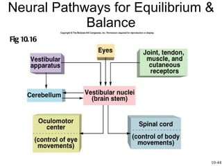
- 24. Neural Pathways for Equilibrium & Balance Fig 10.16 10-44
- 25. Nystagmus & Vertigo Vestibular nystagmus is involuntary oscillations of eyes that occurs when spinning person stops Eyes continue to move in direction opposite to spin, then jerk rapidly back to midline Vertigo is loss of equilibrium Natural response of vestibular apparatus Pathologically, may be caused by anything that alters firing rate of 8th nerve Often caused by viral infection 10-45