Histiocytic sarcoma or Osteosarcoma? That is the question.
- 1. Histiocytic ┬Āsarcoma ┬Ā or ┬Āosteosarcoma... That is the question
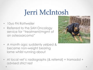
- 2. Jerri ┬ĀMcIntosh ŌĆóŌĆ» 10yo FN Rottweiler ŌĆóŌĆ» Referred to the SAH Oncology service for ŌĆ£treatment/mgmt of an osteosarcomaŌĆØ ŌĆóŌĆ» A month ago: suddenly yelped & became non-weight bearing lame whilst running about ŌĆóŌĆ» At local vetŌĆÖs: radiographs (& referral) + tramadol + advised strict rest
- 3. History ŌĆóŌĆ» EDDU all fine ŌĆóŌĆ» had adhered to rest instructions - only let out to garden ŌĆóŌĆ» left forelimb lameness improved but not entirely ŌĆóŌĆ» generally happy at home though the tramadol made her quite sedated ŌĆóŌĆ» known right forelimb osteoarthritis of 3 years duration ŌĆóŌĆ» has meloxicam for that
- 4. Clinical ┬Āexam ŌĆóŌĆ» bright & alert ŌĆóŌĆ» visibly lame on left fore but mostly weight-bearing ŌĆóŌĆ» feeling the limb, noted a firm swelling of muscle-&- soft-tissue consistency ŌĆóŌĆ» local draining lymph nodes (prescaps, axillary) not palpably enlarged or firm ŌĆóŌĆ» NAD on chest auscultation and abdominal palpation
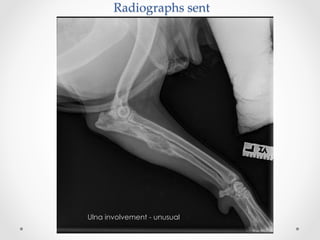
- 5. Radiographs ┬Āsent Ulna involvement - unusual
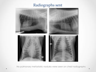
- 6. Radiographs ┬Āsent No pulmonary metastatic nodules were seen on chest radiographs
- 7. Our ┬Āplan ┬Āof ┬Āaction oŌĆ» routine bloodwork (haem & biochem) oŌĆ» CT scan of elbows (both) oŌĆ» CT scan of thorax oŌĆ» Abdominal ultrasound scan oŌĆ» FNA of the swelling ŌĆóŌĆ» in-house + sent up to the clinical pathology unit ŌĆóŌĆ» multiple slides to account for variation within the lesion The obligatory metcheck
- 8. Test ┬Āresults ┬Ā/ ┬Ā’¼ündings ŌĆóŌĆ» Routine blood work oŌĆ» Haematology unremarkable. oŌĆ» Biochemistry mild elevation of AST but no increase in ALP (often seen with OSAs) ŌĆóŌĆ» Abdominal ultrasound scan NAD
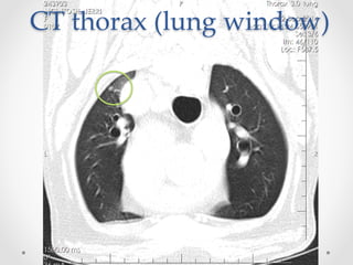
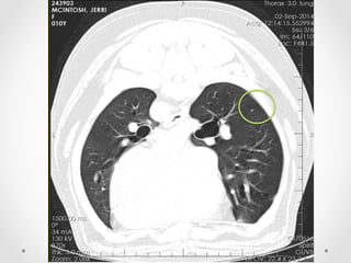
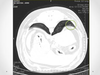
- 9. CT ┬Āthorax ┬Ā(lung ╠²Ę╔Š▒▓į╗Õ┤ŪĘ╔)
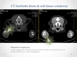
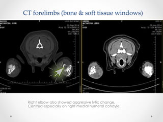
- 12. Diagnostic imaging dx: Large aggressive soft tissue lesion with invasion and destruction of proximal left ulna - likely neoplastic. CT ┬Āforelimbs ┬Ā(bone ┬Ā& ┬Āsoft ┬Ātissue ┬Āwindows)
- 13. Right elbow also showed aggressive lytic change, Centred especially on right medial humeral condyle. CT ┬Āforelimbs ┬Ā(bone ┬Ā& ┬Āsoft ┬Ātissue ┬Āwindows)
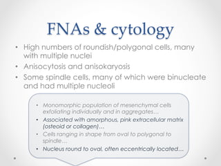
- 14. FNAs ┬Ā& ┬Ācytology ŌĆóŌĆ» High numbers of roundish/polygonal cells, many with multiple nuclei ŌĆóŌĆ» Anisocytosis and anisokaryosis ŌĆóŌĆ» Some spindle cells, many of which were binucleate and had multiple nucleoli ŌĆóŌĆ» Monomorphic population of mesenchymal cells exfoliating individually and in aggregatesŌĆ” ŌĆóŌĆ» Associated with amorphous, pink extracellular matrix (osteoid or collagen)ŌĆ” ŌĆóŌĆ» Cells ranging in shape from oval to polygonal to spindleŌĆ” ŌĆóŌĆ» Nucleus round to oval, often eccentrically locatedŌĆ”
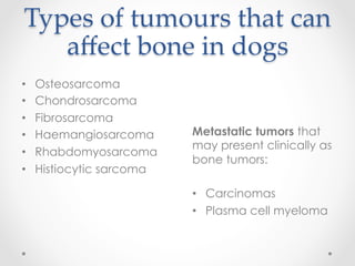
- 15. Types ┬Āof ┬Ātumours ┬Āthat ┬Ācan ┬Ā a’¼Ćect ┬Ābone ┬Āin ┬Ādogs ŌĆóŌĆ» Osteosarcoma ŌĆóŌĆ» Chondrosarcoma ŌĆóŌĆ» Fibrosarcoma ŌĆóŌĆ» Haemangiosarcoma ŌĆóŌĆ» Rhabdomyosarcoma ŌĆóŌĆ» Histiocytic sarcoma Metastatic tumors that may present clinically as bone tumors: ŌĆóŌĆ» Carcinomas ŌĆóŌĆ» Plasma cell myeloma
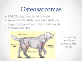
- 16. Osteosarcomas ŌĆóŌĆ» 85-90% of primary bone tumours ŌĆóŌĆ» Appendicular skeleton > axial skeleton ŌĆóŌĆ» large and giant breeds >> small breeds ŌĆóŌĆ» Predilection sites: away from the elbow, towards the knee
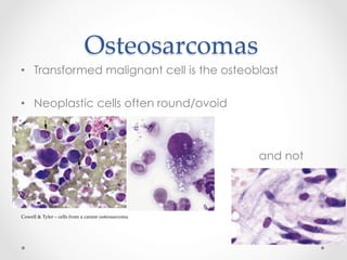
- 17. Osteosarcomas ŌĆóŌĆ» Transformed malignant cell is the osteoblast ŌĆóŌĆ» Neoplastic cells often round/ovoid Cowell ┬Ā& ┬ĀTyler ┬ĀŌĆō ┬Ācells ┬Āfrom ┬Āa ┬Ācanine ┬Āosteosarcoma and not
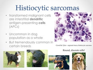
- 18. Histiocytic ┬Āsarcomas ┬Ā ŌĆóŌĆ» transformed malignant cells are interstitial dendritic antigen-presenting cells (APCs) ŌĆóŌĆ» Uncommon in dog population as a whole ŌĆóŌĆ» But tremendously common in certain breeds Cowell ┬Ā& ┬ĀTyler ┬ĀŌĆō ┬Āaspirate ┬Āfrom ┬Āa ┬Āhistiocytic ┬Āsarcoma Round, ┬Ādiscrete ┬Ācells!
- 19. Histiocytic ┬Āsarcomas ŌĆóŌĆ» 2 forms oŌĆ» Localised ŌĆóŌĆ» deep limb musculature and periarticular ŌĆóŌĆ» Highly malignant and metastatic oŌĆ» Disseminated ŌĆóŌĆ» both localised and disseminated forms carry quite a guarded prognosis
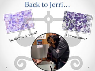
- 20. Back ┬Āto ┬ĀJerriŌĆ” Histiocytic ┬Āsarcoma? Osteosarcoma?
- 21. JerriŌĆÖs ┬ĀFNA ┬Āslides ┬Ā-┬ŁŌĆÉŌĆæ ┬Ā discussion ŌĆóŌĆ» Cytology can be rewarding but also confusing, especially to the unpractised eye ŌĆóŌĆ» Special cytochemical stains available oŌĆ» BCIP/NBT┬Āsolution stains ALP in osteoblasts oŌĆ» ANBE stains intracellular esterase enzymes that are present in cells of dendritic/ monocytic origin ŌĆóŌĆ» Variation amongst different sites of the lesion sampled Provisional diagnosis: Lytic bone tumour of left ulna. Sarcoma.
- 22. Treatment ┬Āoptions ŌĆóŌĆ» Amputation + follow-up with chemo = first-line ŌĆóŌĆ» Radiotherapy ŌĆóŌĆ» More analgesics oŌĆ» e.g. tramadol , fentanyl patches ŌĆóŌĆ» Bisphosphonates oŌĆ» help reduce bone lysis and pain Palliative intent
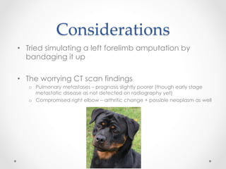
- 23. Considerations ŌĆóŌĆ» Tried simulating a left forelimb amputation by bandaging it up ŌĆóŌĆ» The worrying CT scan findings oŌĆ» Pulmonary metastases ŌĆō prognosis slightly poorer (though early stage metastatic disease as not detected on radiography yet) oŌĆ» Compromised right elbow ŌĆō arthritic change + possible neoplasm as well
- 24. Recommendations ┬Āand ┬Ā ownerŌĆÖs ┬Ādecision ŌĆóŌĆ» Radiotherapy ŌĆóŌĆ» +/- chemotherapy to follow oŌĆ» explained would be much less effective as, even though irradiated, whole mass still there ŌĆóŌĆ» +/- bisphosphonates and additional analgesic medication Jerri went home on metacam SID and sheŌĆÖs scheduled to come in week commencing 15 sept for a 5d course of radiotherapy (palliative intent)
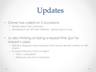
- 25. Updates ŌĆóŌĆ» Owner has called on 2 occasions oŌĆ» Worried about JerriŌĆÖs lameness oŌĆ» Wondering if can XRT both forelimbs ŌĆō going to give it a go ŌĆóŌĆ» Jo also thinking of doing a repeat FNA (just for interestŌĆÖs sake) oŌĆ» Definitive diagnosis more important only if owners decide to follow on with chemo oŌĆ» As would influence choice of agent ŌĆóŌĆ» OSA ├Ā’āĀ carboplatin ŌĆóŌĆ» Histiocytic sarcoma ├Ā’āĀ lomustine
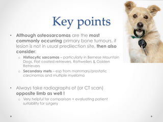
- 26. Key ┬Āpoints ŌĆóŌĆ» Although osteosarcomas are the most commonly occurring primary bone tumours, if lesion is not in usual predilection site, then also consider: oŌĆ» Histiocytic sarcomas ŌĆō particularly in Bernese Mountain Dogs, Flat coated retrievers, Rottweilers & Golden Retrievers oŌĆ» Secondary mets ŌĆō esp from mammary/prostatic carcinomas and multiple myeloma ŌĆóŌĆ» Always take radiographs of (or CT scan) opposite limb as well ! oŌĆ» Very helpful for comparison + evaluating patient suitability for surgery
- 27. Julie, who initially saw Jerri Gawain Hammond, for imaging interpretation advice Jo Morris, for going over the case with me Everyone here, for listening J’üŖ

