


More Related Content
Similar to histology for laboratory students 8.pptx (20)
More from ssuser9976be (20)


Recently uploaded (20)


histology for laboratory students 8.pptx
- 1. Muscle tissue is a soft tissue that consists of elongated cells called muscle fibers that are highly specialized to generate force.
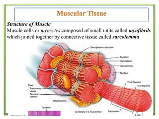
- 2. Structure of Muscle Muscle cells or myocytes composed of small units called myofibrils which joined together by connective tissue called sarcolemma
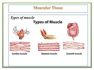
- 4. A) Skeletal Muscle Skeletal muscle is a form of striated muscle tissue which is under the voluntary control of the somatic nervous system. Most skeletal muscles are attached to bones by bundles of collagen fibers known as tendons.
- 7. A skeletal muscle refers to multiple bundles of cells called muscle fibers or muscle cell. Each muscle fiber is covered by a plasma membrane called the sarcolemma (flesh sheath). Multiple nuclei lie at the periphery of the fiber, under the sarcolemma. The muscle fiberŌĆÖs cytoplasm, called sarcoplasm, contains many mitochondria that produce large amounts of ATP during muscle contraction.
- 8. Extending throughout the sarcoplasm is sarcoplasmic reticulum a network of fluid-filled membrane-enclosed tubules (similar to smooth endoplasmic reticulum) that stores calcium ions required for muscle contraction. Also in the sarcoplasm are numerous molecules of myoglobin (stores oxygen until it is needed by mitochondria to generate ATP), a reddish pigment similar to hemoglobin in blood.
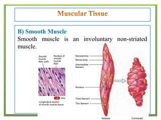
- 9. B) Smooth Muscle Smooth muscle is an involuntary non-striated muscle.
- 10. Smooth muscle fibers are considerably smaller in length and diameter than skeletal muscle fibers and are tapered at both ends. Within each fiber is a single, oval, centrally located nucleus. In addition to thick and thin filaments, smooth muscle fibers also contain intermediate filaments. In smooth muscle fibers, the thin filaments attach to structures called dense bodies, which are functionally similar to Z discs in striated muscle fibers.
- 11. C) Cardiac Muscle Cardiac muscle or heart muscle is an involuntary, striated muscle that constitutes the main tissue of the walls of the heart.
- 13. Cardiac muscle fibers often are branched; are shorter in length and larger in diameter than skeletal muscle fibers; and have a single, centrally located nucleus. Cardiac muscle fibers interconnect with one another by irregular transverse thickenings of the sarcolemma called intercalated discs (insert between). The intercalated discs hold the fibers together and contain gap junctions, which allow muscle action potentials to spread quickly from one cardiac muscle fiber to another.