Human digestion teacher
•Download as PPT, PDF•
34 likes•19,346 views

The document summarizes the key parts and functions of the human digestive system. It describes the main organs of the alimentary canal including the mouth, esophagus, stomach, small intestine, large intestine and anus. It explains the processes of ingestion, digestion, absorption, assimilation and egestion. It provides details on digestion in each part of the alimentary canal and the roles of the liver, gallbladder and pancreas in aiding digestion.


Human digestion teacher
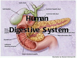
- 1. Human Digestive System
- 2. What you will be learning... (a) identify the main regions of the alimentary canal and the associated organs: mouth, salivary glands, oesophagus, stomach, duodenum, pancreas, gall-bladder, liver, ileum, colon, rectum, anus (b) *describe the functions of these parts in relation to ingestion, digestion, absorption, assimilation and egestion of food, as appropriate (c) explain why most foods must be digested (d) describe: (i) digestion in the alimentary canal (ii) the functions of a typical amylase, protease and lipase, listing the substrate and end-products (e) describe the structure of a villus (including role of capillaries and lacteals) in absorption (f) State the function of the hepatic portal vein as the route taken by most of the most absorbed from the small intestine (g) state the role of the liver in: (i) carbohydrate and fat metabolism (ii) breakdown of red blood cells (iii) metabolism of amino acids and the formation of urea (iv) breakdown of alcohol, including the effects of excessive alcohol consumption
- 3. Recall. . . What are the main organs of the alimentary canal? Mouth Oesophagus Stomach Small intestine Large intestine Anus Although not part of the alimentary canal, the liver, gall bladder and pancreas are closely associated with it. They play an important role in digestion by secreting digestive enzymes.
- 4. 5 Digestion Processes (IDAAE) Ingestion : taking in of food into the body. Digestion : breaking down of food into simpler substances Absorption : diffusion of food from small intestine into the blood Assimilation : using digested nutrients to make new material Egestion : removal of undigested waste material
- 5. Digestion Mechanical / physical digestion physically breaks down the food in the mouth (chewing) . Smaller pieces of food increase surface area for digestion. It also takes place in the stomach (churning of food by the muscular stomach walls) Chemical digestion uses enzymes to chemically break down complex food substances into their simplest form. e.g. Starch maltose amylase
- 6. Chemical digestion: Starch (carbohydrate) digestion: in mouth and small intestine. Protein digestion: in stomach and small intestine Fat digestion: only in small intestine Why must food be digested??? Large molecules of food are unable to pass through cell membranes, thus must be broken down into small molecules so that they can diffuse through cell membranes into the blood stream
- 7. Mouth (Ahhh....) Mouth ingests food Teeth masticates food into small pieces to increase surface area for digestion Saliva (pH 7) moisten and soften food Starch maltose Tongue mixes food with saliva and rolls food into a bolus before swallowing Saliva - water, mucus, salivary amylase Salivary amylase
- 8. Ěý
- 9. Swallowing
- 10. What Happens During Breathing and Swallowing? trachea (windpipe) glottis During breathing, the larynx is lowered and the glottis is open. pharynx oesophagus larynx (voice-box) air Normally, air passes into the trachea ( windpipe ) while food passes into the oesophagus .
- 11. What Happens During Breathing and Swallowing? During swallowing, the larynx is raised and the glottis is covered by the epiglottis . This prevents food particles from entering the trachea. pharynx trachea (windpipe) oesophagus glottis epiglottis food particles larynx (voice-box)
- 12. What Happens During Breathing and Swallowing? Occasionally, small particles of food or water may get into the larynx or trachea. trachea (windpipe) larynx (voice-box) food particles
- 13. What Happens During Breathing and Swallowing? This automatically induces violent coughing to force the food particles or water out and to prevent choking. trachea (windpipe) larynx (voice-box) food particles
- 14. Oesophagus Minimal digestion Carries food from mouth to stomach by peristalsis Oesophagus has circular and longitudinal muscles which are antagonistic. When circular muscles contract, longitudinal muscles relax and vice-versa.
- 15. Peristalsis The two layers of muscles cause rhythmic, wave-like contractions of the gut walls. Such movements are known as peristalsis . Peristalsis: enables food to be mixed with the digestive juices; and moves the food along the gut . Part of the gut wall circular muscles longitudinal muscles
- 16. Wall here constricts. Circular muslces contract; longitudinal muscles relax Wall here dilates Direction of movement of food Circular muscles relax Longitudinal muscles contract
- 17. Peristalsis – Move the food down! When circular muscles contract, longitudinal muscles relax. Gut wall constricts i.e. gut becomes narrower and longer. Food is squeezed or pushed forward. Gravity and slippery mucous lining helps push food down too. http://arbl.cvmbs.colostate.edu/hbooks/pathphys/digestion/basics/peristalsis.html
- 18. Stomach Stores food temporarily Stomach muscles churns and mixes food (also by peristalsis) with gastric juice to form chyme. Gastric juice contains hydrochloric acid (HCl) and enzymes like rennin and pepsin HCl is very acidic ( pH2 ), thus it kills bacteria and other microorganisms , as well as stopping the action of salivary amylase Provides acidic medium for gastric enzymes to work Only protein digestion here
- 19. HCl converts inactive pepsinogen and prorennin to their active forms
- 20. The stomach is “guarded” at the entrance and exit points by sphincter muscles which control the amount of food entering and leaving the stomach.
- 21. Small Intestine Subdivided into duodenum, jejunum and ileum In the small intestine, chyme stimulates Pancreas to secrete pancreatic juice Gall bladder to secrete bile Intestinal glands to secrete intestinal juice All three juices secreted are alkaline, pH 8.5
- 22. bile intestinal juice pancreatic duct pancreatic juice bile duct 1 3 2 Pancreatic and intestinal juice contain many digestive enzymes. Bile does not contain enzymes. Bile emulsifies fats, increasing the surface area for lipase action
- 23. Duodenum Starch maltose Protein polypeptides Fats fatty acids + glycerol Ileum Maltose glucose Polypeptides amino acids Fats fatty acids + glycerol Lactose glucose + galactose Sucrose glucose + fructose Note that the small intestine is the main site of digestion of food and absorption of nutrients. pancreatic amylase proteases lipase maltase protease lipase lactase sucrase
- 24. Ěý
- 25. Ěý
- 26. Large Intestine (colon) Large inverted U shaped tube. No digestion takes place here Absorbs water and minerals salts Stores the faeces (dead cells, mucus, germs, undigested food)
- 27. Is the colon the main region for water absorption? No! About 94% of the total amount of water passing through the alimentary canal is absorbed by the small intestine! The large intestine absorbs most of the remaining 6% of water. Rectum – temporarily stores faeces Anus – egests (= removal of undigested matter) faeces
- 28. Organs associated with the alimentary canal These organs do not digest food but aid in digestion Gall bladder Pancreas Liver
- 29. Gall bladder Temporarily stores bile (smelly green substance) secreted by liver. Secretes bile in the presence of chyme. Bile breaks up large fat droplets into very small fat droplets to increase surface area for lipase action ( Emulsification ) Bile emulsifies fats *Bile is not an enzyme, so it is not affected by temperature
- 30. Pancreas Connects to small intestine by pancreatic duct Produces pancreatic juice Secretes hormones like insulin (controls blood glucose concentration) and glucagon (controls carbohydrate metabolism) Liver Produces bile, which is stored in the gall bladder
- 31. Absorption Adaptations of the small intestine Small intestine is very long (~5 m) Internal surface of the small intestine has many folds . On these folds, there are many finger-like projections called villi These 3 adaptations increase surface area for absorption
- 32. Ěý
- 33. Lacteal – fatty acids and glycerol recombine in the epithelium to form fat which then enters the lacteal as fine fat droplets Blood capillaries – transport sugars and amino acids away from the small intestine One cell thick epithelium – for efficient absorption of food particles This continual transport of digested food substances maintains the concentration gradient for the absorption of digested food substances.
- 34. The concentration of simple nutrients (e.g. glucose, amino acids, fatty acids and glycerol) is higher in the lumen of the small intestine then in the blood capillaries that pass through the villi. Thus, nutrients diffuse across a region of high concentration (lumen of the small intestine) to the bloodstream, which has a lower concentration. Note that absorption by active transport is also possible. The blood capillaries in the small intestine unite to form larger blood vessels, which unite to form the hepatic portal vein , which transports the nutrients to the liver.
- 35. What happens to amino acids and glucose after absorption? Products released from liver into general blood circulation Molecules pass into the epithelial cells Through walls of capillaries in the villus and into bloodstream The capillaries join up to form veins Veins unite to form 1 large vein: Hepatic Portal Vein Hepatic portal vein carries blood to liver Liver stores or alters products of digestion
- 36. Ěý
- 37. Glucose Amino Acids Glucose is used by all cells as a source of energy . Excess glucose returned to liver and stored as glycogen . Insulin stimulates liver to convert glucose into glycogen. When the body needs energy, glycogen is converted back to glucose. Amino acids which enter the cells are converted into new protoplasm that is used for growth and repair . Amino acids used to form enzymes and hormones . Excess amino acids deaminated by liver.
- 38. What happens to fatty acids and glycerol after absorption? Molecules pass into the epithelial cells Recombine into fats again in the epithelial cells Fats enter the lacteals Lymph (fluid in lacteals) + fat = chyle Lymphatic vessels discharge chyle into bloodstream
- 39. Fats Blood carries fats to all parts of the body, especially to the liver. When there is enough glucose, fats are not broken down but are used to build protoplam. When there is insufficient glucose , fats are broken down to provide energy . Excess fats stored in adipose tissues.
- 40. Villi – absorption by diffusion Diffusion From intestine To To liver
- 41. Assimilation After travelling through the blood stream to the rest of the body, cells can now make use of glucose as source of energy amino acids to build new cytoplasm and tissue cells fatty acids to build new cell membranes
- 42. Functions of the Liver Regulation of blood glucose concentration 70-90mg of glucose / 100cm 3 of blood (normal conditions) Production of bile Liver produces bile which is stored in the gall bladder
- 43. Functions of the liver 3. Iron storage Red blood cells are destroyed in the spleen and their haemoglobin is sent to the liver to be broken down. The iron released is then stored in the liver. Bile pigments are also formed from the breakdown of haemoglobin. 4. Protein synthesis Liver synthesizes proteins found in blood plasma, e.g. albumins, globulins, fibrinogen
- 44. Functions of the liver 5. Deamination of amino acids Excess amino acids are transported to the liver, where their amino groups are removed and converted to urea .
- 45. Functions of the liver 6. Detoxification Liver cells contain alcohol dehydrogenase to break down alcohol . Excessive alcohol is harmful. Alcohol stimulates acid secretion in the stomach and increases risk of gastric ulcers . Prolonged alcohol abuse may lead to liver cirrhosis (destruction of liver cells), which can lead to liver failure and death. 7. Heat production
Editor's Notes
- #10: (1)The initial stages of eating and swallowing are under voluntary control. (2)Once food enters the mouth the teeth break it down into smaller and smaller pieces. This has the dual function of making the food easier to swallow and increasing the surface area of food on which the saliva can act. The tongue, lips and cheeks assist the teeth in the process by allowing the food to be "rolled" around the oral cavity. The mechanical action described above produces a softened bolus of food which is now ready to be swallowed. The correct biological term for swallowing is deglutition. (3) shows the voluntary stage of deglutition. Here the bolus is pushed into the upper part of the pharynx (known as the oropharynx ) by the action of the tongue.Ěý The pharyngeal stage of deglutiton is stimulated when the bolus enters the oropharynx. This stage of swallowing is mainly due to a reflex response. Various nerve receptors send messages to the deglutition centre of the brain stem. (see medulla and pons in your notes on the central nervous system). (4)This sets off muscular contractions in the pharynx. The soft palate closes off the nasopharynx. The vocal cords in the larynx are moved up and towards the front of the throat thus closing it off to the passage of food. This is extremely important in preventing food from entering the airway.I am sure we have all experienced the unpleasant feeling of food or drink going the "wrong way"!! Another effect of the process is to widen the opening of the oesophagus thus making the passage of the bolus along the alimentary canal easier. (5) As the bolus pushes it's way into the oesophagus it automatically pushes the epiglottis downwards further closing off the airway. (6) The bolus then enters the oesophagus and the final stage of swallowing begins. This final stage is known as the oesophageal stage of deglutiton. http://greenfield.fortunecity.com/rattler/46/upali4.htm
- #11: Figure 6.4 page 92
- #12: Figure 6.4 page 92
- #13: Figure 6.4 page 92
- #14: Figure 6.4 page 92
- #16: Figure 6.6 page 93
- #21: Compare the stomach wall n oesophagus wall? Which is thinker? Why? ( pro