Immune thrombocytopenia as initial presentation of systemic lupus erythematosus.
- 1. IMMUNE THROMBOCYTOPENIA AS INITIAL PRESENTATION OF SYSTEMIC LUPUS ERYTHEMATOSUS. Presented by The Rheumatology Unit, Dept. of Internal Medicine, DELSUTH, Oghara. 8th June, 2023.
- 2. OUTLINE ’é┤ CASE SUMMARY ’é┤ INTRODUCTION ’é┤ EPIDEMIOLOGY ’é┤ AETIOPATHOGENESIS ’é┤ CLINICAL FEATURES ’é┤ INVESTIGATIONS ’é┤ TREATMENT ’é┤ PREDICTIVE FACTORS FOR PROGRESSION OF ITP TO SLE ’é┤ CONCLUSION
- 3. CASE SUMMARY ’é┤ A 22 year old female, nursing student who was referred to the Rheumatology clinic from Haematology unit with complaints of; ’é┤ recurrent joint pain X 1 year ’é┤ In her usual state of health till one year ago when she developed joint pain of insidious onset involving the shoulders, elbows, and knees predominantly and occasionally the small joints of the hands ’é┤ Recent episode was noticed 1/52 prior to presentation and involved the shoulder and elbows with swelling of the right shoulder joint ’é┤ Joint affectation was of inflammatory pattern and symptoms temporarily relieved by Prednisolone and NSAIDs
- 4. CASE SUMMARY ’é┤ No skin rash, no oral ulcers, no hair loss, no headaches, seizures or psychosis, no chest pain, cough nor difficulty in breathing ’é┤ No leg swelling, no facial puffiness or passage of frothy urine, no dryness of the eyes or mouth, no redness of the eyes , no impaired vision. ’é┤ No dysuria, no preceding fever or diarrhoea and she is not sexually active ’é┤ She is a patient of Haematology unit and was on management for immune thrombocytopenia diagnosed 6 years ago when she presented on account of petechial skin rashes and spontaneously bleeding gums worsened by brushing
- 5. CASE SUMMARY ’é┤ She was then found to have a platelet count of zero with anaemia and has been on intermittent steroid therapy till she developed recurrent joint pain necessitating referral to Rheumatology unit to evaluate for lupus. ’é┤ Not known to be hypertensive or diabetic. ’é┤ Does not take alcoholic beverages or tobacco in any form and there is no family history of similar symptoms, SLE or other autoimmune disorders.
- 6. CASE SUMMARY ’é┤ Physical examination findings were essentially normal except for pallor and tenderness over the elbows and shoulders with swelling over the RT shoulder joint. ’é┤ Investigation Results Results from 6 years ago at point of diagnosis of ITP ’é┤ FBC + ESR ’é┤ ESR 56mm/hr ’é┤ Hb 8.9g/dl ’é┤ PCV 23.9% ’é┤ WBC 4.73X 10⁹/l ’é┤ Lymph 24.2% (1.1 x 10⁹/l) ’é┤ Neut 66.2% ’é┤ Platelet 0
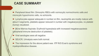
- 7. CASE SUMMARY ’é┤ Peripheral blood film: Dimorphic RBCs with normocytic normochromic cells and microcytic hypochromic cells, no schizonts. ’é┤ Lymphocytes appear adequate in number on film, neutrophils are mostly mature with about 3 segments, platelets appear reduced in number with megakaryocytes, no platelet clumps visualized. ’é┤ Bone Marrow Aspirate: Erythroid hyperplasia with increased megakaryopoiesis (peripheral immune destruction of platelets). ’é┤ Viral serologies were all negative ’é┤ SEUCR, Urinalysis were both normal. ’é┤ The impression for the above patient was ITP R/O Evan's syndrome and myeloproliferative disease.
- 8. CASE SUMMARY ’é┤ Investigation results after review in rheumatology clinic ’é┤ FBC + ESR ’é┤ Hb 10.6g/dl ’é┤ WCC 4.9 X 10⁹/L ’é┤ Lymph 44% (2.1 x 10⁹/l ’é┤ Neut 52% ’é┤ PLT 266X 109/L ’é┤ ESR 105mm/hr ’é┤ Urinalysis: one plus protein ’é┤ uPCR 76.2 mg/day (<150 normal or mild proteinuria) ’é┤ SEUCR, LFT, serum uric acid were all normal
- 9. CASE SUMMARY ENA panel ’é┤ ANA 1:2560 ’é┤ Anti ds DNA +++ ’é┤ Anti RIB ++ ’é┤ Anti Nucleosome ++ ’é┤ The ITP 6 years ago was managed with serial pulse doses of methyl prednisolone and she had marked improvement with occasional flares ’é┤ Last flare was 3 years prior to presentation to the Rheumatology clinic.
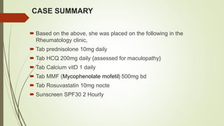
- 10. CASE SUMMARY ’é┤ Based on the above, she was placed on the following in the Rheumatology clinic, ’é┤ Tab prednisolone 10mg daily ’é┤ Tab HCQ 200mg daily {assessed for maculopathy} ’é┤ Tab Calcium vitD 1 daily ’é┤ Tab MMF (Mycophenolate mofetil) 500mg bd ’é┤ Tab Rosuvastatin 10mg nocte ’é┤ Sunscreen SPF30 2 Hourly
- 11. INTRODUCTION ’é┤ Immune thrombocytopenia (ITP), formerly known as immune idiopathic thrombocytopenic purpura, is an immune mediated acquired disease of adults and children characterised by a transient or persistent decrease of platelet counts ’é┤ In ITP, platelets become coated with autoantibodies to platelet membrane antigens, resulting in splenic sequestration and phagocytosis ’é┤ Systemic lupus erythematosus (SLE) is a chronic inflammatory autoimmune disease involving multiple systems, with a remitting or relapsing course ’é┤ It is characterised by the production of various autoantibodies and by several clinical manifestations, some of which are haematological.
- 12. INTRODUCTION ’é┤ Thrombocytopenia (<100├Ś109/L) has been reported in 20% to 40% of patients with SLE and is usually attributed to an autoimmune mechanism similar to that of immune thrombocytopenia (ITP) ’é┤ ITP can occur at any stage of SLE, but it has been observed to occur as an initial manifestation in some patients ’é┤ This can pose a diagnostic challenge, as the symptoms of ITP may be the only presenting feature of SLE, and other clinical features of the disease may not become apparent until later stages
- 13. INTRODUCTION ’é┤ ITP is classified as primary or secondary according to the presence of underlying etiology ’é┤ Thrombocytopenia in SLE is associated with a worse prognosis and higher mortality from the disease ’é┤ It has been linked with severe disease course including neuropsychiatric disorders, renal involvement, hemolytic anemia and anti phospholipid syndrome.
- 14. EPIDEMIOLOGY ’é┤ ITP is generally thought to be a condition that predominantly affects young women. ’é┤ Some studies have corroborated a female predominance in younger adults, ratio of 3:1, others have not, while SLE has a female to male ratio of 9-12:1 ’é┤ Most studies show a similar incidence of ITP in males and females over the age of 60 years ’é┤ Thrombocytopenia (<100├Ś109/L) has been reported in 20% to 40% of patients with SLE and is usually attributed to an autoimmune mechanism similar to that of idiopathic immune thrombocytopenia (ITP) ’é┤ Acute ITP presents in childhood affects both sexes equally, and most have a benign course ’é┤ The chronic form affects individuals between 20-50 years
- 15. EPIDEMIOLOGY ’é┤ In a study in Greece, thrombocytopenia was found to be present at diagnosis in 12% and cummulatively 16% ’é┤ It has been estimated that 3 to 15 percent of patients with apparently isolated ITP go on to develop SLE ’é┤ It may be the first manifestation of lupus in up to 16% of patients, presenting months or as early as 10 years before diagnosis ’é┤ In Nigeria, a study in South South done to show the laboratory and clinical profile of SLE patients identified thrombocytopenia in 34.6% of the subjects
- 16. EPIDEMIOLOGY ’é┤ Retrospective study done in ABUTH Zaria, over a 10 year period identified 9 cases of ITP, 6 females, 3 males, aged 6-20 ’é┤ The commonest manifestation was epistaxis (88.9%) and one of the subjects had ICH ’é┤ All the patients had megakaryocytic hyperplasia ’é┤ A similar study in OAUTH over an 11 year period had 11 cases of ITP, 7 females and 4 males, aged 10-55 ’é┤ Two of the females were positive for SLE test.
- 18. AETIOPATHOGENESIS ’é┤ Inciting events ŌĆö In some patients with ITP, there may appear to be inciting events. ’é┤ Genetic and acquired factors may contribute. ’é┤ Infection ŌĆō Some cases of ITP are associated with a preceding viral infection or, less commonly, bacterial infection. ’é┤ Antibodies against viral antigens may cross-react with normal platelet antigens (a form of molecular mimicry).
- 19. AETIOPATHOGENESIS ’é┤ Immune alteration ŌĆō Alterations in immune homeostasis might induce loss of peripheral tolerance and promote the development of self-reactive antibodies ’é┤ This often occurs in the setting of other autoimmune conditions including the antiphospholipid syndrome (APS), systemic lupus erythematosus (SLE), Evans syndrome, hematopoietic cell transplantation, chronic lymphocytic leukemia (CLL) and other low-grade lymphoproliferative disorders ’é┤ Alternative immunologic mechanisms involving T cells have also been postulated to cause ITP, including T cell-mediated cytotoxicity and defects in the number and/or function of regulatory T cells (Tregs).
- 20. AETIOPATHOGENESIS ’é┤ Antibody production ŌĆö Antibody production in ITP appears to be driven by CD4-positive helper T cells reacting to platelet surface glycoproteins, possibly involving CD40:CD40L co- stimulation. ’é┤ Splenic macrophages appear to be the major antigen- presenting cells. ’é┤ Despite this likely mechanism, antiplatelet antibodies are not demonstrable in close to 50 percent of patients with ITP.
- 21. AETIOPATHOGENESIS ’é┤ Platelet destruction ŌĆö The primary site of platelet clearance for most patients is the spleen, which removes opsonized (antibody coated) cells including platelets. ’é┤ The prominent role of splenic clearance explains the effectiveness of splenectomy in most patients. ’é┤ However, clearance may occur in other tissues as well, such as the liver, bone marrow, lymph nodes, and accessory splenic tissue. This helps explain why ITP can persist or recur post-spleen.ectomy.
- 22. AETIOPATHOGENESIS ’é┤ In SLE thrombocytopenia, the implicated antigens are antigenic glycoproteins on platelet membranes ’é┤ Other autoantibodies include the antiphospholipid antibodies, anti thrombopoietin, anti TPO receptor. ’é┤ Just like ITP, the most frequent antibodies in SLE thrombocytopenia are the anti Iib/IIIa antibodies. ’é┤ Other identified antibodies include; anti CD40 ligand molecule, antibodies against Gp Ia/IIA,HLA I, GpIb/Ix complex, though uncommon.
- 23. AETIOPATHOGENESIS ’é┤ Other causes of thrombocytopenia in SLE patients that should also be considered but are less common than ITP include the following: ’é┤ Drug-induced thrombocytopenia, which may be immune or non immune (eg, due to bone marrow suppression) ’é┤ Heparin-induced thrombocytopenia is a form of drug-induced thrombocytopenia in which drug-dependent antibodies lead to platelet activation and may cause venous or arterial thromboses. ’é┤ Platelet consumption in the setting of a thrombotic microangiopathic process ’é┤ In this setting, thrombocytopenia is typically associated with microangiopathic hemolysis with schistocytes on the peripheral blood smear ’é┤ Platelet consumption in the setting of antiphospholipid syndrome (APS)
- 24. CLINICAL FEATURES ’é┤ ITP manifests as a bleeding tendency, easy bruising (purpura), or extravasation of blood from capillaries into skin and mucous membranes (petechiae) ’é┤ Although most cases of acute ITP, particularly in children, are mild and self-limited, intracranial hemorrhage may occur when the platelet count drops below 10 ├Ś 109/L ’é┤ ITP is a primary illness occurring in an otherwise healthy person. Signs of chronic disease, infection, wasting, or poor nutrition indicate that the patient has another illness ’é┤ Splenomegaly excludes the diagnosis of ITP.
- 25. CLINICAL FEATURES ’é┤ An initial impression of the severity of ITP is formed by examining the skin and mucous membranes, as follows: ’é┤ Widespread petechiae and ecchymoses, oozing from a venipuncture site, gingival bleeding, and hemorrhagic bullae indicate that the patient is at risk for a serious bleeding complication. ’é┤ If the patient's blood pressure was taken recently, petechiae may be observed under and distal to the area where the cuff was placed and inflated. ’é┤ Petechiae over the ankles in ambulatory patients or on the back in bedridden ones suggest mild thrombocytopenia and a relatively low risk for a serious bleeding complication.
- 26. CLINICAL FEATURES ’é┤ Findings suggestive of intracranial hemorrhage include the following: ’é┤ Headache, blurred vision, somnolence, or loss of consciousness ’é┤ Hypertension and bradycardia, which may be signs of increased intracranial pressure ’é┤ On neurologic examination, any asymmetrical finding of recent onset ’é┤ On fundoscopic examination, blurring of the optic disc margins or retinal hemorrhage.
- 27. DIAGNOSIS ’é┤ No single laboratory result or clinical finding establishes a diagnosis of ITP; it is a diagnosis of exclusion. ’é┤ Clinically, ITP is mainly diagnosed based on the following criteria: ’é┤ the reduced platelet count is detected at least twice, and the blood cell morphology is normal ’é┤ the spleen is generally small ’é┤ the number of megakaryocytes in the bone marrow is normal or increased, accompanied by maturation disorders and ’é┤ other secondary thrombocytopaenia is excluded.
- 28. DIAGNOSIS ’é┤ On complete blood cell count, isolated thrombocytopenia is the hallmark of ITP. Anemia and/or neutropenia may indicate other diseases. Findings on peripheral blood smear are as follows: ’é┤ The morphology of red blood cells (RBCs) and leukocytes is normal. ’é┤ The morphology of platelets is typically normal, with varying numbers of large platelets. ’é┤ If most of the platelets are large, approximating the diameter of red blood cells, or if they lack granules or have an abnormal color, consider an inherited platelet disorder.
- 29. DIAGNOSIS ’é┤ Role of bone marrow aspiration and biopsy are as follows: ’é┤ The value of bone marrow evaluation for a diagnosis of ITP is unresolved ’é┤ Biopsy in patients with ITP shows a normal-to-increased number of megakaryocytes in the absence of other significant abnormalities ’é┤ In children, bone marrow examination is not required except in patients with atypical hematologic findings, such as immature cells on the peripheral smear or persistent neutropenia.
- 30. DIAGNOSIS ’é┤ In adults older than 60 years, biopsy is used to exclude myelodysplastic syndrome or leukemia ’é┤ In adults whose treatment includes corticosteroids, a baseline pretreatment biopsy may prove useful for future reference, as corticosteroids can change marrow morphology ’é┤ Biopsy is performed before splenectomy to evaluate for possible hypoplasia or fibrosis ’é┤ Unresponsiveness to standard treatment after 6 months is an indication for bone marrow aspiration.
- 31. DIAGNOSIS ’é┤ Clumps of platelets on a peripheral smear prepared from ethylenediaminetetraacetic acid (EDTA)ŌĆōanticoagulated blood are evidence of pseudo thrombocytopenia. ’é┤ The diagnosis of this type of pseudo thrombocytopenia is established if the platelet count is normal when repeated on a sample from heparin-anticoagulated or citrate-anticoagulated blood. ’é┤ In patients who have risk factors for HIV infection, a blood sample should be tested with an enzyme immunoassay for anti-HIV antibodies.
- 32. DIAGNOSIS ’é┤ ANA screening especially if SLE is suspected ’é┤ If anemia and thrombocytopenia are present, a positive direct antiglobulin (Coombs) test result may help establish a diagnosis of Evans syndrome ’é┤ A negative antiplatelet antibody assay result does not exclude the diagnosis of ITP therefore this test is not recommended as part of the routine evaluation ’é┤ Testing for antiplatelet antibodies is not required to diagnose ITP.
- 33. EVALUATION OF THROMBOCYTOPENIA IN THE SETTING OF SLE Isolated, mild thrombocytopenia ’é┤ For patients with isolated, mild thrombocytopenia who are not acutely ill, the evaluation includes a thorough history for medications and other illnesses that may affect the platelet count, as well as review of the CBC ’é┤ Additional testing may be done sequentially or simultaneously for vitamin B12 and folate deficiencies, liver disease, and coagulation abnormalities, especially antiphospholipid antibodies, as appropriate, based on the history and preliminary laboratory results.
- 34. EVALUATION OF THROMBOCYTOPENIA IN THE SETTING OF SLE Acutely ill, severe thrombocytopenia, or other cytopenias ’é┤ For patients who are acutely ill or have new onset of thrombocytopenia plus other cytopenias (eg, neutropenia, anemia), hematology consultation is recommended ’é┤ The patient should have a thorough history and physical examination; review of medications; review of the blood smear for schistocytes or other abnormal cells, coagulation testing, and testing of renal and hepatic function ’é┤ Disorders to be considered include ITP, thrombotic microangiopathies, severe infections and severe drug reactions.
- 35. EVALUATION OF THROMBOCYTOPENIA IN THE SETTING OF SLE Fanoouriakis A. et al Population-based studies in systemic lupus erythematosus: immune thrombocytopenic purpura or ŌĆśblood- dominantŌĆÖ lupus? Ann Rheum Dis. 2020 Jun;79(6):683ŌĆō4
- 36. TREATMENT ’é┤ Treatment of ITP and immune mediated Thrombocytopenia in SLE bear many similarities because of the similarities in pathophysiology ’é┤ Severe thrombocytopenia requires emergency therapy in order to eliminate hemorrhagic complications and achieve a complete or partial platelet response ’é┤ Maintenance treatment is usually needed to prevent relapse ’é┤ The decision to treat when thrombocytopenia is the sole disorder in SLE depends on the hemorrhagic manifestations and platelet count ’é┤ Generally, patients with a platelet count > 50├Ś109/l without bleeding manifestations do not require treatment in the absence of coexistent hemostatic disorders, anticoagulation treatment, trauma or major surgery.
- 37. TREATMENT ’é┤ Corticosteroids are the cornerstone of initial treatment ’é┤ High dose oral prednisolone or pulse high dose methylprednisolone (MP) with or without intravenous immune globulin (IVIG) are used in the acute phase ’é┤ Second line agents include hydroxychloroquine (HCQ), danazol, immunosuppressive drugs like azathioprine (AZA), cyclosporine (CSA), mycophenolate mofetil (MMF), cyclophosphamide (CYC), and biological therapies such as rituximab ’é┤ The thrombopoietin receptor agonists romiplostim and eltrombopag have been widely used for the treatment of ITP and they also seem to have a role in SLE thrombocytopenia ’é┤ Splenectomy is indicated for recurrent or resistant cases (Controversial in SLE because of the role of the kidneys in immune complex clearance).
- 38. PREDICTIVE FEATURES IN ITP FOR DEVELOPMENT OF SLE ’é┤ Young age (< 40 years), ANA positivity, and organ bleeding ,at the diagnosis of ITP are significantly related to the development of SLE within 1 year following ITP diagnosis ’é┤ Development of anaemia and lymphopenia during the course of management of ITP may also predict a future diagnosis of SLE ’é┤ This suggests that continued follow-up for the detection of SLE development is needed for patients with ITP, particularly those with young age, ANA positivity, or organ bleeding.
- 39. DIFFERENTIATING ITP FROM TTP AND DIC FLASHCARDS (ITP VS TTP) quizlets.com
- 40. CONCLUSION ’é┤ ITP can be an initial presentation of SLE. SLE presents occasionally with non-specific symptoms and ITP can be one of the early manifestations ’é┤ It is important to recognise the association between ITP and SLE , as early dtection and management of SLE can improve patient outcomes ’é┤ The treatment of ITP in SLE patients would require a multidisciplinary approach, including haematologists, rheumatologists and immunologists ’é┤ Proper evaluation for SLE should be carried out in patients with ITP especially children and women of reproductive age.
- 41. REFERENCES ’é┤ Justiz Vaillant AA, Gupta N. ITP-immune thrombocytopenic purpura. (Updated 2022 Dec 23). In:StatPearls (Internet). Treasure Island (FL0: StatPearls Publishing;2023 ’é┤ Galanopoulos N, Christoforidou A, Bezirgiannidou Z. Lupus thrombocytopenia: pathogenesis and therapeutic implications. MJR. 2017 Jan 1;28(1):20ŌĆō6. ’é┤ Zhu FX, Huang JY, Ye Z, Wen QQ, Wei JCC. Risk of systemic lupus erythematosus in patients with idiopathic thrombocytopenic purpura: a population-based cohort study. Ann Rheum Dis. 2020 Jun;79(6):793ŌĆō9. ’é┤ Fanouriakis A, Bertsias G, Boumpas DT. Population-based studies in systemic lupus erythematosus: immune thrombocytopenic purpura or ŌĆśblood-dominantŌĆÖ lupus? Ann Rheum Dis. 2020 Jun;79(6):683ŌĆō4. ’é┤ Emorinken A, Dic-Ijiewere MO, Erameh CO, Ugheoke AJ, Agbadaola OR, Agbebaku FO. Clinical and laboratory profile of systemic lupus erythematosus patients at a rural tertiary centre in South-South Nigeria: Experience from a new rheumatology clinic. ’é┤ Lateef S, Muheez D, Immune thro,bocytopenic purpura :11-year experience in Ile-Ife , Nigeria. African journal of Medicine and med sciences.2001. 20(1-2):99-103 ’é┤ Abdulaziz H, Adeshola A, Mohammed SA. Clinical feature and management of immune thrombocytopenic purpurs in a tertiary hospital in Northwest Nigeria. Nigerian medical journal, 2017 Mar-Apr;58(2):68-71.
- 42. ’é┤THANK YOU
