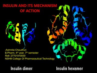
Insulin and its mechanism of action
- 1. INSULIN AND ITS MECHANISM OF ACTION INSULIN AND ITS MECHANISM OF ACTION -Ashmita Chaudhuri B.Pharm, 4th year, 7th semester Roll- 27701910050 NSHM College Of Pharmaceutical Technology
- 2. INTRODUCTION: Insulin is a peptide hormone, produced by beta cells of the pancreas, and is central to regulating carbohydrate and fat metabolism in the body. Insulin causes cells in the liver, skeletal muscles, and fat tissue to absorb glucose from the blood. In the liver and skeletal muscles, glucose is stored as glycogen, and in fat cells (adipocytes) it is stored as triglycerides. When control of insulin levels fails, diabetes mellitus can result. As a consequence, insulin is used medically to treat some forms of diabetes mellitus.
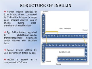
- 3. STRUCTURE OF INSULIN ’üČ Human insulin consists of 51aa in two chains connected by 2 disulfide bridges (a single gene product cleaved into 2 chains during posttranslational modification). ’üČ T1/2~5-10 minutes, degraded by glutathione-insulin transhydrogenase (insulinase) which cleaves the disulfide links. ’üČ Bovine insulin differs by 3aa, pork insulin differs by 1aa. ’üČ Insulin is stored complex with Zn2+ions. in a
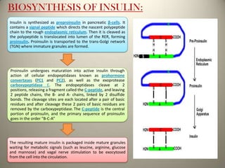
- 4. BIOSYNTHESIS OF INSULIN: Insulin is synthesized as preproinsulin in pancreatic ╬▓-cells. It contains a signal peptide which directs the nascent polypeptide chain to the rough endoplasmic reticulum. Then it is cleaved as the polypeptide is translocated into lumen of the RER, forming proinsulin. Proinsulin is transported to the trans-Golgi network (TGN) where immature granules are formed. Proinsulin undergoes maturation into active insulin through action of cellular endopeptidases known as prohormone convertases (PC1 and PC2), as well as the exoprotease carboxypeptidase E. The endopeptidases cleave at 2 positions, releasing a fragment called the C-peptide, and leaving 2 peptide chains, the B- and A- chains, linked by 2 disulfide bonds. The cleavage sites are each located after a pair of basic residues and after cleavage these 2 pairs of basic residues are removed by the carboxypeptidase. The C-peptide is the central portion of proinsulin, and the primary sequence of proinsulin goes in the order "B-C-AŌĆØ The resulting mature insulin is packaged inside mature granules waiting for metabolic signals (such as leucine, arginine, glucose and mannose) and vagal nerve stimulation to be exocytosed from the cell into the circulation.
- 5. The endogenous production of insulin is regulated in several steps along the synthesis pathway: ’āśAt transcription from the insulin gene ’āśIn mRNA stability ’āśAt the mRNA translation ’āśIn the post translational modifications
- 6. EFFECT OF INSULIN ON GLUCOSE UPTAKE AND METABOLISM Insulin binds to its receptor Starts many protein activation cascades These include translocation of Glut-4 transporter to the plasma membrane and influx of glucose glycogen synthesis glycolysis triglyceride
- 7. MECHANISM OF ACTION: ’üČ Insulin acts on specific receptors located on the cell membrane of practically every cell, but their density depends on the cell type: liver and fat cells are very rich. ’üČ The insulin receptor is a receptor tyrosine kinase (RTK) which is a heterotetrameric glycoprotein consisting of 2 extracellular ╬▒ and 2 transmembrane ╬▓ subunits linked together by disulfide bonds, orienting across the cell membrane as a heterodimer ’üČ It is oriented across the cell membrane as a heterodimer. ’üČ The ╬▒ subunits carry insulin binding sites, while the ╬▓ subunits have tyrosine kinase activity.
- 9. ’ü▒ Insulin stimulates glucose transport across cell membrane by ATP dependent translocation of glucose transporter GLUT4 to the plasma membrane. ’ü▒ The second messenger PIP3 and certain tyrosine phosphorylated guanine nucleotide exchange proteins play crucial roles in the insulin sensitive translocation of GLUT4 from cytosol to the plasma membrane, especially in the skeletal muscles and adipose tissue. ’ü▒ Over a period of time insulin also promotes expression of the genes directing synthesis of GLUT4. ’ü▒ Genes for a large number of enzymes and carriers are regulated by insulin through Ras/Raf and MAP-Kinase as well as through the phosphorylation cascade.
- 10. DEGRADATION OF INSULIN: The internalized receptor-insulin complex is either degraded intercellularly or returned back to the surface from where the insulin is released extracellularly. The relative preponderance of these two processes differs among different tissues: maximum degradation occurs in liver, least in vascular endothelium.
- 11. FATE OF INSULIN Ō¢▓ Insulin is distributed only extracellularly. It is a peptide; gets degraded in the g.i.t. if given orally. Ō¢▓ Injected insulin or that released from the pancreas is metabolized primarily in liver and to a smaller extent in kidney and muscles. Ō¢▓ Nearly half of the insulin entering portal vein from pancreas is inactivated in the first passage through liver. Ō¢▓ Thus, normally liver is exposed to a much higher concentration (4-8 fold) of insulin than other tissues. Ō¢▓ During biotransformation the disulfide bonds are reduced- A and B chains are separated. These are further broken down to the constituent amino acids. Ō¢▓ The plasma t1/2 is 5-9 minutes.
- 12. Different types of Insulin Preparations: Type Appearance Onset (hr) Peak (hr) Duration (hr) Insulin lispro Clear 0.2-0.3 1-1.5 3-5 Insulin aspart Clear 0.2-0.3 1-1.5 3-5 Insulin glulisin Clear 0.2-0.4 1-2 3-5 Clear 0.5-1 2-3 6-8 Insulin zinc suspension or Lente Cloudy 1-2 8-10 20-24 NPH or isophane Insulin Cloudy 1-2 8-10 20-24 Clear Glargine: 2-4 Detemir: 1-4 RAPID ACTING SHORT ACTING Regular (soluble) insulin INTERMEDIATE ACTING LONG ACTING Insulin glargine and Insulin detemir _ _ Glargine: 24 Detemir: 20-24
- 13. CONCLUSION: DIABETES MELLITUS DIABETIC KETOACIDOSIS (DIABETIC COMA) HYPEROSMOLAR (NONKINETIC HYPERGLYCAEMIC COMA) Insulin is effective in all forms of diabetes mellitus and is a must for type 1 cases, as well as for post pancreatectomy diabetes and gestational diabetes. Many type 2 cases can be controlled. Regular insulin is used to rapidly correct the metabolic abnormalities. This usually occurs in elderly type 2 cases. The cause is obscure. Insulin therapy is generally started with regular insulin given s.c. before each major meal. The requirement is assessed by testing urine or blood glucose levels . Usually within 4-6 hours blood glucose reaches 300 mg/dl. Then the rate of infusion is reduced to 2-3 U/hr The general principles of treatment are the same as for ketoacidotic coma, except that faster fluid replacement is to be instituted as alkali is usually not required.
- 14. REFERENCES: ŌŚÖ http://edrv.endojournals.org/content/2/2/210.abstract visited on 10th October, 2013. ŌŚÖ http://en.wikipedia.org/wiki/Insulin visited on 25th September, 2013 ŌŚÖ http://www.authorstream.com/Presentation/saneet-1638200-insulin-inside/ visited on 28th September, 2013. ŌŚÖhttp://www.google.co.in/url?sa=t&rct=j&q=&esrc=/slideshow/insulin-and-its-mechanism-of-action-31298888/31298888/s&source=web&cd=1&ved= 0CC8QFjAA&url=http%3A%2F%2Fwww.calstatela.edu%2Ffaculty%2Fmchen %2F454L%2520lectures%2FAntiDiabeticDrugs.ppt&ei=wzV_UsryE8isrAfXsIH 4Bg&usg=AFQjCNGP7mDC39_aOTodF7_SYzObBpFcPQ visited on 20th October, 2013. ŌŚÖ http://www.medbio.info/horn/time%203-4/homeostasis_2.htm visited on 2nd November, 2013. ŌŚÖ Essentials of Medical Pharmacology- by KD Tripathi, 7th Edition, Jaypee Brothers Medical Publishers (P) Limited, Chapter-19, Page: 258-268.


