Interactive case
4 likes1,004 views

A 5-year-old male patient presented with fever, cough and expectoration. Chest x-rays showed consolidation in the left middle and lower lung zones initially. Follow up x-rays 4 days later revealed no lung abnormalities but detected an expansile bony lesion of the right 3rd rib. Differential diagnoses for expansile rib lesions in pediatric patients were provided, including both benign conditions like fibrous dysplasia, aneurysmal bone cyst, and neurofibromatosis, as well as malignant conditions such as Ewing sarcoma. Proper diagnosis and timely treatment of rib lesions are important given the risk of malignancy.


Interactive case
- 1. Interactive case D Amira El Azab
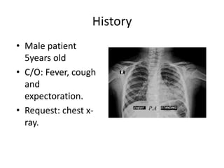
- 2. History ŌĆó Male patient 5years old ŌĆó C/O: Fever, cough and expectoration. ŌĆó Request: chest x- ray.
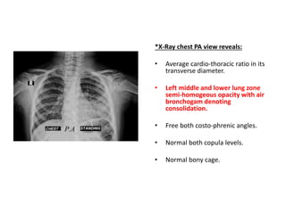
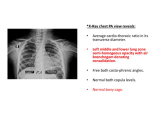
- 3. *X-Ray chest PA view reveals: ŌĆó Average cardio-thoracic ratio in its transverse diameter. ŌĆó Left middle and lower lung zone semi-homogeous opacity with air bronchogam denoting consolidation. ŌĆó Free both costo-phrenic angles. ŌĆó Normal both copula levels. ŌĆó Normal bony cage.
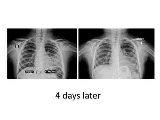
- 4. 4 days later
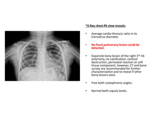
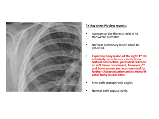
- 5. *X-Ray chest PA view reveals: ŌĆó Average cardio-thoracic ratio in its transverse diameter. ŌĆó No focal pulmonary lesion could be detected. ŌĆó Expansile bony lesion of the right 3rd rib anteriorly, no calcification, cortical destruction, periosteal reaction or soft tissue component, however, CT and bone survey are recommended for further characterization and to reveal if other bony lesions exist. ŌĆó Free both costophrenic angles. ŌĆó Normal both copula levels.
- 6. *X-Ray chest PA view reveals: ŌĆó Average cardio-thoracic ratio in its transverse diameter. ŌĆó No focal pulmonary lesion could be detected. ŌĆó Expansile bony lesion of the right 3rd rib anteriorly, no sclerosis, calcification, cortical destruction, periosteal reaction or soft tissue component, however, CT and bone survey are recommended for further characterization and to reveal if other bony lesions exist. ŌĆó Free both costophrenic angles. ŌĆó Normal both copula levels.
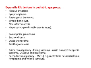
- 7. Expansile Rib Lesions in pediatric age group: ŌĆó Fibrous dysplasia ŌĆó Lymphangioma. ŌĆó Aneurysmal bone cyst ŌĆó Simple bone cyst. ŌĆó Neurofibromatosis ŌĆó Hyperparathyroidism (brown tumors). ŌĆó Eosinophilic granuloma ŌĆó Enchondroma. ŌĆó Osteochondroma ŌĆó Xanthogranuloma ŌĆó Primary malignancy ŌĆōEwing sarcoma - Askin tumor Osteogenic sarcoma, Osseous angiosarcoma. ŌĆó Secondary malignancy ŌĆō Mets ( e.g. metastatic neuroblastoma, lymphoma and WilmŌĆÖs tumour).
- 8. ŌĆó Rib tumors are rare entities in the pediatric population. However, a significant number of rib lesions are malignant. Therefore, proper diagnosis and expeditious treatment are critical.
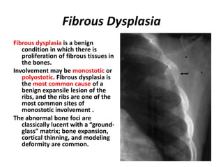
- 9. Fibrous Dysplasia Fibrous dysplasia is a benign condition in which there is proliferation of fibrous tissues in the bones. Involvement may be monostotic or polyostotic. Fibrous dysplasia is the most common cause of a benign expansile lesion of the ribs, and the ribs are one of the most common sites of monostotic involvement . The abnormal bone foci are classically lucent with a ŌĆ£ground- glassŌĆØ matrix; bone expansion, cortical thinning, and modeling deformity are common.
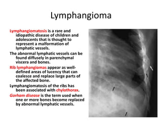
- 10. Lymphangioma Lymphangiomatosis is a rare and idiopathic disease of children and adolescents that is thought to represent a malformation of lymphatic vessels. The abnormal lymphatic vessels can be found diffusely in parenchymal viscera and bones. Rib lymphangiomas appear as well- defined areas of lucency that can coalesce and replace large parts of the affected bone. Lymphangiomatosis of the ribs has been associated with chylothorax. Gorham disease is the term used when one or more bones become replaced by abnormal lymphatic vessels.
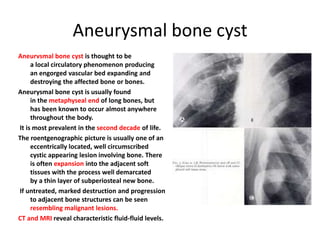
- 11. Aneurysmal bone cyst Aneurvsmal bone cyst is thought to be a local circulatory phenomenon producing an engorged vascular bed expanding and destroying the affected bone or bones. Aneurysmal bone cyst is usually found in the metaphyseal end of long bones, but has been known to occur almost anywhere throughout the body. It is most prevalent in the second decade of life. The roentgenographic picture is usually one of an eccentrically located, well circumscribed cystic appearing lesion involving bone. There is often expansion into the adjacent soft tissues with the process well demarcated by a thin layer of subperiosteal new bone. If untreated, marked destruction and progression to adjacent bone structures can be seen resembling malignant lesions. CT and MRI reveal characteristic fluid-fluid levels.
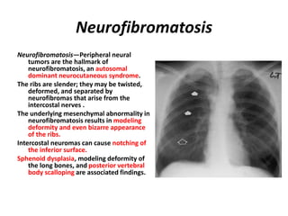
- 12. Neurofibromatosis NeurofibromatosisŌĆöPeripheral neural tumors are the hallmark of neurofibromatosis, an autosomal dominant neurocutaneous syndrome. The ribs are slender; they may be twisted, deformed, and separated by neurofibromas that arise from the intercostal nerves . The underlying mesenchymal abnormality in neurofibromatosis results in modeling deformity and even bizarre appearance of the ribs. Intercostal neuromas can cause notching of the inferior surface. Sphenoid dysplasia, modeling deformity of the long bones, and posterior vertebral body scalloping are associated findings.
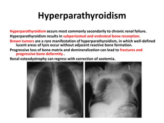
- 13. Hyperparathyroidism Hyperparathyroidism occurs most commonly secondarily to chronic renal failure. Hyperparathyroidism results in subperiosteal and endosteal bone resorption. Brown tumors are a rare manifestation of hyperparathyroidism, in which well-defined lucent areas of lysis occur without adjacent reactive bone formation. Progressive loss of bone matrix and demineralization can lead to fractures and progressive bone deformity . Renal osteodystrophy can regress with correction of azotemia.
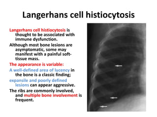
- 14. Langerhans cell histiocytosis Langerhans cell histiocytosis is thought to be associated with immune dysfunction. Although most bone lesions are asymptomatic, some may manifest with a painful soft- tissue mass. The appearance is variable: A well-defined area of lucency in the bone is a classic finding; expansile and poorly defined lesions can appear aggressive. The ribs are commonly involved, and multiple bone involvement is frequent.
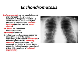
- 15. Enchondromatosis Enchondromatosis are a group of disorders characterized by the presence of medullary cartilaginous bone tumors, which are further subdivided by the presence of hemangiomas (Maffucci syndrome) or their absence (Ollier disease). Rib lesions are common in enchondromatosis. Inheritance is sporadic. At radiography, enchondromas appear as areas of lucency in the bones, occasionally with areas of calcification in the cartilaginous matrix. Modeling deformity is not uncommon. The appearance is similar to that of fibrous dysplasia. Enchondromas are associated with a 25% prevalence of malignant degeneration.
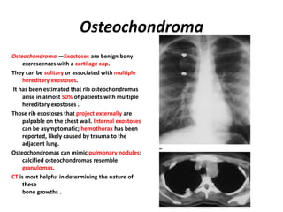
- 16. Osteochondroma Osteochondroma.ŌĆöExostoses are benign bony excrescences with a cartilage cap. They can be solitary or associated with multiple hereditary exostoses. It has been estimated that rib osteochondromas arise in almost 50% of patients with multiple hereditary exostoses . Those rib exostoses that project externally are palpable on the chest wall. Internal exostoses can be asymptomatic; hemothorax has been reported, likely caused by trauma to the adjacent lung. Osteochondromas can mimic pulmonary nodules; calcified osteochondromas resemble granulomas. CT is most helpful in determining the nature of these bone growths .
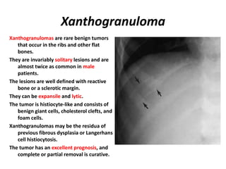
- 17. Xanthogranuloma Xanthogranulomas are rare benign tumors that occur in the ribs and other flat bones. They are invariably solitary lesions and are almost twice as common in male patients. The lesions are well defined with reactive bone or a sclerotic margin. They can be expansile and lytic. The tumor is histiocyte-like and consists of benign giant cells, cholesterol clefts, and foam cells. Xanthogranulomas may be the residua of previous fibrous dysplasia or Langerhans cell histiocytosis. The tumor has an excellent prognosis, and complete or partial removal is curative.
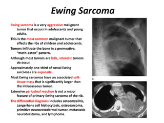
- 18. Ewing Sarcoma Ewing sarcoma is a very aggressive malignant tumor that occurs in adolescents and young adults. This is the most common malignant tumor that affects the ribs of children and adolescents. Tumors infiltrate the bone in a permeative, ŌĆ£moth-eatenŌĆØ pattern. Although most tumors are lytic, sclerotic tumors do occur. Approximately one-third of costal Ewing sarcomas are expansile. Most Ewing sarcomas have an associated soft- tissue mass that is significantly larger than the intraosseous tumor. Extensive periosteal reaction is not a major feature of primary Ewing sarcoma of the rib. The differential diagnosis includes osteomyelitis, Langerhans cell histiocytosis, osteosarcoma, primitive neuroectodermal tumor, metastatic neuroblastoma, and lymphoma.
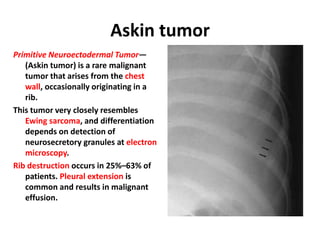
- 19. Askin tumor Primitive Neuroectodermal TumorŌĆö (Askin tumor) is a rare malignant tumor that arises from the chest wall, occasionally originating in a rib. This tumor very closely resembles Ewing sarcoma, and differentiation depends on detection of neurosecretory granules at electron microscopy. Rib destruction occurs in 25%ŌĆō63% of patients. Pleural extension is common and results in malignant effusion.
- 20. *X-Ray chest PA view reveals: ŌĆó Average cardio-thoracic ratio in its transverse diameter. ŌĆó Left middle and lower lung zone semi-homogeous opacity with air bronchogam denoting consolidation. ŌĆó Free both costo-phrenic angles. ŌĆó Normal both copula levels. ŌĆó Normal bony cage.
- 21. Keep Your Eyes On The Ribs Thank you