
1 of 19
Downloaded 214 times


Recommended
PHYSIOLOGY OF SPINAL ANAESTHESIA.pptx
PHYSIOLOGY OF SPINAL ANAESTHESIA.pptxpugalrockzz1
?
This document discusses the physiology of nerve fibers and spinal anesthesia. It begins by describing the different types of nerve fibers (A, B, C fibers), their functions, and myelination. It then explains the mechanism of spinal anesthesia, including how local anesthetics act in the subarachnoid space to block nerve fibers. Finally, it discusses factors that can affect the level and duration of the spinal blockade, such as drug properties, patient factors, and procedural details.Caeliac plexus block.dr quiyum
Caeliac plexus block.dr quiyumMD Quiyumm
?
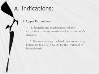
CPB is one the preferable method of pain alleviation in chronic pancreatitis and unresectable pancreatic malignancyBrachial plexus block
Brachial plexus blockSami Ur Rehman
?
Brachial Plexus Block, Anatomy, Relations, Formation of brachial plexus, Cutaneous branches, Nerve branches from each cord, Anatomic variations, Anesthetic implications, Inter-scalene block, Supraclavicular block, Infraclavicular block, Axillary block, local anesthetic drugs25820 - Cervical Plexus Block (1).pptx
25820 - Cervical Plexus Block (1).pptxTiktokethiodaily
?
This document summarizes a technique for performing local/regional anesthesia for thyroid surgery. It begins by describing the patient who will undergo a parathyroidectomy due to hypercalcemia from hyperparathyroidism. It then provides background on the history and development of performing thyroid surgery under local/regional anesthesia. It proceeds to describe the relevant anatomy of the cervical plexus and its branches. It concludes by outlining the technique for performing a superficial cervical plexus block, including patient position, landmarks, local anesthetic used, and injection points along the posterior border of the sternocleidomastoid muscle at the level of the external jugular vein. The summary is provided in 3 sentences or less as requested.regional anesthesia and beir block
regional anesthesia and beir blockAhmed Almumtin
?
This document provides an overview of regional anesthesia techniques including spinal anesthesia, epidural anesthesia, and Bier block. Spinal anesthesia involves injecting local anesthetic into the subarachnoid space, while epidural anesthesia involves injection into the epidural space. Bier block, also called intravenous regional anesthesia, involves exsanguinating a limb and injecting local anesthetic near the tourniquet. The document describes anatomy, procedures, indications, advantages, complications, and pharmacology for each technique.Regional Anaesthesia for Neck surgeries 
Regional Anaesthesia for Neck surgeries Abhinav Gupta
?
Regional anaesthesia involves blocking sensation to a large area of the body like a limb or lower body. It can be divided into central techniques like epidural and spinal anaesthesia, and peripheral techniques like brachial plexus and single nerve blocks. Regional anaesthesia of the neck can provide operative or postoperative analgesia and is useful for many neck surgeries. The superficial and deep cervical plexus blocks involve injecting anaesthetic around the cervical plexus to numb the skin of the neck. Care must be taken to avoid complications like phrenic nerve blockade, hematoma, or local anesthetic toxicity.Anesthetic management of facio maxillary trauma
Anesthetic management of facio maxillary traumaMadhan Chandramohan
?
will be useful for anesthesia interns regarding various methods available for securing airway in patients with severe facio-maxillary injuriesAwake intubation distribution
Awake intubation distributionNC Association of Nurse Anesthetists
?
The document discusses awake intubation, including indications, patient preparation, pharmacological considerations like using lidocaine to anesthetize the airway via various methods to block different nerves, equipment needs, and personnel requirements to safely perform an awake intubation. It also reviews closed claims analyses related to airway management and difficult intubation, and the ASA's difficult airway algorithm.Peripheral Nerve block(ankle block,wrist block, digital block)
Peripheral Nerve block(ankle block,wrist block, digital block)Lih Yin Chong
?
This document provides information on peripheral nerve blocks of the ankle, wrist, and digits. It describes the anatomy and techniques for ankle blocks of the posterior tibial, deep peroneal, superficial peroneal, sural, and saphenous nerves. Wrist blocks are outlined for the radial, ulnar, and median nerves. Digital nerve blocks involve blocking the dorsal and volar branches at the base of each finger. Complications of nerve blocks and management of local anesthetic systemic toxicity are also reviewed.Caudal anesthesia
Caudal anesthesiaArjun Chhetri
?
Caudal anesthesia involves needle penetration through the sacral hiatus into the sacral canal. In adults, the sacrum is a triangular bone formed from the fusion of five sacral vertebrae. It differs in neonates and infants due to delayed myelination and fusion of vertebrae. The sacral hiatus is wider in children, allowing easier identification and catheter insertion for caudal anesthesia. Regional techniques require lower approaches in pediatrics due to the lower termination of the spinal cord and dural sac.Ultrasound Guided Transversus Abdominis Plane (TAP) Block
Ultrasound Guided Transversus Abdominis Plane (TAP) BlockSaeid Safari
?
This document provides background information and details on the technique of performing a transversus abdominis plane (TAP) block. It begins with an overview of the TAP block, including its original description and subsequent modifications using ultrasound guidance. Next, it discusses the indications for TAP blocks, including postoperative analgesia for various abdominal surgeries. The anatomy section describes the layers and nerves of the abdominal wall targeted by the block. Finally, the technique section outlines the materials, patient positioning, ultrasound probe placement, needle insertion, and local anesthetic injection steps to perform a TAP block.Bier block (intravenous regional anesthesia)
Bier block (intravenous regional anesthesia)Komal Haleem
?
This presentation details the indications, mechanism, procedure, complications and drugs used for bier block anesthesia.Extubation problems and its management
Extubation problems and its managementDr Kumar
?
Dr. Kumar presented on extubation problems and their management. Some key points:
1. Tracheal extubation requires careful planning and preparation to prevent complications like laryngospasm, laryngeal edema, and pulmonary aspiration.
2. Patients should generally be extubated awake to allow for airway protection, but deep extubation may be considered to reduce cardiovascular stimulation.
3. Potential problems include mechanical issues removing the tube, cardiovascular changes, respiratory complications, and airway obstruction. Management depends on the specific issue but may include medications, positioning, or alternative extubation techniques.
4. Careful evaluation of each patient's risk factors and planning is necessary to safely perform extubation and preventcaudal anesthesia.pdf
caudal anesthesia.pdfKhodifadVijay
?
Caudal anesthesia involves injecting local anesthetic into the caudal canal of the sacrum to provide pain relief below the umbilicus. It can be used alone or with general anesthesia for surgeries involving the perineum, anus, rectum, or lower extremities. The technique involves identifying the sacral hiatus and inserting a needle, with ultrasound or fluoroscopy guidance available. Potential complications include dural puncture, nerve injury, and local anesthetic toxicity. The level of pain relief varies significantly among patients.Lower limb blocks
Lower limb blocksgaganbrar18
?
1) Lower limb nerve blocks provide post-operative pain relief and are safer than complete sympathectomy, though they are technically more difficult than upper limb blocks or central neuraxial blocks.
2) The lumbar and sacral plexuses give rise to various nerves that can be blocked individually or as combinations to anesthetize the lower limb, including the femoral, lateral femoral cutaneous, and obturator nerves.
3) The 3-in-1 block anesthetizes the femoral, obturator, and lateral femoral cutaneous nerves via a single injection into the femoral sheath above the inguinal ligament.Management of intraoperative bronchospasm
Management of intraoperative bronchospasmChaithanya Malalur
?
The document provides information on the management of intra-operative bronchospasm, including risk factors, triggers, diagnosis, prevention, and treatment approaches. Bronchospasm can be caused by airway irritation or anaphylaxis and presents with signs of wheezing, increased airway pressures, and falling oxygen saturation. Differential diagnoses must be ruled out. Management involves deepening anesthesia, administering bronchodilators, optimizing ventilation, and considering anaphylaxis or postponing surgery. A case example demonstrates treatment of bronchospasm potentially caused by succinylcholine-induced anaphylaxis.Presentation on intravenous regional anaesthesia
Presentation on intravenous regional anaesthesiapriadharshini31
?
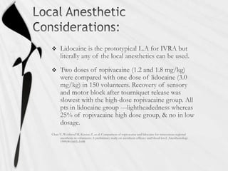
1) Intravenous regional anesthesia (IVRA), also known as Bier block, was first introduced in 1908 by German surgeon August Bier. It involves injecting local anesthetic into the venous system of an extremity that has been isolated from circulation using a tourniquet.
2) The mechanism of action is that the local anesthetic diffuses extravascularly to block peripheral nerve branches and into vasa nervorum and ciliary plexuses of nerves, producing a peripheral and conduction block.
3) IVRA is commonly used for short surgical procedures below the elbow or knee, such as carpal tunnel release or hand surgery. It provides rapid onset anesthesia within 5 minutes when 30-50mL of 0Anatomy of epidural space
Anatomy of epidural spaceKarthavya S L
?
This document provides an overview of the anatomy of the epidural space. It discusses the boundaries, contents, size, and structures that must be penetrated to access the epidural space. Key points include that the epidural space lies between the spinal meninges and vertebral canal, contains connective tissue, fat, blood vessels and spinal nerves. It varies in size from 1-6mm depending on the spinal region. To access it requires penetrating the skin, ligaments and ligamentum flavum in the midline.brachial plexus blocks
brachial plexus blocksanaesthesiology-mgmcri
?
The document provides information about brachial plexus anatomy and different approaches for brachial plexus block, including interscalene, supraclavicular, infraclavicular, and axillary approaches. It discusses the anatomy relevant to each approach, positioning and needle placement techniques, methods for localizing nerves, injection procedures, expected durations and volumes of local anesthetic, and potential complications.Awake craniotomy 
Awake craniotomy samaresh Drsamareshdas
?
1. Awake craniotomy is a surgical procedure performed with the patient awake to allow mapping of brain functions while removing a brain tumor.
2. During surgery, a neurosurgeon performs cortical mapping to identify vital brain areas that should not be disturbed while removing the tumor.
3. Awake craniotomy provides benefits over surgery under general anesthesia such as higher rates of total tumor resection, fewer permanent neurological deficits, and shorter hospital stays. However, it requires careful patient selection and management of anesthesia to balance pain and cooperation.Post dural puncture headache
Post dural puncture headacheKIMS
?
Post dural puncture headache (PDPH) occurs when the dura mater is punctured during a spinal tap, causing cerebrospinal fluid (CSF) to leak out. This can lead to a decrease in CSF volume and pressure, causing headaches. PDPH presents with a postural headache that worsens when upright and improves when lying down. The incidence of PDPH depends on the size and design of the spinal needle used, with smaller pencil-point needles having lower rates. Treatment focuses on bed rest, hydration, caffeine, and analgesics. An epidural blood patch, where the patient's own blood is injected into the epidural space, is an effective treatment when headaches are disabling or persist beyondBrachial plexus block
Brachial plexus blockDhritiman Chakrabarti
?
The brachial plexus is formed by the ventral rami of cervical and thoracic spinal nerves C5-T1. It provides innervation to the upper limb. The document describes the anatomy and formation of the brachial plexus in detail. It also discusses various techniques for brachial plexus block, including interscalene, supraclavicular, infraclavicular, and axillary approaches. Ultrasound guidance is recommended to visualize the relevant anatomy and spread of local anesthetic.Hypothermia and anaesthesia implication
Hypothermia and anaesthesia implicationdrriyas03
?
This document discusses hypothermia, including its definition, causes, risk factors, effects on the body, diagnosis, and treatment methods. Hypothermia is defined as an unintentional drop in core body temperature below 35ˇăC or 95ˇăF and can be caused by direct cold exposure or as a complication of disease. Risk factors include extremes of age, environmental factors, medical conditions, medications, and injuries or illnesses that increase heat loss or impair thermoregulation. As body temperature drops, effects progress from mild cognitive changes to loss of consciousness and life-threatening arrhythmias. Treatment focuses on rewarming patients either passively or actively depending on severity and involves external or internal heating methods.Delayed recovery from anaesthesia.ppt
Delayed recovery from anaesthesia.pptShaiq Hameed
?
Delayed recovery from anesthesia can have multiple contributing factors and causes. It is important to consider potential drug interactions, metabolic abnormalities, and organic causes that may cause prolonged unconsciousness and have serious health implications. Signs and symptoms of metabolic issues may not present normally in an anesthetized patient. The Glasgow Coma Scale provides an objective measure of conscious state regardless of cause.Capnography
Capnographylarryide
?
This document provides an overview of capnography including:
1) The objectives of describing ventilation, perfusion, and their relationship as assessed by capnography.
2) A description of the normal capnogram waveform and factors that can cause abnormal waveforms related to airway, breathing, and circulation problems.
3) Clinical applications of capnography including confirming endotracheal tube placement, assessing ventilation status, and predicting outcomes of cardiac arrest resuscitation.LMA (1).ppt
LMA (1).pptMSrujanaDevi
?
The document provides information on the Laryngeal Mask Airway (LMA). It discusses the key features and design of the LMA, how to select the appropriate size, how to properly insert and secure the LMA, and how to manage the airway intraoperatively. Some potential complications are also outlined. In summary, the LMA is a supraglottic airway device that provides an alternative to face masks or endotracheal tubes to manage the airway during procedures requiring anesthesia. Proper technique and monitoring are important to ensure effective ventilation and patient safety.Anaesthesia for posterior fossa surgery
Anaesthesia for posterior fossa surgeryDhritiman Chakrabarti
?
The document discusses the posterior fossa, including its boundaries, contents, blood supply, clinical presentation of lesions, and considerations for anesthesia. The posterior fossa is bounded anteriorly by the clivus and petrous bone, posteriorly by the occipital bone, and laterally by the temporal bone. It contains the cerebellar hemispheres, brainstem, and cranial nerves III-XII. Lesions can cause a variety of signs and symptoms depending on location, including ataxia, nystagmus, limb weakness, and cranial nerve deficits. Anesthesia for posterior fossa surgery requires careful monitoring and positioning to maintain stability while allowing surgical access.Intraoperative awareness
Intraoperative awarenessHimanshu Jangid
?
This document provides information about general anesthesia and intraoperative awareness. It discusses what general anesthesia is, how its depth is determined, and the different stages of awareness and memory formation. It also covers risk factors for awareness, its incidence and impact on patients. Monitoring techniques like BIS, entropy and EEG patterns are described. Finally, it discusses approaches to preventing and managing cases of intraoperative awareness.Regional anesthesia 
Regional anesthesia Mohammed Dabbour
?
Regional anesthesia can be divided into neuraxial blocks and peripheral nerve blocks. Neuraxial blocks include subarachnoid, epidural and caudal anesthesia. Neuraxial blocks have specific anatomy, indications, contraindications, safety precautions, equipment, and techniques that must be followed. The document outlines the key anatomical structures involved in neuraxial blocks, when they are indicated, potential risks, how to prepare the patient, types of needles used, and proper positioning and aseptic techniques.Regional Anesthesia
Regional Anesthesiameducationdotnet
?
Regional anesthesia is a technique that induces loss of sensation in part of the body using local anesthetics. It has benefits like lower costs, high patient satisfaction, and decreased risks of DVT and PE compared to general anesthesia. However, it requires skills and may cause issues like hypotension. The main types are topical, intravenous, peripheral nerve blocks, plexus blocks, and neuro-axial blocks. Regional anesthesia can provide anesthesia for surgery, post-op analgesia, or chronic pain treatment. Factors like the anesthetic used, patient position, and injection speed affect its spread. Spinal and epidural blocks involve injecting anesthetic into the subarachnoid or epidural space and have risks like anaphylMore Related Content
What's hot (20)
Peripheral Nerve block(ankle block,wrist block, digital block)
Peripheral Nerve block(ankle block,wrist block, digital block)Lih Yin Chong
?
This document provides information on peripheral nerve blocks of the ankle, wrist, and digits. It describes the anatomy and techniques for ankle blocks of the posterior tibial, deep peroneal, superficial peroneal, sural, and saphenous nerves. Wrist blocks are outlined for the radial, ulnar, and median nerves. Digital nerve blocks involve blocking the dorsal and volar branches at the base of each finger. Complications of nerve blocks and management of local anesthetic systemic toxicity are also reviewed.Caudal anesthesia
Caudal anesthesiaArjun Chhetri
?
Caudal anesthesia involves needle penetration through the sacral hiatus into the sacral canal. In adults, the sacrum is a triangular bone formed from the fusion of five sacral vertebrae. It differs in neonates and infants due to delayed myelination and fusion of vertebrae. The sacral hiatus is wider in children, allowing easier identification and catheter insertion for caudal anesthesia. Regional techniques require lower approaches in pediatrics due to the lower termination of the spinal cord and dural sac.Ultrasound Guided Transversus Abdominis Plane (TAP) Block
Ultrasound Guided Transversus Abdominis Plane (TAP) BlockSaeid Safari
?
This document provides background information and details on the technique of performing a transversus abdominis plane (TAP) block. It begins with an overview of the TAP block, including its original description and subsequent modifications using ultrasound guidance. Next, it discusses the indications for TAP blocks, including postoperative analgesia for various abdominal surgeries. The anatomy section describes the layers and nerves of the abdominal wall targeted by the block. Finally, the technique section outlines the materials, patient positioning, ultrasound probe placement, needle insertion, and local anesthetic injection steps to perform a TAP block.Bier block (intravenous regional anesthesia)
Bier block (intravenous regional anesthesia)Komal Haleem
?
This presentation details the indications, mechanism, procedure, complications and drugs used for bier block anesthesia.Extubation problems and its management
Extubation problems and its managementDr Kumar
?
Dr. Kumar presented on extubation problems and their management. Some key points:
1. Tracheal extubation requires careful planning and preparation to prevent complications like laryngospasm, laryngeal edema, and pulmonary aspiration.
2. Patients should generally be extubated awake to allow for airway protection, but deep extubation may be considered to reduce cardiovascular stimulation.
3. Potential problems include mechanical issues removing the tube, cardiovascular changes, respiratory complications, and airway obstruction. Management depends on the specific issue but may include medications, positioning, or alternative extubation techniques.
4. Careful evaluation of each patient's risk factors and planning is necessary to safely perform extubation and preventcaudal anesthesia.pdf
caudal anesthesia.pdfKhodifadVijay
?
Caudal anesthesia involves injecting local anesthetic into the caudal canal of the sacrum to provide pain relief below the umbilicus. It can be used alone or with general anesthesia for surgeries involving the perineum, anus, rectum, or lower extremities. The technique involves identifying the sacral hiatus and inserting a needle, with ultrasound or fluoroscopy guidance available. Potential complications include dural puncture, nerve injury, and local anesthetic toxicity. The level of pain relief varies significantly among patients.Lower limb blocks
Lower limb blocksgaganbrar18
?
1) Lower limb nerve blocks provide post-operative pain relief and are safer than complete sympathectomy, though they are technically more difficult than upper limb blocks or central neuraxial blocks.
2) The lumbar and sacral plexuses give rise to various nerves that can be blocked individually or as combinations to anesthetize the lower limb, including the femoral, lateral femoral cutaneous, and obturator nerves.
3) The 3-in-1 block anesthetizes the femoral, obturator, and lateral femoral cutaneous nerves via a single injection into the femoral sheath above the inguinal ligament.Management of intraoperative bronchospasm
Management of intraoperative bronchospasmChaithanya Malalur
?
The document provides information on the management of intra-operative bronchospasm, including risk factors, triggers, diagnosis, prevention, and treatment approaches. Bronchospasm can be caused by airway irritation or anaphylaxis and presents with signs of wheezing, increased airway pressures, and falling oxygen saturation. Differential diagnoses must be ruled out. Management involves deepening anesthesia, administering bronchodilators, optimizing ventilation, and considering anaphylaxis or postponing surgery. A case example demonstrates treatment of bronchospasm potentially caused by succinylcholine-induced anaphylaxis.Presentation on intravenous regional anaesthesia
Presentation on intravenous regional anaesthesiapriadharshini31
?
1) Intravenous regional anesthesia (IVRA), also known as Bier block, was first introduced in 1908 by German surgeon August Bier. It involves injecting local anesthetic into the venous system of an extremity that has been isolated from circulation using a tourniquet.
2) The mechanism of action is that the local anesthetic diffuses extravascularly to block peripheral nerve branches and into vasa nervorum and ciliary plexuses of nerves, producing a peripheral and conduction block.
3) IVRA is commonly used for short surgical procedures below the elbow or knee, such as carpal tunnel release or hand surgery. It provides rapid onset anesthesia within 5 minutes when 30-50mL of 0Anatomy of epidural space
Anatomy of epidural spaceKarthavya S L
?
This document provides an overview of the anatomy of the epidural space. It discusses the boundaries, contents, size, and structures that must be penetrated to access the epidural space. Key points include that the epidural space lies between the spinal meninges and vertebral canal, contains connective tissue, fat, blood vessels and spinal nerves. It varies in size from 1-6mm depending on the spinal region. To access it requires penetrating the skin, ligaments and ligamentum flavum in the midline.brachial plexus blocks
brachial plexus blocksanaesthesiology-mgmcri
?
The document provides information about brachial plexus anatomy and different approaches for brachial plexus block, including interscalene, supraclavicular, infraclavicular, and axillary approaches. It discusses the anatomy relevant to each approach, positioning and needle placement techniques, methods for localizing nerves, injection procedures, expected durations and volumes of local anesthetic, and potential complications.Awake craniotomy
Awake craniotomy samaresh Drsamareshdas
?
1. Awake craniotomy is a surgical procedure performed with the patient awake to allow mapping of brain functions while removing a brain tumor.
2. During surgery, a neurosurgeon performs cortical mapping to identify vital brain areas that should not be disturbed while removing the tumor.
3. Awake craniotomy provides benefits over surgery under general anesthesia such as higher rates of total tumor resection, fewer permanent neurological deficits, and shorter hospital stays. However, it requires careful patient selection and management of anesthesia to balance pain and cooperation.Post dural puncture headache
Post dural puncture headacheKIMS
?
Post dural puncture headache (PDPH) occurs when the dura mater is punctured during a spinal tap, causing cerebrospinal fluid (CSF) to leak out. This can lead to a decrease in CSF volume and pressure, causing headaches. PDPH presents with a postural headache that worsens when upright and improves when lying down. The incidence of PDPH depends on the size and design of the spinal needle used, with smaller pencil-point needles having lower rates. Treatment focuses on bed rest, hydration, caffeine, and analgesics. An epidural blood patch, where the patient's own blood is injected into the epidural space, is an effective treatment when headaches are disabling or persist beyondBrachial plexus block
Brachial plexus blockDhritiman Chakrabarti
?
The brachial plexus is formed by the ventral rami of cervical and thoracic spinal nerves C5-T1. It provides innervation to the upper limb. The document describes the anatomy and formation of the brachial plexus in detail. It also discusses various techniques for brachial plexus block, including interscalene, supraclavicular, infraclavicular, and axillary approaches. Ultrasound guidance is recommended to visualize the relevant anatomy and spread of local anesthetic.Hypothermia and anaesthesia implication
Hypothermia and anaesthesia implicationdrriyas03
?
This document discusses hypothermia, including its definition, causes, risk factors, effects on the body, diagnosis, and treatment methods. Hypothermia is defined as an unintentional drop in core body temperature below 35ˇăC or 95ˇăF and can be caused by direct cold exposure or as a complication of disease. Risk factors include extremes of age, environmental factors, medical conditions, medications, and injuries or illnesses that increase heat loss or impair thermoregulation. As body temperature drops, effects progress from mild cognitive changes to loss of consciousness and life-threatening arrhythmias. Treatment focuses on rewarming patients either passively or actively depending on severity and involves external or internal heating methods.Delayed recovery from anaesthesia.ppt
Delayed recovery from anaesthesia.pptShaiq Hameed
?
Delayed recovery from anesthesia can have multiple contributing factors and causes. It is important to consider potential drug interactions, metabolic abnormalities, and organic causes that may cause prolonged unconsciousness and have serious health implications. Signs and symptoms of metabolic issues may not present normally in an anesthetized patient. The Glasgow Coma Scale provides an objective measure of conscious state regardless of cause.Capnography
Capnographylarryide
?
This document provides an overview of capnography including:
1) The objectives of describing ventilation, perfusion, and their relationship as assessed by capnography.
2) A description of the normal capnogram waveform and factors that can cause abnormal waveforms related to airway, breathing, and circulation problems.
3) Clinical applications of capnography including confirming endotracheal tube placement, assessing ventilation status, and predicting outcomes of cardiac arrest resuscitation.LMA (1).ppt
LMA (1).pptMSrujanaDevi
?
The document provides information on the Laryngeal Mask Airway (LMA). It discusses the key features and design of the LMA, how to select the appropriate size, how to properly insert and secure the LMA, and how to manage the airway intraoperatively. Some potential complications are also outlined. In summary, the LMA is a supraglottic airway device that provides an alternative to face masks or endotracheal tubes to manage the airway during procedures requiring anesthesia. Proper technique and monitoring are important to ensure effective ventilation and patient safety.Anaesthesia for posterior fossa surgery
Anaesthesia for posterior fossa surgeryDhritiman Chakrabarti
?
The document discusses the posterior fossa, including its boundaries, contents, blood supply, clinical presentation of lesions, and considerations for anesthesia. The posterior fossa is bounded anteriorly by the clivus and petrous bone, posteriorly by the occipital bone, and laterally by the temporal bone. It contains the cerebellar hemispheres, brainstem, and cranial nerves III-XII. Lesions can cause a variety of signs and symptoms depending on location, including ataxia, nystagmus, limb weakness, and cranial nerve deficits. Anesthesia for posterior fossa surgery requires careful monitoring and positioning to maintain stability while allowing surgical access.Intraoperative awareness
Intraoperative awarenessHimanshu Jangid
?
This document provides information about general anesthesia and intraoperative awareness. It discusses what general anesthesia is, how its depth is determined, and the different stages of awareness and memory formation. It also covers risk factors for awareness, its incidence and impact on patients. Monitoring techniques like BIS, entropy and EEG patterns are described. Finally, it discusses approaches to preventing and managing cases of intraoperative awareness.Viewers also liked (20)
Regional anesthesia
Regional anesthesia Mohammed Dabbour
?
Regional anesthesia can be divided into neuraxial blocks and peripheral nerve blocks. Neuraxial blocks include subarachnoid, epidural and caudal anesthesia. Neuraxial blocks have specific anatomy, indications, contraindications, safety precautions, equipment, and techniques that must be followed. The document outlines the key anatomical structures involved in neuraxial blocks, when they are indicated, potential risks, how to prepare the patient, types of needles used, and proper positioning and aseptic techniques.Regional Anesthesia
Regional Anesthesiameducationdotnet
?
Regional anesthesia is a technique that induces loss of sensation in part of the body using local anesthetics. It has benefits like lower costs, high patient satisfaction, and decreased risks of DVT and PE compared to general anesthesia. However, it requires skills and may cause issues like hypotension. The main types are topical, intravenous, peripheral nerve blocks, plexus blocks, and neuro-axial blocks. Regional anesthesia can provide anesthesia for surgery, post-op analgesia, or chronic pain treatment. Factors like the anesthetic used, patient position, and injection speed affect its spread. Spinal and epidural blocks involve injecting anesthetic into the subarachnoid or epidural space and have risks like anaphylAdjuncts to local anesthetics
Adjuncts to local anestheticsNin Thitayaporn
?
This document discusses common adjuncts and additives used with local anesthetics for nerve blocks and spinal anesthesia. It describes how epinephrine prolongs the duration and intensity of nerve blocks by causing vasoconstriction. Alkalinization can increase the effectiveness of local anesthetics but risks of precipitation limit its usefulness. Opioids and alpha-2 adrenergic agonists provide analgesic effects when added to local anesthetics by binding to receptors in the spinal cord. Fentanyl and morphine are commonly used opioid adjuncts. Dexamethasone may also prolong the duration of local anesthetic nerve blocks when used, though the mechanism is unknown.Transfusion related acute lung injury
Transfusion related acute lung injury9857038254
?
Transfusion-related acute lung injury (TRALI) is a potentially fatal syndrome characterized by acute respiratory distress within 6 hours of blood transfusion. It is believed to be caused by anti-leukocyte antibodies in plasma products that cause leukocyte aggregation in the lungs, inflammatory injury, and non-cardiogenic pulmonary edema. TRALI has an incidence of 1 in 5000 transfusions and mortality rate of 5-25%. Risk factors include plasma-rich products, multiparous donors, and underlying patient conditions. Diagnosis involves new pulmonary edema within 6 hours of transfusion in the absence of circulatory overload. Treatment focuses on supportive care, with most patients recovering within 72 hours. Prevention strategies include leukoreduction of blood productsTrali
TraliRHMBONCO
?
Transfusion-related acute lung injury (TRALI) is a potentially fatal pulmonary complication of blood transfusion. It is caused by antibodies and bioactive substances in blood products that activate neutrophils in the lungs. TRALI accounts for 13% of transfusion-related fatalities. Risk factors include plasma-containing products and antibodies from female donors who have been pregnant. Studies show implementing male-predominant plasma transfusion strategies and HLA antibody screening can reduce TRALI cases and fatalities. However, screening also reduces the available donor pool and platelet availability. Further research is still needed to balance TRALI mitigation and adequate blood supply.Andrea TRESCOT.ppt
Andrea TRESCOT.pptsobramid
?
This document discusses post-herpetic neuralgia (PHN), a chronic neuropathic pain syndrome that persists after an outbreak of herpes zoster (shingles). PHN is caused by damage to large myelinated sensory nerves during a shingles outbreak, resulting in pain sensations like burning, shooting, and pressure. Treatment involves multimodal approaches like drugs, nerve blocks, and in rare cases surgery. The document focuses on intercostal nerve blocks and other nerve targets in the thoracic region that may be blocked to provide pain relief for PHN patients suffering pain in that dermatome.Laryngectomy and laryngeal cancer
Laryngectomy and laryngeal cancersahughes
?
This document discusses voice therapy considerations following laryngeal cancer treatment. It covers the impact of radiation, chemotherapy, and surgery on the vocal folds. For non-laryngectomy patients, therapy may focus on techniques like inhalation phonation to address stiffness. Post-laryngectomy, patients lose their larynx and ability to speak normally. Communication options include electrolarynx, esophageal speech, or tracheoesophageal puncture. Support groups can help patients cope with the physical and emotional impacts of losing their voice box and learning alternative communication methods.Dr. ms goud management of forearm fractures
Dr. ms goud management of forearm fracturesvaruntandra
?
The document discusses the anatomy, biomechanics, classification systems, treatment options, and complications of forearm fractures. It provides details on the bones, joints, ligaments, and muscles of the forearm. Furthermore, it examines various forearm fracture patterns and treatments such as plating, intramedullary nailing, and external fixation. Proper treatment aims to restore alignment, length, rotation, and blood supply to promote healing.Forearm shaft fractues
Forearm shaft fractuesOrthosurg2016
?
The document discusses fractures of the forearm and their treatment. It summarizes that the forearm functions as a joint with six articulations. Forearm fractures can result in deformities like shortening, angulation, and loss of alignment if not treated properly. Treatment goals are anatomical reduction, restoration of length and rotation, and early return of function. Plate fixation is the gold standard and provides stable fixation, allowing early motion to restore function with high union rates over 95%.Elbow and forearm fractures
Elbow and forearm fracturesLouis law Mwadziwana
?
This document discusses fractures of the elbow and forearm. It describes the anatomy of the elbow joint and various types of fractures that can occur in the distal humerus, radial head, coronoid process, and olecranon. Treatment options for different fracture patterns include closed reduction, open reduction and internal fixation using plates, screws and tension band wiring. Complications like stiffness, non-union and nerve injuries are also discussed. Physiotherapy management aims to regain range of motion, muscle strength, and function.Laryngectomy post op
Laryngectomy post opmderami
?
After a laryngectomy, patients require careful monitoring during a 7-9 day hospital stay and follow-up visits over several years. They must carefully manage their airway through breathing exercises, humidification, and tracheostomy tube care to prevent complications like pneumonia. Strict wound care is also needed to avoid infections, hematomas, or pharyngocutaneous fistulas forming in the first few weeks of recovery. A multidisciplinary team approach involving doctors, nurses, dietitians, and speech therapists helps laryngectomy patients safely progress from tube feeding to oral intake and rehabilitation.SNAKE AND SCORPION
ENVENOMATION
SNAKE AND SCORPION
ENVENOMATIONshashank sunny
?
This document summarizes the epidemiology, signs and symptoms, clinical syndromes, investigations, and management of snake bites. It notes that India has the highest snakebite mortality in the world, with estimates of 83,000 bites and 11,000 deaths annually. Signs and symptoms vary depending on the type of snake but can include local swelling, pain, bleeding, shock, paralysis, and kidney injury. Investigations include blood clotting tests and urine analysis. Antivenom treatment is the primary therapy and should be given for systemic signs of envenoming like bleeding, paralysis, shock, or kidney injury.Xenon anaesthesia
Xenon anaesthesiameducationdotnet
?
Xenon is a gaseous anesthetic agent with several potential advantages over traditional inhaled anesthetics. It has minimal environmental impact, provides cardiovascular stability, and may have neuroprotective and renoprotective effects. Studies in animal models and some human trials suggest xenon can reduce neuronal injury from ischemia and hypoxia. It also appears to reduce delayed graft function in renal transplant patients. However, xenon is very expensive compared to other agents. Further research is still needed to fully evaluate its clinical benefits and cost-effectiveness.Diagnosis and treatment of carcinoma of larynx by nitesh Kr.
Diagnosis and treatment of carcinoma of larynx by nitesh Kr.Nitesh Kr
?
The document discusses the diagnosis and clinical features of laryngeal cancer. It outlines the various steps involved in diagnosis which include collecting a history, physical examination, laryngoscopy, endoscopy, biopsy, radiological imaging and staging. Specific symptoms associated with glottic, supraglottic and subglottic cancers are provided. Treatment options discussed include radiotherapy, conservative and total laryngectomy surgery, and combined therapy. Complications, disabilities after total laryngectomy and vocal rehabilitation methods are also summarized.Preemptive analgesia
Preemptive analgesiaMounika Thommandru
?
Preemptive analgesia is an antinociceptive treatment that prevents the establishment of altered processing of afferent input which amplifies postoperative pain. It was first formulated by Crile who advocated regional blocks in addition to general anesthesia to prevent intraoperative nociception and formation of painful scars. There are three definitions of preemptive analgesia: treatment starting before surgery to prevent central sensitization caused by incisional injury; treatment preventing central sensitization caused by incisional and inflammatory injuries; and treatment covering the period of surgery and initial postoperative period. While some studies found no difference between preincisional and postincisional treatment, others reported modest benefits with preincisional analgesia.Peripheral nerve blocks
Peripheral nerve blocksAmit Lall
?
Peripheral nerve blocks are gaining popularity for pain management due to advantages like less nausea, hemodynamic stability, and ability to perform surgery in patients with cardiovascular or bleeding risks. Specific nerve blocks include interscalene, supraclavicular, infraclavicular, axillary, and others. Ultrasound is often used to identify nerves and nearby structures before injection of local anesthetic, allowing effective pain relief with fewer side effects compared to general or epidural anesthesia. Proper technique and understanding of anatomy are required to perform safe and effective peripheral nerve blocks.Forearm fractures
Forearm fracturesMohamed Fazly
?
The document discusses the anatomy and classification of forearm fractures. It describes the radius and ulna bones of the forearm and their articulations. Forearm fractures can be classified as proximal, middle, or distal, and can affect one or both bones. Common types include radial shaft fractures, Galeazzi fractures, and Monteggia's fractures. Assessment involves neurovascular and range of motion exams. Treatment depends on the fracture type but may include immobilization, closed reduction, open reduction and internal fixation, or external fixation.ANAESTHETIC CONSIDERATIONS IN AIDS PATIENTS
ANAESTHETIC CONSIDERATIONS IN AIDS PATIENTSSelva Kumar
?
1) HIV/AIDS patients present special challenges for anesthesia due to the virus' effects on multiple body systems and interactions with antiretroviral drugs.
2) Evaluation of patients should assess organ involvement and potential for drug interactions. Anesthetic plans must be tailored to individual patients while minimizing interruptions to antiretroviral therapy.
3) Strict universal precautions including protective equipment, careful handling of sharps and contaminated materials, and safety equipment help minimize risk of infection to hospital staff from needle sticks or exposure to bodily fluids.Brain death and organ donation
Brain death and organ donationDr Praveen kumar tripathi
?
This document discusses brain death and organ donation. It begins by outlining the history of defining brain death from 1959 to present. It then explains how brain death is determined, including establishing the cause of coma, performing a clinical examination to demonstrate signs of brain death like coma, brainstem areflexia and apnea, and confirming with ancillary tests if needed. The document provides details on specific tests like the apnea test and discusses special considerations for determining brain death in children. It also outlines conditions that must be excluded before declaring brain death. Finally, it discusses the importance of organ donation and the types of donations possible.Anaesthesia challenges in Organ Retrieval
Anaesthesia challenges in Organ RetrievalDr Jayashree Patki
?
Introduction of organ donation .
Introduction of brain death and pathophysiology following it.
Perioperative problems in organ retrieval .
Goals of management of these patients .
Anesthetic management of the cadaver during organ harvesting.More from Dr. Shaheer Haider (18)
Perioperative EKG in Cardiothoracic Anesthesia
Perioperative EKG in Cardiothoracic AnesthesiaDr. Shaheer Haider
?
1. The document discusses various abnormalities that can be seen on preoperative electrocardiograms (EKGs), including chamber hypertrophies, conduction defects, arrhythmias, and signs of ischemia/infarction.
2. Chamber hypertrophies covered include left and right atrial and ventricular hypertrophies, each with characteristic EKG patterns.
3. Conduction defects discussed are right and left bundle branch blocks, along with their typical EKG presentations and common causes.Enteral nutrition finall
Enteral nutrition finallDr. Shaheer Haider
?
The document provides information on calculating calorie requirements, indications for tube feeding, types of enteral formulas, methods of enteral feeding administration, and potential complications. It discusses formulas for different clinical conditions including renal, hepatic, diabetic and pulmonary. Continuous and bolus feeding methods are described. Common gastrointestinal complications like diarrhea, constipation and nausea are outlined along with potential causes and treatments. Electrolyte imbalances from enteral feeding and their management are also summarized.Anaesthetic plan
Anaesthetic planDr. Shaheer Haider
?
This document outlines an anesthetic plan with sections discussing the case scenario, anesthetic concerns, preoperative management, monitoring standards, choice of anesthesia, drugs and dosages, intraoperative fluids, airway management, maintenance, postanesthesia plan, and postop pain management. However, most of the document consists of blank fields to be filled in, so it does not provide much contextual information on its own.Thyroid disease in pregnancy
Thyroid disease in pregnancyDr. Shaheer Haider
?
This document summarizes thyroid disease in pregnancy. It discusses how thyroid function changes normally during pregnancy, with relative iodine deficiency and increased levels of thyroid binding globulin and T4 in early gestation. It notes that hyperthyroidism in pregnancy is usually caused by Graves' disease. Left untreated, it can lead to risks for both mother and fetus, including heart failure, thyroid storm, growth restriction and preterm labor. Management involves achieving an euthyroid state through medications like thionamides or propranolol, with close monitoring of thyroid function tests during pregnancy and treatment of any thyroid storm that may occur during labor and delivery.Pv loops
Pv loopsDr. Shaheer Haider
?
The document discusses three key determinants of cardiac output: preload, afterload, and contractility. Preload refers to the presystolic stretch of the heart and is reflected by the end-diastolic pressure and volume. Afterload is the pressure the left ventricle must overcome during systole and is often represented by systolic pressure. Contractility determines the strength of the heart's contraction and can be measured by the left ventricular ejection fraction.Postop carotid endarterectomy
Postop carotid endarterectomyDr. Shaheer Haider
?
The document provides guidelines for postoperative care after carotid endarterectomy, noting that patients should be closely monitored for complications like hypertension, hypotension, hematoma, and cardiac issues in the first 48 hours after surgery. It also discusses potential complications like hyperperfusion syndrome, intracerebral hemorrhage, and seizures that require strict blood pressure control. The guidelines emphasize vigilant monitoring and management of hemodynamics and neurological symptoms in the postoperative period to optimize outcomes and prevent complications.Evidence Based Medicine (Anesthesiology)
Evidence Based Medicine (Anesthesiology)Dr. Shaheer Haider
?
1. The document provides guidelines from the American Society of Anesthesiologists for managing difficult airways. It defines different types of difficult airways and levels of evidence for various airway management techniques.
2. Key recommendations include conducting an airway evaluation, preparing basic airway equipment, having a pre-planned intubation strategy with non-invasive options, considering awake versus induced intubation, and careful planning for extubation with criteria for awake versus induced extubation.
3. The guidelines provide evidence-based recommendations for each step of difficult airway management - evaluation, preparation, intubation strategy, extubation strategy, and follow-up care - to assist anesthesiologists in decision-making.Recognition And Management Of Difficult Airway
Recognition And Management Of Difficult AirwayDr. Shaheer Haider
?
This document discusses recognition and management of difficult airways. It defines difficult airway as difficulty with mask ventilation or tracheal intubation. It discusses incidence, risk factors, assessment techniques like Mallampati classification and thyromental distance, and management strategies including awake intubation, use of airway adjuncts like LMA, and the ASA difficult airway algorithm which provides a structured approach. The key points are assessing risk factors, having proper equipment and backup plans, and pursuing oxygenation throughout management of a difficult airway.B R A I N D E A T H
B R A I N D E A T HDr. Shaheer Haider
?
The document discusses brain death, its diagnosis and pathophysiology. It defines brain death as the complete and irreversible loss of brain function. The diagnosis involves meeting strict clinical criteria demonstrating the absence of brainstem reflexes as well as confirmatory tests like EEG. Brain death results in no prospect of survival without life support or recovery of brain function. Proper diagnosis is important for organ donation where brain death constitutes legal death.Anti Coagulation In Pregnancy
Anti Coagulation In PregnancyDr. Shaheer Haider
?
1) The patient requires careful management of anticoagulation for her mechanical heart valve during pregnancy and delivery. She should receive adjusted-dose low molecular weight heparin throughout pregnancy.
2) A regional anesthetic technique could be used for her planned c-section, but her coagulation status and platelet count must be checked closely both before and after the procedure due to her anticoagulation.
3) After delivery, she will need to resume anticoagulation while monitoring closely for any signs of spinal hematoma due to her recent regional block and anticoagulated state.S C O L I O S I S
S C O L I O S I SDr. Shaheer Haider
?
Scoliosis can cause restrictive lung defects, ventilation-perfusion mismatch, and hypoxemia due to thoracic deformity. It can also involve the cardiovascular system by raising right heart pressures or causing mitral valve prolapse. Careful pre-anesthetic evaluation should assess the respiratory, cardiovascular, and neurological systems. Intraoperatively, temperature, fluids, positioning, spinal cord monitoring, and blood conservation are important considerations, as is post-operative respiratory therapy and pain management.Patient Ventilator Synchrony & Successful WeaningÖvÁx
Patient Ventilator Synchrony & Successful WeaningÖvÁxDr. Shaheer Haider
?
This document discusses patient-ventilator synchrony and successful weaning. It defines weaning as the gradual decrease of ventilatory support to prepare for extubation. Optimal synchrony depends on factors like trigger sensitivity, ventilator response time, appropriate tidal volume, and complete expiration to minimize work of breathing. Various ventilator modes and settings can be adjusted to improve synchrony and reduce the risk of reintubation during weaning and extubation.Optimal Patient Vent Synchrony
Optimal Patient Vent SynchronyDr. Shaheer Haider
?
This document discusses obtaining optimal patient-ventilator synchrony. It describes three phases of patient-ventilator interaction: triggering, breath delivery, and cycling. Trigger asynchrony can occur if the ventilator does not respond quickly enough to the patient's effort. Breath delivery asynchrony may happen if the ventilator does not provide enough flow. Cycling asynchrony can result if the neural inspiration time does not match the mechanical inspiration time. The document provides tips for recognizing and addressing asynchrony issues, such as adjusting ventilator settings and modes. The overall goal is to minimize patient discomfort and optimize lung protection and recovery.Interpretation Of Arterial Blood Gas
Interpretation Of Arterial Blood GasDr. Shaheer Haider
?
This document provides information about interpreting arterial blood gases, including:
1. It discusses acid-base balance and how the respiratory and renal systems work to maintain pH.
2. It describes the four main acid-base disorders: respiratory acidosis, respiratory alkalosis, metabolic acidosis, and metabolic alkalosis.
3. It explains how to interpret the components of an arterial blood gas like pH, PaCO2, PaO2, and bicarbonate levels.Sepsis Guidelines 2007
Sepsis Guidelines 2007Dr. Shaheer Haider
?
Early recognition and treatment of sepsis is important to improve outcomes. The guidelines recommend initially evaluating airway, breathing, and circulation. Goals of early resuscitation include restoring central venous pressure, blood pressure, urine output, and central venous oxygen saturation through aggressive fluid administration and vasopressors if needed within 6 hours of recognition. Intravenous antibiotics should also be administered within 1 hour, and cultures obtained, to treat the underlying infection.Obesity
ObesityDr. Shaheer Haider
?
This document provides clinical guidelines for the identification, evaluation, and treatment of overweight and obesity in adults. An expert panel was convened by the National Heart, Lung, and Blood Institute and the National Institute of Diabetes and Digestive and Kidney Diseases to develop the guidelines based on a thorough review of scientific evidence. The guidelines examine the health risks of overweight and obesity and various treatment strategies, providing recommendations to help practitioners effectively assess and treat overweight and obese patients.P R E G N A N C Y I N D U C E D H Y P E R T E N S I O N
P R E G N A N C Y I N D U C E D H Y P E R T E N S I O NDr. Shaheer Haider
?
Pregnancy-induced hypertension (PIH) is defined as new hypertension developing after 20 weeks of gestation. It affects 5-8% of pregnancies and can range from mild to severe, including pre-eclampsia and eclampsia. The exact cause is unknown but may involve immunological and endothelial dysfunction factors. Treatment aims to prevent complications and involves bed rest, magnesium sulfate, antihypertensive drugs, and delivery if gestation reaches term or the mother/baby's condition deteriorates.P O N V
P O N VDr. Shaheer Haider
?
An anxious 52-year-old man is presenting for revision of a previous tympano-mastoidectomy surgery. He has a history of postoperative nausea and vomiting after prior procedures. Monitored anesthesia care with sedation may be suitable for this patient's surgery given his anxiety. General anesthetic techniques that minimize postoperative nausea and vomiting risks include the use of antiemetics like ondansetron and dexamethasone. Regional anesthesia may further reduce postoperative nausea and vomiting risks compared to general anesthesia. Control of blood loss is important during middle ear surgery to maintain a bloodless surgical field. Long-acting neuromuscular blocking agents should be avoided due to the delicate nature of middle ear surgery.