
![Disclosures
â–Ş Our imaging project is proudly sponsored by the Emergency Medicine and
Neurosurgery Residency Programs at Carolinas Medical Center.
â–Ş The goal is to promote widespread mastery of imaging interpretation.
â–Ş There is no personal health information [PHI] within, and all ages have been
changed to protect patient confidentiality.](https://image.slidesharecdn.com/intraventricularruptureofbrainabscessivroba-250214211133-2263e30d/85/Intraventricular-Rupture-of-Brain-Abscess-IVROBA-pdf-2-320.jpg)


![The Case Continues
After Returning From CT The Patient Became Progressively More Obtunded
And Had A Brief, Self-Terminating Seizure.
Immediate Management
• The patient was intubated
• Antibiotics: cefepime + vancomycin + metronidazole
• The patient was loaded with levetiracetam 60 mg/kg
• Neurosurgery placed an external ventricular drain at bedside
CSF: Glucose 14 mg/dl, protein 358 g/L, 4172 nucleated cells with 95%
neutrophilic predominance. Gram stain: gram [+] cocci in pairs and chains
• A STAT MRI of the brain was performed](https://image.slidesharecdn.com/intraventricularruptureofbrainabscessivroba-250214211133-2263e30d/85/Intraventricular-Rupture-of-Brain-Abscess-IVROBA-pdf-13-320.jpg)


![Clinical features and predictive factors of intraventricular rupture in patients
who have bacterial brain abscesses.
Journal of Neurology Neurosurgery & Psychiatry 2007;78:303-309.
Objective
To identify potential risk factors predictive of intraventricular ruptures in hospitalized patients
with brain abscess without intraventricular rupture upon presentation.
Design
20-year retrospective review of 179 patients brain abscess. 45 had IVRBA on admission (initial
IVRBA) and 17 had the episode during hospitalization (subsequent IVRBA).
Results
• Multiloculated abscess vs. non-loculated abscess had a higher risk of intraventricular
rupture [OR 4.2 (95% CI 2.4-14.3); p = 0.020].
• A shorter distance between the abscess and ventricle was associated with a higher risk of
rupture each 1 mm decrease of this distance increased the risk of rupture by 10% per mm.](https://image.slidesharecdn.com/intraventricularruptureofbrainabscessivroba-250214211133-2263e30d/85/Intraventricular-Rupture-of-Brain-Abscess-IVROBA-pdf-24-320.jpg)


More Related Content
More from Sean M. Fox (20)
Recently uploaded (20)


Intraventricular Rupture of Brain Abscess (IVROBA).pdf
- 1. L. Erin Miller, MD2, Teresa Crow, MD1, Troy Carnwath, MD1, Scott DiMeo, MD1, Natalie Rall, MD1 Carolinas Medical Center & Levine Children’s Hospital Department of Emergency Medicine1 Carolina Neurosurgery & Spine Associates2 Michael Gibbs, MD, Imaging Mastery Project Lead Editor Neuroimaging Mastery Project Case Study #2 Intraventricular Rupture Of Brain Abscess (IVROBA)
- 2. Disclosures â–Ş Our imaging project is proudly sponsored by the Emergency Medicine and Neurosurgery Residency Programs at Carolinas Medical Center. â–Ş The goal is to promote widespread mastery of imaging interpretation. â–Ş There is no personal health information [PHI] within, and all ages have been changed to protect patient confidentiality.
- 3. Meet Our Neuroimaging Editorial Team Andrew Asimos, MD, FACEP Medical Director, Atrium Health Stroke Network Neurosciences Institute Professor, Department of Emergency Medicine Jonathan Clemente, MD, FACR Chief, Department of Radiology, Carolinas Medical Center Charlotte Radiology, Neuroradiology Section Adjunct Clinical Associate Professor, Department of Radiology Andrew Perron, MD, FACEP Associate Dean of Graduate Medical Education and DIO Professor of Emergency Medicine Department of Graduate Medical Education Dartmouth Hitchcock Medical Center
- 4. Meet Our Neuroimaging Editorial Team Christa Swisher, MD, FNCS, FACNS Neurocritical Care/Pulmonary Critical Care Consultants Department of Medicine, Atrium Health Clinical Assistant Professor, Department of Neurology Scott Wait, MD, FAANS Chief, Pediatric Neurosurgery, Levine Children’s Hospital Carolina Neurosurgery & Spine Associates Adjunct Clinical Associate Professor, Department of Neurosurgery
- 5. Meet Our Medical Illustrator Anne Olson Anne Olson has shared her expert Medical Illustrator skills with the CMC Department of Emergency Medicine for the past 30 years. As co-founder of Blazon Productions, LLC, Anne’s ongoing efforts to support the Imaging Mastery Project and a growing number of presentations dedicated to Neuroimaging elevate the quality and impact of this work. We are very grateful for her talents!
- 6. Visit Our Website www.EMGuidewire.com For A Complete Archive Of Imaging Presentations And Much More!
- 7. Case A 49-Year-Old Male With No Known Medical History Presented To The ED With Confusion And Agitation. Co-Worker Shared That For The Past Week He Complained Of Headache And Fever. The Initial ED Examination: Vital Signs: BP 151/96, HR 113, RR 16, SAO2 100%, T 101.5° • Confused and restless but able to follow commands • Equal pupils and normal eye movement • Pain with neck movement • No focal neurological deficits
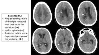
- 8. STAT Head CT
- 9. → STAT Head CT • Ring-enhancing lesion of the right temporal lobe that abuts the ventricles (→)
- 10. STAT Head CT • Ring-enhancing lesion of the right temporal lobe that abuts the ventricles (→) • Hydrocephalus (⇒) →
- 11. STAT Head CT • Ring-enhancing lesion of the right temporal lobe that abuts the ventricles (→) • Hydrocephalus (⇒) • Scattered debris in the dependent portions of the ventricles (➤) ➤ ➤ ➤ →
- 12. STAT Head CT • Ring-enhancing lesion of the right temporal lobe that abuts the ventricles (→) • Hydrocephalus (⇒) • Scattered debris in the dependent portions of the ventricles (➤) ➤ ➤ ➤ → These Findings Are Suggestive Of IVROBA
- 13. The Case Continues After Returning From CT The Patient Became Progressively More Obtunded And Had A Brief, Self-Terminating Seizure. Immediate Management • The patient was intubated • Antibiotics: cefepime + vancomycin + metronidazole • The patient was loaded with levetiracetam 60 mg/kg • Neurosurgery placed an external ventricular drain at bedside CSF: Glucose 14 mg/dl, protein 358 g/L, 4172 nucleated cells with 95% neutrophilic predominance. Gram stain: gram [+] cocci in pairs and chains • A STAT MRI of the brain was performed
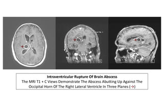
- 14. Intraventricular Rupture Of Brain Abscess The MRI T1 + C Views Demonstrate The Abscess Abutting Up Against The Occipital Horn Of The Right Lateral Ventricle In Three Planes (→) → → →
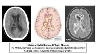
- 15. Intraventricular Rupture Of Brain Abscess The MRI FLAIR Image Demonstrates Confluent Subependymal Hyperintensity And Dependent /Layering Intraventricular Debris. FLAIR
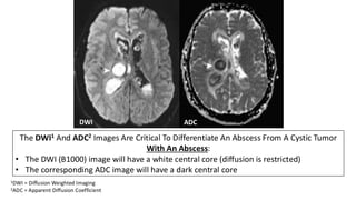
- 16. The DWI1 And ADC2 Images Are Critical To Differentiate An Abscess From A Cystic Tumor With An Abscess: • The DWI (B1000) image will have a white central core (diffusion is restricted) • The corresponding ADC image will have a dark central core DWI ADC 1DWI = Diffusion Weighted Imaging 2ADC = Apparent Diffusion Coefficient ➤ ➤ DWI ADC ➤ ➤
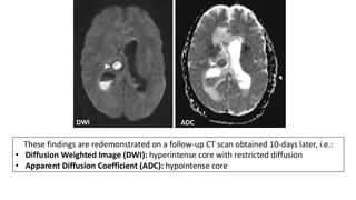
- 17. These findings are redemonstrated on a follow-up CT scan obtained 10-days later, i.e.: • Diffusion Weighted Image (DWI): hyperintense core with restricted diffusion • Apparent Diffusion Coefficient (ADC): hypointense core DWI ADC ➤ ➤
- 18. New England Journal of Medicine 2014;4(371):447-456. From: www.EMGuidewire.com
- 19. New England Journal of Medicine 2014;4(371):447-456. From: www.EMGuidewire.com
- 20. New England Journal of Medicine 2014;4(371):447-456. From: www.EMGuidewire.com
- 21. IVROBA: Pathophysiology • Spontaneous intraventricular rupture of brain abscess (IVROBA) is a potentially fatal complication of pyogenic brain abscess. • IVROBA results in severe ventriculitis, which may progress to widespread pan-meningo-encephalitis if not treated promptly. • In early studies the mortality rate of IVROBA was reported to be over 80%, however, with the advent of better diagnostic imaging and aggressive antibiotic therapy, mortality has come down to below 40%. • Young patients and those presenting in good neurological status are predicted to have better outcomes.
- 22. IVROBA: Imaging Recommendations • A CT without contrast is still first-line for neuroimaging in the ED • If a brain abscess is suspected clinically, ordering a CT with contrast as the first study is reasonable • Avoid ordering a CT “with and without” contrast to minimize the radiation dose • If the patient has a known brain abscess, then ordering an MRI of the brain with and without contrast is appropriate
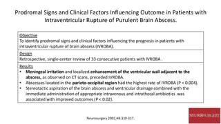
- 23. Prodromal Signs and Clinical Factors Influencing Outcome in Patients with Intraventricular Rupture of Purulent Brain Abscess. Neurosurgery 2001;48:310-317. Objective To identify prodromal signs and clinical factors influencing the prognosis in patients with intraventricular rupture of brain abscess (IVROBA). Design Retrospective, single-center review of 33 consecutive patients with IVROBA . Results • Meningeal irritation and localized enhancement of the ventricular wall adjacent to the abscess, as observed on CT scans, preceded IVROBA. • Abscesses located in the parieto-occipital region had the highest rate of IVROBA (P < 0.004). • Stereotactic aspiration of the brain abscess and ventricular drainage combined with the immediate administration of appropriate intravenous and intrathecal antibiotics was associated with improved outcomes (P < 0.02).
- 24. Clinical features and predictive factors of intraventricular rupture in patients who have bacterial brain abscesses. Journal of Neurology Neurosurgery & Psychiatry 2007;78:303-309. Objective To identify potential risk factors predictive of intraventricular ruptures in hospitalized patients with brain abscess without intraventricular rupture upon presentation. Design 20-year retrospective review of 179 patients brain abscess. 45 had IVRBA on admission (initial IVRBA) and 17 had the episode during hospitalization (subsequent IVRBA). Results • Multiloculated abscess vs. non-loculated abscess had a higher risk of intraventricular rupture [OR 4.2 (95% CI 2.4-14.3); p = 0.020]. • A shorter distance between the abscess and ventricle was associated with a higher risk of rupture each 1 mm decrease of this distance increased the risk of rupture by 10% per mm.
- 25. Journal of Neurology Neurosurgery & Psychiatry 2007;78:303-309. IVROBA1 (n=62) Non-IVROBA2 (n=117) Fever/Chills 69% 55% Altered Mental Status 56% 41% Headache 48% 55% Hemiparesis 43% 46% Nausea/Vomiting 32% 21% Stiff Neck 27% 26% Seizure 10% 17% Facial Palsy 3% 7% Mean Age 47 yrs Male Gender 70% Duration Of Symptoms 14 days Underlying Condition (n=179) Post-Neurosurgical/TBI 26% Diabetes 17% Congenital Heart 13% Chronic Otitis Media 11% Alcoholism/Cirrhosis 11% Neoplasm 8% 1Intraventricular rupture of brain abscess 2These patients had a brain abscess without IVROBA
- 26. Journal of Neurology Neurosurgery & Psychiatry 2007;78:303-309. Neuroimaging (n=62) Ependymal Enhancement 90% Ventricular Debris 71% Hydrocephalus 48% Meningeal Enhancement 45%
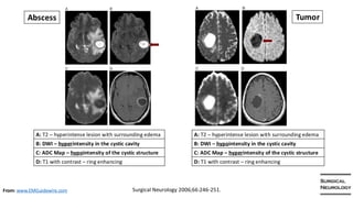
- 27. Surgical Neurology 2006;66:246-251. The Role Of Diffusion-Weighted Imaging In The Differential Diagnosis Of Intracranial Cystic Mass Lesions: A Report Of 147 Lesions. Objective To evaluate the sensitivity and specificity of diffusion-weighted imaging (DWI) in differentiating brain abscesses from other intracranial cystic masses. Methods 115 patients with 147 cystic lesions were prospectively studies with MRI diffusion weighted images. Lesions appearing hyperintense on DWI were considered as brain abscess, whereas hypointense lesions were categorized as non-abscess cystic lesions. The Apparent Density Coefficient (ADC) was also assessed. Results The sensitivity of DWI for the differentiation of brain abscesses from non-abscesses was 96%, specificity 96%, positive predictive value 98%, negative predictive value 92%, and accuracy of the test 96%. Conclusions Diffusion-weighted imaging has high sensitivity and specificity for the differentiation of brain abscesses from other non-abscess cystic brain lesions. From: www.EMGuidewire.com
- 28. Surgical Neurology 2006;66:246-251. A: T2 – hyperintense lesion with surrounding edema B: DWI – hyperintensity in the cystic cavity C: ADC Map – hypointensity of the cystic structure D: T1 with contrast – ring enhancing A: T2 – hyperintense lesion with surrounding edema B: DWI – hypointensity in the cystic cavity C: ADC Map – hyperintensity of the cystic structure D: T1 with contrast – ring enhancing Abscess Tumor From: www.EMGuidewire.com
- 29. The Case Continues The patient was admitted to the ICU and was managed with bilateral external ventricular drains and IV antibiotics. • Final CSF cultures grew Streptococcus intermedius • The antibiotic regimen included ceftriaxone, metronidazole, and intrathecal vancomycin • His hospital course was complicated by saddle pulmonary embolus requiring catheter- directed thrombectomy and therapeutic heparin • Additional Neurosurgical interventions included endoscopic ventricular lavage and ventriculoperitoneal shunt placement • One month after presentation, the patient developed a devastating intracranial hemorrhage and the family elected for comfort measures and a palliative extubation • No definitive source of the original infection was ever identified.
- 30. Ventriculoperitoneal Shunt Placement CT without contrast demonstrating the course of the ventricular catheter entering the occipital horn of right lateral ventricle, traversing the septum, and terminating in the frontal horn of left lateral ventricle.
- 31. Right Parietal Intraparenchymal Hemorrhage CT head without contrast demonstrating a large parenchymal hemorrhage with associated uncal herniation (➤) and obstruction of CSF outflow resulting in transependymal flow (→). ➤ →
- 32. Summary • Spontaneous intraventricular rupture of brain abscess (IVROBA) is a devastating complication of pyogenic brain abscess associated with high rates of morbidity and mortality. • A CT without contrast is first line neuroimaging in the ED. If there is a high clinical suspicion for brain abscess, then performing a CT with contrast is appropriate. • Important CT clues of IVROBA include: • Presence of a ring-enhancing structures adjacent to the ventricles • Ependymal enhancement • Intraventricular debris • Hydrocephalus
- 33. Summary • Early identification and prompt initiation of appropriate treatment is crucial to provide the best opportunity for improved long-term outcomes. • If a diagnosis of IVROBA is suspected based on the clinical history and initial CT finds, antibiotics should be given at once, i.e.: before MRI. • It is important to differentiate a ring-enhancing abscess from a cystic tumor. On MRI abscesses have restricted diffusion and demonstrate a white (hyperintense) central core on DWI and a dark (hypointense) central core on ADC. The reverse is true for cystic tumors. • Definitive treatment involves a combination of surgical EVD drainage and targeted antibiotic therapies.
- 34. References Brain Abscess. New England Journal of Medicine 2014;4(371):447-456. Prodromal Signs and Clinical Factors Influencing Outcome in Patients with Intraventricular Rupture of Purulent Brain Abscess. Neurosurgery 2001; 48:310-317. Clinical features and predictive factors of intraventricular rupture in patients who have bacterial brain abscesses. Journal of Neurology Neurosurgery & Psychiatry 2007;78:303- 309. The Role Of Diffusion-Weighted Imaging In The Differential Diagnosis Of Intracranial Cystic Mass Lesions: A Report Of 147 Lesions. Surgical Neurology 2006;66:246-251.