This document discusses various aspects of visual acuity and accommodation. It defines visual acuity as the ability to clearly see details and outlines of objects. It describes methods of testing visual acuity including Snellen's chart and acuity in infants. Factors that affect visual acuity like contrast sensitivity and color vision are also examined. Accommodation is defined as the ability of the eye to focus on near objects and factors like presbyopia that affect accommodation are discussed.
Physiology of lens and CataractogenesisSristiThakur
油
This document discusses the physiology of the lens and the process of cataractogenesis. It begins by describing the biochemical composition and metabolism of the lens, which is important for maintaining transparency. It then discusses how cataracts form, including congenital and acquired cataracts. The key factors involved in cataractogenesis are any disturbances that disrupt the critical intracellular and extracellular balance in the lens, deranging the colloid system and resulting in opacification.
Subjective refraction techniques rely on the patient's response to determine the refractive correction that provides the best visual acuity. Determining the astigmatic correction is more complex than just the spherical error. Different techniques are used, including the cross-cylinder method and clock dial method. For the cross-cylinder method, the axis is refined first followed by the cylinder power to get the best visual acuity. For children, cycloplegic refraction is important and the full refractive error should typically be corrected, though sometimes undercorrection may be used initially.
There are four axes of the human eye: the optical axis, visual axis, fixation axis, and pupillary axis. There are also three angles of the eye: angle kappa is formed between the pupillary axis and visual axis, angle alpha is formed between the optical axis and visual axis, and angle gamma is formed between the optical axis and fixation axis.
The document discusses Pentacam, a diagnostic tool that uses a rotating Scheimpflug camera to capture 50 images in 2 seconds and create a 3D model of the anterior eye segment. It has applications in assessing corneal ectasia, refractive surgery, corrected intraocular pressure, corneal aberrations, IOL power calculation, and densitometry. The Pentacam provides curvature, pachymetry, and elevation maps. It can detect ectasia by identifying if the highest curvature, thinnest thickness, and steepest elevation points coincide. The Pentacam is also used to measure corneal aberrations via Zernike analysis and calculate accurate IOL power for patients with previous refractive surgery or cataracts.
The document discusses the Jackson Cross Cylinder (JCC) test, which is used during refraction to detect and refine astigmatism. The JCC is a combination of two cylinders of equal strength but opposite signs, placed at right angles to each other. During the test, the JCC is held in different positions before the eye to see if there is a change in visual acuity. If a position is clearer, it indicates the axis of astigmatism. The test is then used to refine the axis and power of any astigmatic correction.
The document summarizes the boxing system used for measuring eyeglass frames. The boxing system imagines drawing a box around the lens shape with the sides tangent to the edges. Key measurements include the A measurement (eye size), B measurement (vertical size), geometric center, distance between lenses, distance between centers, effective diameter, seg height/drop, temple length, length to bend, and front to bend. These standardized measurements are used to specify frame sizes and fit lenses.
The document provides an overview of optical dispensing. It discusses defining optical dispensing and the steps involved, including frame selection based on facial shape, frame measurements, lens measurements, counseling patients on lens materials and coatings, and the process of fitting lenses into frames which involves marking, cutting, and edging lenses.
The document describes the use of various Pentacam maps and indices for screening patients for keratoconus, including:
1) The standard 4-map composite report, keratoconus map, Holladay report, and Belin/Ambrosio Enhanced Ectasia Display.
2) Key features to examine on each map include anterior and posterior elevation maps, pachymetry maps, curvature maps, and indices values.
3) The Belin/Ambrosio Enhanced Ectasia Display aims to improve sensitivity by calculating an "enhanced" best fit sphere reference surface that excludes the thinnest corneal region, highlighting differences between normal and ectatic corneas.
The document discusses various types of optical aberrations that can occur in the eye. It describes monochromatic aberrations, which are caused by the geometry of the lens, and chromatic aberrations, which are caused by dispersion and the variation of the lens refractive index with wavelength. It also discusses how wavefront aberrometry can be used to measure aberrations by analyzing the distortion of reflected light to generate a map of the optical system of the eye. Common higher-order aberrations measured include coma, spherical aberration, and trefoil.
The cornea is the transparent front part of the eye that covers the iris, pupil, and anterior chamber. The cornea, with the anterior chamber and lens, refracts light, with the cornea accounting for approximately two-thirds of the eye's total optical power.
magnification, It's definition, types, clinical uses, Uses in Optical instruments like microscopes, telescopes, Uses in Optical instruments like direct Ophthalmoscopes, indirect ophthalmoscopes and slit lamps, In low vision
The Maddox rod test is used to detect heterophoria or heterotropia. It consists of a series of cylindrical lenses mounted in a trial frame that produces an elongated streak of light. When viewed through the Maddox rod, a spot of light appears as a streak. The orientation of the streak indicates whether the eye is deviated vertically or horizontally. The test is easy for patients to understand and perform, and useful for detecting vertical deviations.
This document provides an overview of corneal topography. It begins by defining corneal topography as the study of the shape of the corneal surface. It then describes several techniques for evaluating corneal topography including keratometry, keratoscopy using Placido discs and photokeratoscopy, rasterstereography, and interferometry. Computerized topography systems that provide detailed maps of the corneal surface are also discussed. The document outlines clinical applications of corneal topography and variations in topographic patterns seen in normal and diseased corneas.
- A schematic eye is a mathematical model that represents the basic optical features of the real eye by using spherical surfaces and constant refractive indices.
- The first schematic eyes were developed in the 17th-19th centuries by scientists like Huygens, Smith, Le Grand, and Listing. Gullstrand further improved the model with four lens surfaces and refractive index gradients.
- Modern schematic eyes like Gullstrand's simplified model with three surfaces are commonly used for calculations involving refraction, image size and location, and the effects of refractive errors. While approximations, schematic eyes provide a framework for understanding ocular optics.
Indirect ophthalmoscopy and fundus drawingSonali Singh
油
This document provides an overview of binocular indirect ophthalmoscopy and fundus drawing techniques. It discusses the history and working principles of the indirect ophthalmoscope. Examination techniques are described, including positioning, use of the condensing lens, and scleral indentation. Fundus drawing methods such as color coding retinal findings are also outlined. Proper cleaning and sterilization of equipment is emphasized.
The document describes the components and uses of a trial box, which is a set of lenses, frames, and accessories used to test vision. It contains trial frames that hold spherical, cylindrical, and prismatic lenses in various diopters for refraction testing. Accessories include occluders, filters, charts, and tools like Maddox rods and cross cylinders. The trial box is used for objective and subjective refraction, diagnosing conditions like squint, and assessing binocular vision.
This document discusses different types of best form lenses, which aim to minimize optical aberrations. It describes the history of efforts to improve lens design, dating back to Huygens' proposal in the 17th century. The ideal best form lens is described as being aberration-free, easy to manufacture, and inexpensive. Four main types are covered: aspheric lenses, deep meniscus lenses, lenticular lenses, and periscopic lenses. Aspheric lenses modify the lens curvature to reduce aberrations. Deep meniscus lenses have a base curve of 6.00 diopters or greater. Lenticular lenses have a smaller powered central area within a larger unpowered peripheral frame. Periscopic lenses have
This document discusses anomalies of accommodation, including diminished or deficient accommodation (presbyopia, cycloplegia, insufficiency), and increased accommodation (excessive accommodation, spasm). Presbyopia is an age-related decrease in accommodation due to hardening of the lens or weakening of the ciliary muscles. Treatment includes optical corrections like glasses or surgery. Insufficiency of accommodation can be due to premature lens sclerosis or ciliary muscle weakness from various systemic or local causes. Treatment addresses the underlying cause and includes near vision corrections. Excessive accommodation can occur in young hyperopes or myopes doing excessive near work, while spasm of accommodation is an abnormal excessive accommodation outside voluntary control.
Astigmatic lens used in ophthalmology and eyeRACHANA KAFLE
油
different types and classifications of astigmatic lens used
availability of astigmatic lens
uses of astigmatic lens
advantages and disadvantages of astigmatic lens
The aqueous humor is a thin, watery fluid located in the anterior chamber of the eye between the cornea and iris. It is produced by the ciliary body and nourishes the cornea and lens while maintaining the shape and intraocular pressure of the eye. The aqueous humor is composed primarily of water along with proteins, glucose, ions, ascorbate and other nutrients. It flows continuously from the ciliary body into the anterior chamber and exits through the trabecular meshwork. Glaucoma is caused by increased pressure from blocked aqueous humor flow. The vitreous humor is a clear gel located between the lens and retina that provides structure and contains few cells.
This lecture includes anatomy, Physiology of Sclera, if u like it kindly share it with colleagues and like it. I will share more lectures related to eye anatomy and optometry.
Thank You.
Why we prescribe glass
Able to detect hypermetropia & myopia
Subjective correction child patient
The sequence of prescription writing
Practical events of glass prescription writing procedure
Anatomy of crystalline lens by Dr. Aayush Tandon Aayush Tandon
油
The document summarizes the anatomy of the crystalline lens. It discusses the lens's structure, composition, dimensions, and surgical anatomy. Key points include:
- The lens is a transparent biconvex structure composed mainly of specialized cells and proteins. It helps focus light onto the retina to allow vision.
- Structurally, it has an outer lens capsule enclosing lens epithelium cells and elongated lens fibers in concentric layers. The fibers are arranged in a nucleus and surrounding cortex.
- Dimensions vary with age but the lens is roughly 10mm in diameter and weighs around 258mg in adults. It provides around 16-17 diopters of refractive power and accommodates vision changes.
- Surgically
Anisometropia is a condition where the two eyes have unequal refractive power. It can be congenital due to unequal eyeball growth, or acquired such as after cataract surgery. There are different types including simple where one eye is normal and the other myopic or hyperopic, compound where both eyes are myopic or hyperopic but to different degrees, and mixed where one eye is myopic and the other hyperopic. Treatments include spectacles up to 4 diopters of difference, contact lenses for higher degrees, and refractive surgery for high unilateral refractive errors.
The document discusses accommodation and its mechanism in the human eye. It defines accommodation as the mechanism that allows the eye to focus on near objects by changing the shape of the lens. The key elements that stimulate accommodation are the apparent size of the object, chromatic aberration, blurred retinal image, oscillation of accommodation, and scanning eye movements. Accommodation is measured by its near point, far point, range, and amplitude. Conditions like presbyopia, paralysis, insufficiency, and spasms of accommodation can cause abnormalities in the eye's accommodative ability.
This document discusses various anomalies of accommodation that can occur, including accommodative fatigue, presbyopia, insufficiency of accommodation, inertia of accommodation, paralysis of accommodation, and spasm of accommodation. It describes the symptoms, causes, assessments, and management approaches for each condition. The key message is that anomalies of accommodation are common and their management is an important part of optometric practice.
This document discusses measuring and classifying accommodative convergence/accommodation (AC/A) ratios. It defines the AC/A ratio as the change in accommodative convergence per diopter of accommodation. Abnormal AC/A ratios can cause strabismus. There are several methods described for measuring the AC/A ratio clinically, including the heterophoria, gradient, and graphical methods. The document outlines treatments for different AC/A ratio abnormalities like convergence excess, convergence insufficiency, divergence excess, and divergence insufficiency.
Accommodation anomalies can occur due to various causes and present with different symptoms. Assessment involves dynamic retinoscopy and measuring accommodation amplitudes. Accommodative fatigue can result from overuse and be treated by correcting refractive errors and discussing visual hygiene. Presbyopia is age-related and treated with near vision correction. Other failures of accommodation include insufficiency, paralysis, spasm, and sustained accommodation, each with different etiologies, signs, and treatments.
It is essential to assess vergence during an eye exam. There are many aspects of vergence response including near point of convergence, jump vergence, and fusional vergence. Near point of convergence is normally less than 12cm and indicates the point at which one eye turns out. Fusional vergence keeps phorias latent using the fusion reflex, and it is important to know a patient's fusional reserves. Clinical values for distance phoria and positive fusional reserves are used to evaluate if a patient is using an appropriate amount of vergence. Accommodation and vergence are related, and relative accommodation and the zone of clear vision should also be considered during assessment.
The document discusses various aspects of the near triad response including accommodation, vergence, and pupil response. It focuses on the AC/A ratio, which represents the ratio of accommodative convergence to accommodation. The AC/A ratio is an important clinical measurement that provides insight into a patient's binocular vision and can help with understanding and managing binocular vision problems. The document discusses several methods for measuring the AC/A ratio in clinical practice.
The AC/A ratio measures the amount of accommodative convergence induced per diopter of accommodation. It can be calculated using phorias at distance and near or measured using the gradient method. A normal AC/A ratio is 4:1 with a range of 2-6:1. An elevated or reduced AC/A ratio can indicate different binocular vision dysfunctions and influence treatment decisions.
The document discusses rigid gas permeable contact lenses, including their benefits, applications, fitting process, and lens design considerations. Some key points covered include:
1. RGP lenses can automatically correct astigmatism, provide good vision and eye health benefits like increased oxygen transmission.
2. The fitting process involves evaluating the lens-cornea relationship using fluorescein dye to identify any bearing, clearance or sealing issues.
3. Important lens design factors are the overall diameter, optical zone size, base curve, thickness, and peripheral curve to achieve a proper alignment fit.
This document discusses various theories and anomalies of accommodation. It begins by defining accommodation and related terms. It then discusses several theories of the accommodation mechanism, including Helmholtz's relaxation theory, Gullstrand's mechanical model, and Schachar's, Tsherning's, and Cotenary's theories. It also covers types of accommodation and anomalies such as presbyopia, insufficiency/ill-sustained accommodation, paralysis, excess accommodation, and spasm. Presbyopia is discussed in detail regarding pathophysiology, causes, symptoms, and treatment options like optical correction and surgery. Other anomalies are summarized briefly regarding their etiology, clinical features, and management.
The document provides information about the Jackson Crossed-Cylinder (JCC) technique for determining astigmatism during eye exams. It discusses the optics and proper use of the JCC. It describes the historical origins of the JCC, how it works, and the step-by-step procedure for using it to refine the axis and power of astigmatic corrections. Common sources of error are also outlined. The JCC is presented as an important tool for optometrists to accurately measure and correct astigmatism in clinical practice.
The document discusses accommodation and its anomalies. It begins by explaining how accommodation allows the eye to focus on near objects by increasing the curvature of the crystalline lens. It then discusses components of accommodation including reflex, vergence, proximal, and tonic accommodation. Various conditions that can affect accommodation are explained such as presbyopia, insufficiency of accommodation, paralysis of accommodation, spasm of accommodation, and inertia of accommodation. Treatment options for these conditions include lenses, vision therapy, exercises, and in some cases surgery.
The document discusses several topics related to vision and the visual system:
1. It describes the primary visual cortex and several other areas of the brain involved in vision like the frontal eye field and superior colliculi.
2. It explains visual pathways and how lesions in different areas can cause quadrantanopia.
3. It discusses factors that affect visual acuity like refractive errors, retinal issues, and stimulus factors as well as how acuity is measured.
PHSIO OF VISION in Ophthalmology in detailManjunathN95
油
1. The document describes the physiology of vision and the rhodopsin cycle. Light is converted to electrical signals in the photoreceptors through phototransduction.
2. Phototransduction involves rhodopsin bleaching where light exposure converts the retinal component of rhodopsin. This triggers biochemical reactions generating a receptor potential.
3. The receptor potential is transmitted through the visual pathway to the visual cortex for perception through serial and parallel processing.
This document summarizes accommodation and its anomalies. Accommodation allows the eye to focus on near objects by changing the lens shape. With age, the lens loses elasticity causing presbyopia. Anomalies include decreased accommodation like insufficiency and inertia, as well as increased accommodation like spasm. Accommodation insufficiency results in blurred near vision. Treatments include lenses, vision therapy, and ciliary muscle relaxation for spasm.
This document discusses accommodation and presbyopia. It defines accommodation as the ability of the eye to change focus from distant to near objects by changing the shape of the lens. Accommodation decreases with age in a process called presbyopia as the lens becomes less flexible. Presbyopia symptoms include difficulty seeing close objects clearly and can be treated with reading glasses, bifocal glasses, or multifocal contact lenses to restore near vision. The document also covers topics like ciliary muscle function, amplitude of accommodation measurement, and factors affecting presbyopia.
This document provides an overview of optics and refraction for 5th year medical students. It defines key terms related to light, refraction, the eye, and refractive errors. It describes how the eye focuses light onto the retina using the cornea and lens. Refractive errors like myopia, hyperopia, and astigmatism occur when light is not correctly focused on the retina. Methods for correcting refractive errors include glasses, contact lenses, and refractive surgery procedures.
The human eye allows us to see by forming an inverted real image on the light-sensitive retina. The main parts of the eye and their functions are: the cornea refracts light, the iris controls pupil size to regulate light, the lens focuses light onto the retina, and the retina contains light-sensitive cells that send signals to the brain for vision. The pupil regulates the amount of light entering by contracting or expanding. Common vision defects include near-sightedness, far-sightedness, and astigmatism, which can be corrected using lenses. The eye's ability to focus on near and far objects is called accommodation.
Visual acuity refers to the sharpness and clarity of vision. It is affected by factors like the size and contrast of the object, as well as optical and retinal factors in the eye. The normal visual acuity allows discrimination of objects spaced 1 minute of arc apart. Visual acuity declines in the peripheral vision due to the dense concentration of photoreceptors in the fovea. Common refractive errors that impair visual acuity include myopia, hyperopia, and astigmatism. These errors occur when the eye is not the proper length or shape to focus light correctly on the retina.
The document discusses the physiology of image formation in the eye and principles of optics. It covers topics like the schematic eye model, visual acuity, optical aberrations and defects in image formation. Specifically, it explains how light is refracted by the cornea and lens to form an image on the retina. It also describes common vision conditions like myopia, hyperopia and astigmatism caused by defects in the eye's optical system and how they can be corrected.
This document discusses various aspects of visual field defects and ocular reflexes. It describes hemianopia, which is the loss of vision in half of the visual field, and its two types: homonymous and heteronymous. It also explains the pupillary light reflex, including the direct and consensual responses when light enters the eye. The document outlines the six extraocular muscles that control eye movement and their functions. It discusses accommodation, convergence, and pupillary constriction as the three components of near response. Various methods for testing visual acuity and mapping the visual field are also summarized.
Pupil size and reactivity are tested clinically to evaluate the eye and brain. The normal pupil constricts to light (direct and consensual response) and accommodation. Pupil size is controlled by the iris sphincter and dilator muscles innervated by the parasympathetic and sympathetic nervous systems. Pupillary reflexes like the light and accommodation reflexes are tested to localize lesions. Abnormal pupil size or reactivity can indicate conditions like Horner's syndrome or third nerve palsy. An afferent pupillary defect detected by the swinging flashlight test indicates optic nerve dysfunction. Pharmacologic testing can further localize lesions in the pupillary pathway.
The pupil is a circular opening in the iris that controls the amount of light entering the eye. It constricts (miosis) and dilates (mydriasis) under autonomic nervous system influence. The iris contains two muscle groups - the sphincter pupillae and dilator pupillae - that regulate pupil size. Abnormal pupils may be unequal in size (anisocoria), irregularly shaped, or have abnormal reactions to light or accommodation. Various diseases and drugs can cause pupil abnormalities.
This document discusses pupil size and reaction as it relates to attraction and interest between individuals. It notes that pupil size increases when looking at someone we find attractive due to an involuntary physiological response. It also states that observing whether a person's pupils expand, contract, or do nothing can provide clues as to whether they are attracted to or actively dislike the observer. The document then provides details on pupil anatomy, size, shape, location, and neural pathways controlling pupil size.
This document discusses pupil size and reaction as it relates to attraction and interest between individuals. It notes that pupil size increases when looking at someone we find attractive due to an involuntary physiological response. It also states that observing whether a person's pupils expand, contract, or do nothing can provide clues as to whether they are attracted to or actively dislike the observer. The document then provides further information on pupil anatomy and control, as well as abnormal pupil responses and conditions that cause them.
Physiolology of Eye: Power of Accommodation and PerimetryShishirBadave
油
Points covered-
Physiology of eye
Power of accommodation
Perimetry/ Visual field tests
Focusing powers of eye lenses
M.Tech Medical Device Presentation
2. It is complex integration of light sense, from sense, sense of
contrast and colour sense
1.The Light sense
Its is awareness of the light. The minimum brightness
required to evoke a sensation of light is called the light
minimum. It should be measured when the eye is dark
adapted for atleast 20-30 minutes.
The Process of visual adaptaion primarly involves:
1. Dark adaption (adjustment in dim illumination) and
2. Light adaption (adjustment to bright illumination)
Visual Perception
3. It is the ability of the eye to adapt itself to decreasing illumination.
The time taken to see in dim illumination is called Dark
adaptation time
The rods are much more sensitive to low illumination than the
cones.
Rods are used more in dim light (scotopic vision) and cones in
bright light (photopic vision)
Dark adaptation curve consists of two parts: the initial small curve
represents the adaptation of cones and the remainder of the
curve represents the adaptation of rods.
Delayed dark adaptation occurs in diseases of rods e.g. retinitis
pigmentosa and vitamin A deficiency
DARK ADAPTATION
5. The Process by mean of which retina adapts itself
to bright light is called light adaptation process is
very quick and occurs over a period of 5 minutes
Result in :
Increased spatial acuity
Increased temporal acuity
Decreased sensitivity
LIGHT ADAPTATION
6. It is ability to discriminate between the shapes of
the objects.
The from sense is most acute at the fovea, where
there are maximum number of cones and decrease
very rapidly towards the periphery.
Visual acuity recorded by snellens test chart is a
measure of Form sense.
The Form sense
7. Minimum visible
Determine presence or absence of target in visual field without
resolving them.
Threshold is 1 second of arc.
Components of Visual Acuity
8. Minimum resolvable ( ordinary visual acuity)
Determines presence of identifying/ distinguishing feature in visible
target
Threshold 60 sec of arc.
Snellens visual acuity measure Minimum resolvable ( ordinary
visual acuity)
The test targets may either consists of letters (Snellens Chart) or
broken circle (Landolts ring)
Minimum discriminable(hyper acuity/vernier acuity)
Determines relative location of 2 or more visible features with
respect to each other
Determines whether or not two parallel and straight lines are aligned
in the frontal plane.
Thresold 3-5 sec of arc.
9. It consists of a series of black capital letters on a
white board, arranged in lines, Diminishing in
size,
The each part of the letters subtend an angle of
1 min at the nodal point
Thus, at the given distance, each letter subtends
as angle of 5 min at the nodal point of the eve.
SNELLEN S TEST TYPES
The Chart should be properly illuminated
The numerator being the distance of the patient
from the letters, and the denominator being the
smallest letters accurately read.
10. Simple picture chart: used for children
Landolts C-chart used for illiterate
patients
E-chart: used for illiterate patients
OTHER TESTS BASED ON
SNELLENS TEST TYPES
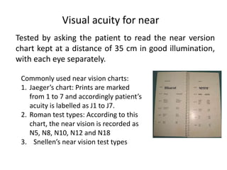
11. Tested by asking the patient to read the near version
chart kept at a distance of 35 cm in good illumination,
with each eye separately.
Visual acuity for near
Commonly used near vision charts:
1. Jaegers chart: Prints are marked
from 1 to 7 and accordingly patients
acuity is labelled as J1 to J7.
2. Roman test types: According to this
chart, the near vision is recorded as
N5, N8, N10, N12 and N18
3. Snellens near vision test types
13. VISUAL MILESTONES :
Very soon after birth - Can fix and follow a light source, face or large, colorful
toy.
1 months - Fixation is central, steady and maintained, can follow a slow target,
and converge, preference of looking at face.
3 months - binocular vision and eye cordination, eyes follow a moving light or
face, responsive smile.
6 months - Reaches out accurately for toys.
9 months look for hidden toys.
2 years - Picture matching
3 years - Letter matching of single letters (e.g., Sheridan Gardiner)
5 years - Snellen chart by matching or naming
14. VISUAL ACUITY OF INFANT EYES
Test 2Months 4Months 6Months 1Year Attainment
(months)
Opticokinetic
nystagmus
test
20/400 20/400 20/200 20/80 2430
Forced
choice
preferential
looking test
20/400 20/200 20/200 20/50 1824
Visual
evoked
response test
20/200 20/80 20/6020/20 20/4020/20 612
15. Tests for indirect assessment of vision :
a) Historical and observational tests,
b) Binocular fixation preference and fixation
targets,
c) CSM method.
24. Ability of the eye to perceive slight changes in the
luminance between regions which are not separated by
definite borders.
Contrast sensitivity is affected by various factors like age,
refractive errors, glaucoma, amblyopia, diabetes, optic
nerve disease and lenticular changes.
Impaired even in the presence of normal visual acuity.
Snellens chart is a measure of visual acuity under 100%
contrast.
Clinical measurement targets at various spatial
frequencies or at various peak contrast
Sense of contrast
26. Ability of the eye to discriminate between
different colour excited by light of different
wavelenghts
Is a function of the cones
In dim light all colours are seen as grey
(purkinje shift).
Colour sense
27. Trichromatic theory (young Helmholtz)
Existence of three kinds of cones, each containing a different
photopigment which is maximallly sensitive to one of the three
primary colours viz. red, green and blue.
Each having different absorption spectrum as below
Red sensitive cone pigment, long wave length sensitive (LWS)
cone pigment, absorbs maximally with a peak at 565 nm.
Green sensitive cone pigment, also known medium wavelength
sensitive (MWS) cone with a peak at 535 nm.
Blue Sensitive cone pigment, short wave length sensitive (SWS)
cone pigment absorbs maximally in the blue-violet with a peak
at440 nm.
The gene for human rhodopsin is located on chromosome 3, and
the gene for the blue-sensitive cone is located on choromosome 7
The Red and green sensitive cones q arm of the X choromosomes
Theories of colour vision
28. Opponent colour theory of Hering some
colours appear to be mutually exclusive
Two main types of colour opponent
ganglion Cells:
Red green opponent colour cells
Blue yellow opponent colour cells
THEORIES OF COLOUR VISION
31. ACCOMMODATION
In an emmetropic eye, parallel rays of light coming
from infinity are brought to focus on the retina, with
accommodation being at rest. The mechanism to
focus diverging rays coming from a near object on the
retina, is called accommodation . In this there occurs
increase in the power of crystalline lens due to
increase in the curvature of its surface
At rest the anterior radius of curvature of lens is 10
mm and in accommodation changes to 6 mm and
posterior radius of curvature of lens is 6 mm and in
accommodation changes to 6 mm
.
33. The nearest point at which small objects can be seen
clearly is near point and the farthest point at which
they are seen clearly is the far point
Far Point and near point of the eye vary with the static
refraction of the eye
1. In an emmetropic eye far point is infinity and near
point varies with age.
2. In hypermetropic eye far point is virtual and lies
behind the eye.
3. In myopic eye, it is real and lies in front of the eye
Far Point and near point
36. Presbyopia condition of falling near vision due to age
related decrease in the amplitude of
accommodation or increase in near point.
Causes
1. Age related change in the lens
i. Decrease in the elasticity of lens capsule,
ii. Progressive, increase in size and hardness of
lens substance.
2. Age related decrease in ciliary muscle power
Symptoms
i. Difficulty in near vision
ii. Asthenopia
37. Treatment
i. Prescription of convex glasses
Basic principle for presbyopic correction
i. Correct distance refractive error
ii. Find near correction for each eye separately and add
to the distance correction
iii. Consider patients profession for fixing the near point
iv. Prescribe the weakest lens
38. Insufficiency of Accoomdation
Accomondative power is significantly less than the normal limits
for the patients age
Causes
1. Premature sclerosis of lens
2. Weakness of ciliary muscle due to systemic causes
3. Weakness of ciliary muscle associated with POAG
Symptoms
1. Asthenopia
2. Blurring of vision
Treatment
1. Treat the systemic cause
2. Near vision spectacles
3. Accomondation exercises
39. PARALYSIS OF ACCOMMODATION
Complete absence of accommodation
Causes
1. Drug induced cycloplegia
2. Internal ophthalmoplegia
3. Third nerve paralysis
Symptoms
1. Blurring of near vision
2. Photophobia
3. Abnormal receding of near point and decreased range of
accommodation
Treatment
1. Self recovery
2. Dark Glasses
3. Convex lenses for near vision
40. SPASM OF ACCOMMODATION
Abnormally excessive accomodation
Causes
1. Drug Induced
2. Spontaneous spasm
Symptoms
1. Defective vision due to induced myopia
2. Asthenopia
Diagnosis
1. Refraction under atropine
Treatment
1. Relaxtation of ciliary muscle by atropine
2. Prohibition of near work
3. Correction of associated causative factors
4. Assurance and psychotherapy
41. CONVERGENCE
Simultaneous adduction (Inward turning)
1. Voluntary
2. Reflex
Reflex Convergence
1. Tonic Inherent innervational tone to the MR
2. Proximial Psychological awareness of near object
3. Fusional Maintains BSV by insuring that similar images
are projected on to corresponding retinal areas of each
eye.
4. Accommodative Induced by act of accommodation as
part of synkinetic near reflex