Leptospirosis
•Download as PPT, PDF•
1 like•1,712 views

Leptospirosis is a zoonotic bacterial disease caused by Leptospira interrogans bacteria transmitted from infected animals to humans through contact with water or soil contaminated by animal urine. It has a wide range of clinical manifestations from a mild flu-like illness to potentially fatal Weil's disease affecting the liver and kidneys. High risk groups include agricultural, sewer and military workers exposed to contaminated environments. Diagnosis involves serological tests or culture of the bacteria from blood or urine, with antibiotics like doxycycline used for treatment. Prevention focuses on minimizing exposure through protective equipment or chemoprophylaxis for high risk groups.


Leptospirosis
- 1. Leptospirosis Mohd Zaim bin Abdullah Zawawi
- 2. Leptospirosis • Leptospirosis is an infectious disease caused by pathogenic spirochete bacteria of the genus leptospira that are transmitted directly or indirectly from animals to human (i.e., a zoonotic disease). • Pathogenic leptospires belong to the species Leptospira interrogans, which is subdivided into more than 200 serovars with 25 serogroups . • The leptospiral serovars are naturally carried in the renal tubules of rodents, wild and domestic animals.
- 3. • Leptospirosis is usually a seasonal disease that starts at the onset of the rainy season and declines as the rainfall recedes. • Sporadic cases may occur throughout the year with outbreaks associated with extreme changing weather events such as heavy rainfall and flooding
- 5. FACTORS RESPONSIBLE FOR THE EMERGENCE OF LEPTOSPIROSIS • a) Reservoir and carrier hosts • b) Flooding, drainage congestion • c) Animal-Human Interface • d) Human host risk factors
- 6. MODES OF TRANSMISSION • Contact through skin, mucosa, conjunctiva • Ingestion of contaminated water
- 7. HIGH RISK GROUPS • Workers in the agricultural sectors • Sewerage workers • Livestock handlers • Pet shops workers • Military personnel • Search and rescue workers in high risk environment • Disaster relief workers (e.g.during floods) • People involved with outdoor/recreational activities such as water recreational activities, jungle trekking, etc. • Travelers who are not previously exposed to the bacteria in their environment especially those travelers and/or participants in jungle adventure trips or outdoor sport activities • People with chronic disease and open skin wounds.
- 8. CLINICAL MANIFESTATIONS • The incubation period is usually 10 days, with a range of 2 to 30 days • The clinical manifestations are highly variable. Typically, the disease presents in four broad clinical categories • (i) a mild, influenza-like illness (ILI); • (ii) Weil's syndrome characterized by jaundice, renal failure, haemorrhage and myocarditis with arrhythmias; • (iii) meningitis / meningoencephalitis; • (iv) pulmonary haemorrhage with respiratory failure. • DDX: dengue, malaria, typhoid, meliodosis, influenza
- 9. 1. Clinical case Acute febrile illness with history of exposure to water and/or environment possibly contaminated with infected animal urine with ANY of the following symptoms: • Headache • Myalgia particularly associated with the calf muscles and lumbar region • Arthralgia • Conjunctival suffusion • Meningeal irritation • Anuria or oliguria and/or proteinuria • Jaundice • Hemorrhages (from the intestines and lungs) • Cardiac arrhythmia or failure • Skin rash • Gastrointestinal symptoms such as nausea, vomiting, abdominal pain, diarrhea
- 10. 2. Probable Case • A clinical case AND positive ELISA/other Rapid tests
- 11. 3. Confirmed case • A confirmed case of leptospirosis is a suspected OR probable case with any one of the following laboratory tests: • Microscopic Agglutination Test (MAT), • For single serum specimen - titre ≥1:400 • For paired sera - four fold or greater rise in titre • Positive PCR (samples should be taken within 10 days of disease onset) • Positive culture for pathogenic leptospires (blood samples should be taken within 7 days of onset and urine sample after the 10th day) • Demonstration of leptospires in tissues using immunohistochemical staining (e.g. in post mortem cases) • In places where the laboratory capacity is not well established, a case can be considered as confirmed if the result is positive by two (2) different rapid diagnostic tests.
- 12. Laboratory Diagnosis • Leptospira MAT • Leptospira serology IgM antibodies • Urine for leptospira • CSF for leptospira • Tissue for leptospira
- 13. Figure 1: Leptospiremic phases in conjunction with the laboratory methods of diagnosis
- 14. Laboratory Investigations • FBC • RP/LFT/CK • Blood Culture X2 • Coagulation Profile • UFEME • Urine C+S • Leptospira serology • Leptospira MAT
- 15. NOTIFICATION • For the purpose of notification, all probable and confirmed cases must be notified to the nearest Health District Office within 1 week of the date of diagnosis.
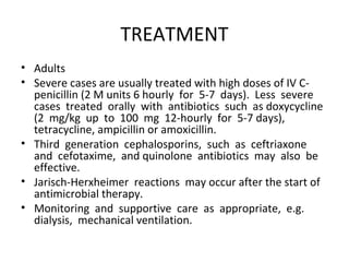
- 16. TREATMENT • Adults • Severe cases are usually treated with high doses of IV C- penicillin (2 M units 6 hourly for 5-7 days). Less severe cases treated orally with antibiotics such as doxycycline (2 mg/kg up to 100 mg 12-hourly for 5-7 days), tetracycline, ampicillin or amoxicillin. • Third generation cephalosporins, such as ceftriaxone and cefotaxime, and quinolone antibiotics may also be effective. • Jarisch-Herxheimer reactions may occur after the start of antimicrobial therapy. • Monitoring and supportive care as appropriate, e.g. dialysis, mechanical ventilation.
- 17. Pediatrics
- 18. PROPHYLAXIS • Preexposure Prophylaxis: • Doxycycline 200mg stat dose then weekly throughout the stay OR • Azithromycin 500mg stat dose then weekly throughout the stay (For pregnant women and those who are allergic to Doxycycline) • Empirical treatment for Post-Exposure: • Doxycycline 200mg stat dose then followed by 100mg BD for 5 – 7 days for those symptomatic with the first onset of fever. OR • Azithromycin 1gm on Day-1, followed by Azithromycin 500mg daily for 2 days (For pregnant women and those who are allergic to Doxycycline)
- 19. Clinical sample Collection and Transportation
- 21. References: • Leptospirosis CPG Malaysia 1st edition 2011 • Sarawak Handbook of Medical Emergencies 3rd edition 2011
Editor's Notes
- #3: Gram negative organism Leptospira interrogans main types affecting humans are L. Interrogans icterohaemorrhagie, Canicola, hardjo and pommona Need High index of clinical suspicion
- #6: The conditions that are favourable for maintenance and transmission of Leptospirosis are: a) Reservoir and carrier hosts Leptospirosis has a very wide range of natural rodent, and non-rodent reservoir hosts especially rats, cattle, dogs, foxes, rabbits, etc. The animals act as carriers of the leptospires and excrete large number of leptospires in their urine, thus responsible for the contamination of large and small water bodies as well as soil. b) Flooding, drainage congestion Flooding and drainage congestion may be risk factors for contamination of water bodies with infected animal urine. Water logged areas may force rodent population to abandon their burrows and contaminatethe stagnant water by their urine. c) Animal-Human Interface The potential for infection increases through exposure from occupational or recreational activities without proper protection. Poor cleanliness/sanitation in recreational areas may attract animal host such as rodent thus increases the risk of contamination. These may be due to poor maintenance of facilities, improper disposal of waste and public attitude/ apathy. d) Human host risk factors Several sections of the population are more susceptible to infection such as those not previously exposed to the bacteria in their environment (naĂŻve immunities), and those with chronic disease and open skin wounds.
- #13: Notes on Laboratory Diagnosis • In cases which needed to be confirmed (hospitalized and suspected death cases), serum samples should be sent for confirmation by MAT. The MAT is considered the "gold standard" or cornerstone of serodiagnosis because of its unsurpassed diagnostic (serovar/serogroup) specificity in comparison with other currently available tests. Second serum samples must be taken to detect fourfold or greater rise in titre. • Simple serological screening method can be done using the rapid test kit for Leptospira. Reminder: any leptospirosis rapid test kit to be used must be validated/approved by IMR. • The ELISA/other rapid tests detect IgM antibodies. The presence of IgM antibodies may indicate current or recent leptospirosis. A patient’s serum may be positive 5 to 10 days after onset of symptoms but not usually before this. Reminder: IgM-class antibodies may remain detectable for several years. • If the initial sample was taken at an early stage in the infection, the ELISA test may be positive but MAT negative. Therefore a follow-up sample is required. Test may be negative if the serogroup of the infecting strain does not react with the Patoc 1 serovar strain used as the antigen. If antibiotics are given from the beginning of the illness, the immune and antibody response may be delayed. • The diagnosis is also confirmed by isolation of Leptospires from blood (first 7 days) or CSF (days 4-10) during the acute illness, from urine (days ≥7) and from tissue samples, by using special media. Inoculation of young guinea pigs, hamsters or gerbils can also be carried out for isolation of leptospires. Leptospires die quickly in urine. Clean urine sample should be inoculated into appropriate culture medium not more than 2 hours after voiding. Survival in acid urine may be increased by making it neutral. For postmortem diagnosis, in addition to serology and culture, leptospires can be demonstrated in tissues using PCR or immunohistochemical staining, notably by direct immunofluorescence.
- #14: Figure 1: Leptospiremic phases in conjunction with the laboratory methods of diagnosis (4). Note: Biphasic nature of leptospirosis and relevantinvestigations at different stages of disease. Specimens 1 and 2 for serology are acute-phase specimens, 3 is a convalescent-phase sample which may facilitate detection of a delayed immune response, and 4 and 5 are follow-up samples which can provide epidemiological information, such as the presumptive infecting serogroup (4).
- #15: Total WBC maybe normal or high as 50 000 per microliter Thrombocytopenia is common Urine may contain bile, protein, casts and red cells Elevated bilirubin and liver enzymes seen in 75 percent cases Elevated creatinine in 50% CSF may show polymorphonuclear of lymphocytic pleocytosis with minimal or moderately elevated protein concentrations and normal glucose CK elevated in 50% of cases
- #17: The Jarisch-Herxheimer reaction is a reaction to endotoxins released by the death of harmful organisms within the body Presentation It resembles bacterial sepsis and can occur after initiation of antibacterials, such as penicillin or tetracycline, for the treatment of louse-borne relapsing fever (80-90% of patients) and in tick-borne relapsing fever (30-40%). An association has been found between the release of heat-stable proteins from spirochetes and the reaction. Typically, the death of these bacteria and the associated release of endotoxins or lipoproteins occurs faster than the body can remove the substances. It usually manifests within a few hours of the first dose of antibiotic as fever, chills, rigor, hypotension, headache, tachycardia, hyperventilation, vasodilation with flushing, myalgia (muscle pain), exacerbation of skin lesions and anxiety. The intensity of the reaction indicates the severity of inflammation. Reaction commonly occurs within two hours of drug administration, but is usually self-limiting. Treatments Prophylaxis and treatment with an anti-inflammatory agent may stop progression of the reaction. Oral aspirin every four hours for 1–2 days, or 60 mg of prednisone orally or intravenously has been used as an adjunctive treatment[citation needed]. However, steroids are generally of no benefit. Patients must be closely monitored for the potential complications (collapse and shock) and may require i.v. fluids to maintain adequate blood pressure. If available, meptazinol, an opioid antagonist, should be administered to reduce the severity of the reaction. Anti TNF-a may also be effective.[citation needed]
- #19: The cost effectiveness and risk versus benefits of antibiotic prophylaxis for leptospirosis remains unclear. If prophylaxis is considered, the possible options include: Pre-exposure Prophylaxis • May be considered for people at high risk of exposure to potentially contaminated sources e.g. soldiers going into jungles, rescue team, persons involved in activities in possible high risk areas e.g. adventurous sports. • Dose: Doxycycline 200mg stat dose then weekly throughout the stay OR Azithromycin 500mg stat dose then weekly throughout the stay (For pregnant women and those who are allergic to Doxycycline) • However the benefit of pre-exposure prophylaxis remains controversial where possible benefits need to be balanced with potential side effects (e.g. doxycycline induced photosensitivity, nausea, etc.) Empirical treatment for Post-Exposure • In an outbreak, there may be a role for post exposure prophylaxis for those exposed to a common source as the index case. Dose: • Doxycycline 200mg stat dose then followed by 100mg BD for 5 – 7 days for those symptomatic with the first onset of fever. OR • Azithromycin 1gm on Day-1, followed by Azithromycin 500mg daily for 2 days (For pregnant women and those who are allergic to Doxycycline) Note: The role of prophylaxis in children has not been adequately studied