Leukaemia i
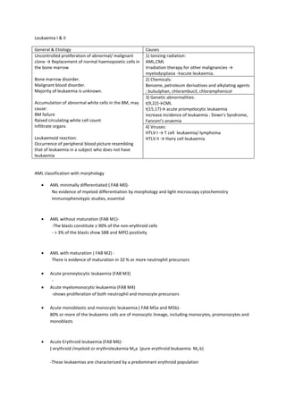
- 1. Leukaemia I & II General & Etiology Causes Uncontrolled proliferation of abnormal/ malignant clone â Replacement of normal haemopoietic cells in the bone marrow Bone marrow disorder. Malignant blood disorder. Majority of leukaemia is unknown. Accumulation of abnormal white cells in the BM, may cause: BM failure Raised circulating white cell count Infiltrate organs Leukaemoid reaction: Occurrence of peripheral blood picture resembling that of leukaemia in a subject who does not have leukaemia 1) Ionizing radiation: AML,CML Irradiation therapy for other malignancies â myelodysplasia âacute leukaemia. 2) Chemicals: Benzene, petroleum derivatives and alkylating agents ; bulsulphan, chlorambucil, chloramphenicol 3) Genetic abnormalities: t(9,22)âCML t(15,17)â acute promyelocytic leukaemia increase incidence of leukaemia : Down's Syndrome, Fanconi's anaemia 4) Viruses: HTLV I â T cell leukaemia/ lymphoma HTLV II â Hairy cell leukaemia AML classification with morphology AML minimally differentiated ( FAB M0)- No evidence of myeloid differentiation by morphology and light microscopy cytochemistry Immunophenotypic studies, essential AML without maturation (FAB M1)- -The blasts constitute âĨ 90% of the non-erythroid cells - > 3% of the blasts show SBB and MPO positivity AML with maturation ( FAB M2) - There is evidence of maturation in 10 % or more neutrophil precursors Acute promeylocytic leukaemia (FAB M3) - Acute myelomonocytic leukaemia (FAB M4) -shows proliferation of both neutrophil and monocyte precursors Acute monoblastic and monocytic leukaemia ( FAB M5a and M5b)- 80% or more of the leukaemic cells are of monocytic lineage, including monocytes, promonocytes and monoblasts Acute Erythroid leukaemia (FAB M6)- (-erythroid /myeloid or erythroleukemia M6a (pure erythroid leukaemia M6 b) -These leukaemias are characterized by a predominant erythroid population
- 2. Acute Leukaemia Clinical features Diagnosis & laboratory investigation Management AML. 1. Pallor, lethargy and dyspnoea due to anaemia 2. Fever, malaise, features of mouth, throat, skin, respiratory, perianal or other infections, including septicemia due to neutropenia 3.Spontaneous bruises, purpura, bleeding gums, menorrhagia and bleeding from venepuncture sites due to thrombocytopenia 4.A bleeding tendency due to thrombocytopenia and disseminated intravascular coagulation (DIC) is characteristic of AML M3 Due to organ infiltration 1.Moderate hepatomegaly, splenomegaly 2.Gum hypertrophy and infiltration (M4 & 5) 3.Skin infiltration, meningeal syndrome (AML M4 & 5) 4.Lysosymes released by the blast cells may cause renal damage in AML M5 .In AML M6 (erythroleukaemia), many erythroblasts may be found in the blood film .In AML M3, tests for DIC are positive .Serum uric acid and LDH may be raised -A normochromic normocytic anaemia -TLC may be decreased, normal or increased -Thrombocytopenia -Blood film shows variable numbers of blast cells, the blasts may contain auer rods Cytochemistry: ï§ Myeloperoxidase ( MPO) activity is specific for myeloid differentiation ï§ Sudan Black B (SBB ) reactivity is similar to MPO in myeloblasts and monoblasts ï§ Non-specific esterase ( NSE) reactivity is diffuse in the cytoplasm of monoblasts Immunophenotype ï§ Immunophenotypic analysis has a central role to differentiate between AML- M0 and ALL ï§ It may be performed ï§ By flow cytometry or immunohistochemistry on the slides -Inform the patient / family -Start treatment ASAP -Supportive care / associated problems -Remission Induction cytotoxic chemotheray -Remission maintenance therapy
- 3. ALL Clinical features Laboratory investigation Management -Common form of leukemia in children -incidence is highest at, 3-4 years -The common (CD10+ ) precursor B type, has an equal sex incidence ALL, L1: blast cells are small, uniform, high N:C ratio, inconspicuous nucleoli ALL, L2: heterogeneous population, some blast cells larger with lower N:C ratio some are like those in L1with high N:C ratio ALL, L3: large cells having vacuolated and basophilic cytoplasm ( usually B-ALL ), nucleoli prominent Symptoms due to bone marrow failure 1.Bone pain and arthralgia 2.Lymphadenopathy, hepatomegaly and splenomegaly are frequent 3.Meningeal syndrome: - headache, nausea, vomiting , blurring of vision and diplopia 4.Testicular swelling ï§ Peripheral blood film and CBC/FBC ï§ Bone marrow examination, which in case of acute leukaemias, is hypercellular with marked proliferation of blasts ï§ Lumbar puncture in patients with meningeal leukaemias X-ray - may reveal lytic bone lesions mediastinal mass due to enlargement of thymus/ and or mediastinal lymph nodes -Immunological markers and chromosome analysis ï§ Supportive care: ï§ (Metabolic complications, hyperleucytosis, infection control, haematologic support) ï§ Risk assessment ï§ Induction chemotherapy, CNS prophylaxis ï§ Consolidation cemotherapy ï§ Maintenance chemotherapy . Chronic lymphocytic leukaemia Proliferation and accumulation of a monoclonal population of abnormal lymphocyte Express CD5 and CD23 Common in west Elderly >50 y/o Male> female Majority: B-cell type (95%) Most patient asymptomatic -Anaemia -Lymphadenopathy (symmetrical and painless) -Immunological failure -Splenomegaly & hepatomegaly - autoimmune hemolytic anaemia (10%) -immune thrombocytopenia (ITP) - 5% - haemorrhagic manisfestation FBC: lymphocytosis Smudge or smear cells Normochromic normocytic anaemia BM biopsy: increase lymphocytes Clonality study: -immunotyping -molecular analysis (IgG/TCR gene rearrangement studies) -chemotherapy - monoclonal antibodies
- 4. Chronic Leukaemia Clinical features Laboratory investigation Management Chronic Myeloid Leukaemia A clonal myeloproliferative disorder ârise from an acquired genetic change in pluripotent stem cell -overproduction of neutrophils and its precursor Philadelphia chromosome -t(9,22)(q34,11) 95% CML; Ph' +ve Fusion BCR-ABL genes Has greater tyrosine kinase 1) Chronic phase - adult (40-60) - anaemia -splenomegaly - hepatomegaly - gout (hyperuricaemia) - hyperviscosity syndrome (due to leucocytosis) - neutropenia, thrombocytopenia (not common) 2) Blast crisis Transform â acute leukaemia, mostly AML Chronic phase FBP: Leucocytosis (increased WBC) Usually >100 Ã 109 /l Morphology: Myeloid cells at all stages of differentiation Bone Marrow: Hypercellular Myelopoiesis is increased (with few blast is < 5%) Neutrophil alkaline phosphatase (NAP) score : reduced Cytogenetic analysis: Ph-chromosome Molecular analysis: BCR-ABL fusion gene. Chronic phase: Hydorxyurea, Glivec/ Imatinib Transplantation - bone marrow / peripheral blood stem cell transplant. Chronic lymphocytic leukaemia Proliferation and accumulation of a monoclonal population of abnormal lymphocyte Express CD5 and CD23 Common in west Elderly >50 y/o Male> female Majority: B-cell type (95%) Most patient asymptomatic -Anaemia -Lymphadenopathy (symmetrical and painless) -Immunological failure -Splenomegaly & hepatomegaly - autoimmune hemolytic anaemia (10%) -immune thrombocytopenia (ITP) -5% - haemorrhagic manisfestation FBC: lymphocytosis Smudge or smear cells Normochromic normocytic anaemia BM biopsy: increase lymphocytes Clonality study: -immunotyping -molecular analysis (IgG/TCR gene rearrangement studies) -chemotherapy - monoclonal antibodies


