LLTech -Imaging C Elegans Worm in 3D with Light-CT
ŌĆóDownload as PPTX, PDFŌĆó
1 likeŌĆó635 views

The document describes a new tomographic imaging technique called Light-CT that allows for fast, non-invasive 3D imaging of the C. Elegans worm. Worms are anesthetized, encapsulated in a gel, and placed between a slide and coverslip before being imaged with the Light-CT scanner at a resolution of 1um. The full 3D imaging of the worm can be acquired in about 2 minutes, allowing identification of internal anatomy like the pharynx, isthmus, and gonad arms. Light-CT provides optical sectioning and volumetric imaging capabilities superior to standard microscopy.


LLTech -Imaging C Elegans Worm in 3D with Light-CT
- 1. Tomographic non-invasive imaging of C. Elegans worm: LLTech Light-CTŌäó scannerSample preparation: worms are anesthetized and encapsulated in a gel
- 2. a gel layer is placed between slide and coverslip
- 3. the sample is imaged using standard Light-CT scanner procedureImaging:3Dtomographic resolution: Ōēł 1┬Ąm, non invasive, non destructive
- 4. 3D acquisition of whole worm: 2minutes (about 60 images, thickness 1┬Ąm), allowing fast 3D optical biopsycoverslipEncapsulated wormsMicroscope slide
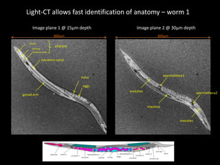
- 5. Light-CT allows fast identification of anatomy ŌĆō worm 1Image plane 1 @ 15┬Ąm depthImage plane 2 @ 30┬Ąm depth800┬Ąm800┬ĄmCorpuspharynxIsthmusPosterior bulbExcretory canalspermatheca1vulvaeggsovocytesspermatheca2gonad armintestineovocytes
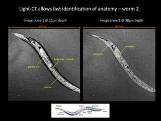
- 6. Light-CT allows fast identification of anatomy ŌĆō worm 2Image plane 1 @ 15┬Ąm depthImage plane 2 @ 30┬Ąm depth800┬Ąm800┬ĄmovocytesintestineBody wall - cuticlegonad armuterus
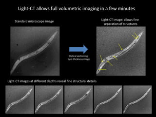
- 7. Light-CT allows full volumetric imaging in a few minutesLight-CT image: allows fine separation of structuresStandard microscope imageOptical sectioning:1┬Ąm thickness imageLight-CT images at different depths reveal fine structural details
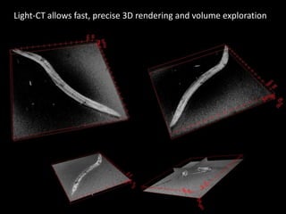
- 8. Light-CT allows fast, precise 3D rendering and volume exploration
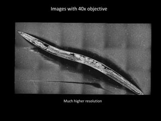
- 9. Images with 40x objectiveMuch higherresolution
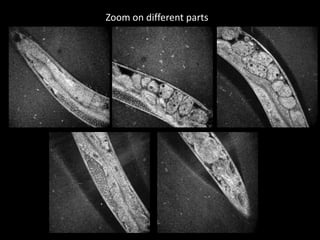
- 10. Zoom on different parts
- 11. Scanning through the sampleMovieobtainedwith 3 microns steps
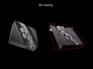
- 12. 3D viewing