Lymphatic - Prac. Histology
Download as PPTX, PDF14 likes4,826 views
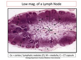
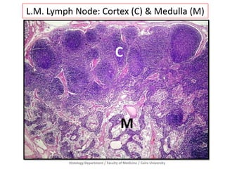
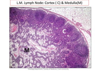
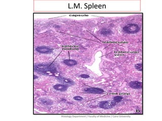
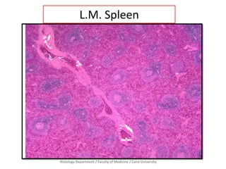
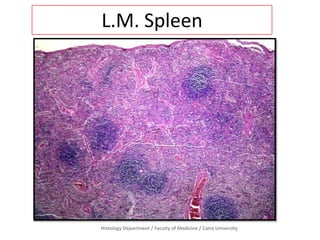
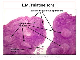
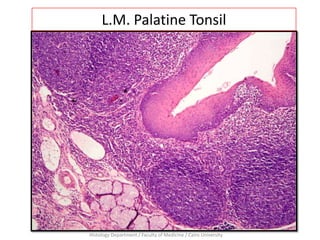
The document describes low magnification microscope images of lymph node tissue showing the cortex and medulla, as well as low magnification images of spleen and palatine tonsil tissue. The images were produced by the Histology Department of the Faculty of Medicine at Cairo University.
1 of 8
Downloaded 87 times
More Related Content
What's hot (20)
PPTX
Lymphatic drainage of head and neck by DR. C.P. ARYA ( B.Sc. ;B.D.S. ;M.D.S. ...DR. C. P. ARYAĚý
More from CU Dentistry 2019 (20)
Ad
Recently uploaded (20)
Ad
Lymphatic - Prac. Histology
- 1. Low mag. of a Lymph Node Cx = cortex / lymphatic nodules (F); M = medulla; C = CT capsule Histology Department / Faculty of Medicine / Cairo University
- 2. Histology Department / Faculty of Medicine / Cairo University L.M. Lymph Node: Cortex (C) & Medulla (M) C M
- 3. Histology Department / Faculty of Medicine / Cairo University L.M. Lymph Node: Cortex ( C) & Medulla(M) C M
- 4. Histology Department / Faculty of Medicine / Cairo University L.M. Spleen
- 5. Histology Department / Faculty of Medicine / Cairo University L.M. Spleen
- 6. Histology Department / Faculty of Medicine / Cairo University L.M. Spleen
- 7. Histology Department / Faculty of Medicine / Cairo University L.M. Palatine Tonsil
- 8. Histology Department / Faculty of Medicine / Cairo University L.M. Palatine Tonsil
