LASIK or Lasik (laser-assisted in situ keratomileusis), commonly referred to as laser eye surgery or laser vision correction, is a type of refractive surgery for the correction of myopia, hyperopia, and an actual cure for astigmatism, since it is in the cornea. LASIK surgery is performed by an ophthalmologist who uses a laser or microkeratome to reshape the eye's cornea in order to improve visual acuity. For most people, LASIK provides a long-lasting alternative to eyeglasses or contact lenses.
The planning and analysis of corneal reshaping techniques such as LASIK have been standardized by the American National Standards Institute, an approach based on the Alpins method of astigmatism analysis. The FDA website on LASIK states,
"Before undergoing a refractive procedure, you should carefully weigh the risks and benefits based on your own personal value system, and try to avoid being influenced by friends that have had the procedure or doctors encouraging you to do so."
The procedure involves creating a thin flap on the eye, folding it to enable remodeling of the tissue beneath with a laser and repositioning the flap.
This document discusses visual acuity testing in children of different age groups. It begins by defining visual acuity and its components. It then outlines the normal milestones of visual development in infants and children from birth through age 5. The document provides details on different vision tests appropriate for various age ranges, including tests of visual reflexes in infants, preferential looking tests for babies 6-12 months, and picture and letter matching tests for older children. It concludes by emphasizing the importance of comprehensive vision assessment and discussing results with parents.
This document provides an overview of indocyanine green angiography (ICGA). It discusses that ICGA uses the near-infrared dye indocyanine green to image the choroidal and retinal vasculature. The dye is injected intravenously and imaged over time using a fundus camera with infrared filters. ICGA allows better visualization of the choroid compared to fluorescein angiography. It has applications in diagnosing conditions like age-related macular degeneration, choroidal neovascularization, and tumors. The document outlines the properties, principles, procedures, indications, limitations and comparisons to fluorescein angiography of ICGA.
Filamentary keratopathy - moc - 2014, pages 63-65Socrates Narvaez
油
Filamentary keratopathy is caused by degenerated epithelial cells and mucus forming filaments on the cornea. It is associated with conditions that cause an irregular ocular surface and altered tear composition. Filaments are diagnosed based on their appearance upon staining and their association with underlying ocular surface diseases. Risk factors include any condition that causes ocular surface irregularity or desiccation. Treatment focuses on managing the underlying cause through measures like artificial tears, punctal occlusion, or lid repair, while filaments may be removed or dissolved. Filaments will resolve once the underlying disease process is effectively treated.
Gonioscopy refers to techniques for examining the anterior chamber angle of the eye to evaluate and classify normal and abnormal angle structures. It involves using specialized lenses and lights to view the iridocorneal angle. There are two main methods: direct gonioscopy uses large contact lenses requiring saline, while indirect uses smaller lenses with mirrors or prisms that utilize tear film. Gonioscopy is used to diagnose angle-closure glaucoma and other conditions by allowing visualization of the trabecular meshwork, scleral spur, and other angle structures. Indentation gonioscopy, where the lens is pressed against the cornea, can differentiate appositional from synechial angle closure. Proper technique and
Intracorneal ring segments, such as INTACS, are thin plastic rings that are implanted into the corneal stroma to flatten the cornea and reduce myopia. They are placed in a lamellar channel using either a mechanical or laser procedure. Thicker rings provide greater flattening and myopia correction. Potential risks include visual disturbances and complications requiring removal. Intracorneal rings have been used off-label to treat conditions like post-LASIK ectasia and keratectasia with some success in improving vision.
This document discusses primary congenital glaucoma, including its history, terminology, classification, management, and associated anomalies. It notes that primary congenital glaucoma is a leading cause of childhood blindness and can be inherited in an autosomal recessive pattern. The classical signs are epiphora, blepharospasm, and photophobia. Examination involves measuring intraocular pressure and corneal diameter. Surgical options for treatment include goniotomy and trabeculotomy to lower IOP and clear the cornea. Developmental glaucomas can be associated with conditions like Axenfeld-Rieger syndrome, aniridia, Peters anomaly, and Sturge Weber syndrome.
Glaucoma drainage devices are implants used to drain aqueous humor from the anterior chamber to control intraocular pressure. They consist of a silicone tube extending from the anterior chamber to a plate beneath the conjunctiva. Open tube designs like Molteno and Baerveldt and flow-restricted designs like Ahmed are commonly used. GDDs are generally used when filtering surgeries have failed or are likely to fail. While they effectively lower IOP, complications can include hypotony, elevated IOP, migration or erosion of the device, and diplopia. Long-term studies show success rates of 65-85% in maintaining IOP control.
Branch retinal vein occlusion case presentation shine.pptxShineNS2
油
A 44-year-old diabetic male presented with decreased vision in his right eye for 2 months. Examination found vision in the right eye was 3/60 and left eye was 6/6. Anterior segment examination was normal in both eyes. Fundus examination of the right eye revealed flame shaped hemorrhages along the superotemporal arcade, cotton wool spots, macular edema, and hard exudates. The left eye fundus appeared normal. The provisional diagnosis was branch retinal vein occlusion with macular edema in the right eye.
1) Toric IOLs are used to correct corneal astigmatism during cataract surgery. They have a cylindrical optic to neutralize corneal astigmatism.
2) The material and design of toric IOLs affect their postoperative rotational stability, with acrylic IOLs showing the highest stability. Larger diameter and loop haptic designs also increase stability.
3) Proper patient selection, preoperative measurements, surgical technique, and IOL alignment are important for achieving optimal visual outcomes with toric IOL implantation. Accurate axis alignment is critical to achieve the intended astigmatic correction.
A cataract is a clouding of the lens in the eye that leads to a decrease in vision. As the lens becomes more opaque, light cannot pass through clearly and vision is impaired. There are different types of cataracts including age-related, congenital, secondary, and traumatic. Cataracts develop as proteins in the lens clump together due to aging, injury, or other health conditions. Symptoms include blurry or foggy vision that worsens over time. Cataracts are typically treated by surgically removing the clouded lens and replacing it with an artificial lens. The surgery is highly effective but does carry risks of complications. Maintaining good eye health through regular exams and lifestyle habits can help reduce
The document discusses visual field testing in glaucoma. It defines the visual field and perimetry, and describes the major types of clinical perimetry tests including full threshold, SITA standard, and SITA fast on Humphrey and normal, dynamic, and TOP strategies on Octopus. It explains parameters such as test patterns, reliability, age-corrected plots, tests like GHT and Bebie curve, and global indices including MD, PSD, SF, and CPSD. The purpose of visual field testing in glaucoma is to detect and monitor disease by measuring light sensitivity across the retinal field.
This document discusses the pupil in health and disease. It begins by describing the normal anatomy and function of the pupil, including its size, location, shape, and role in regulating light entry. It then covers various pupil reflexes and abnormalities such as anisocoria, mydriasis, miosis, light-near dissociation, Argyll Robertson pupils, and disorders of the third cranial nerve and sympathetic pathway. Causes, signs, and diagnostic tests for various pupil abnormalities are provided.
Corneal degeneration refers to conditions where the normal cells of the cornea undergo degenerative changes due to age or pathology. There are several types of corneal degenerations that can be classified based on location (axial or peripheral) and etiology (age-related or pathological). Common age-related peripheral degenerations seen in elderly individuals include arcus senilis, Vogt's white limbal girdle, and Hassall-Henle bodies. Arcus senilis appears as a grey or white ring around the peripheral cornea and is non-pathological. Vogt's white limbal girdle is similar but can occur in younger people and may be associated with hypercholesterolemia. Hassall-
Basic overview of phaco dynamics along with all the Newer phacoemulsification techniques available in current practice - a video-assisted the presentation
The optom faslu muhammed is a haploscopic device used to assess binocular vision. It consists of two tubes mounted on a base with a chin rest and forehead rest. Each tube contains a light source, slide carrier, reflecting mirror, and +6.50D eye piece. It is used to test various grades of binocular vision like simultaneous macular perception, fusion, and stereopsis using different slides. It can also be used to measure the inter-pupillary distance, angle of deviation, and range of fusion.
This document discusses various squint surgery procedures. It begins by introducing the main aims of extraocular muscle surgery, which are to correct misalignment, improve appearance, and restore binocular single vision. It then describes the main types of procedures - muscle weakening, strengthening, and vector adjustment procedures. The document goes into detail about specific muscle recession, resection, and strengthening techniques. It discusses pre-operative assessment and surgical considerations for different muscles. Special procedures like posterior fixation sutures, transpositions, and adjustable sutures are also summarized.
This document summarizes a study on the incidence and types of ocular injuries in patients with head injuries. The study was conducted over 18 months at a hospital in Nanded, India. Of the 200 head injury patients studied, 68% had ocular manifestations. The most common ocular manifestation was lid ecchymosis, seen in 50% of patients. Road traffic accidents accounted for 54% of head injuries. The most commonly affected age group was 21-30 years. Traumatic optic neuropathy was a common cause of visual loss after head injury.
This document discusses the differences between DSAEK and DMEK endothelial keratoplasty procedures. DMEK, where the donor tissue is thinner than DSAEK, provides better visual outcomes but is more technically challenging to perform due to the delicate tissue handling required. DSAEK is easier to perform but results in slightly worse vision initially. Long term graft survival is similar between the two procedures, though DMEK has lower rejection rates while DSAEK has fewer intraoperative complications. The document examines factors related to tissue preparation, surgical technique, patient anatomy, and postoperative outcomes when comparing DSAEK to DMEK.
This document provides information on anterior ischemic optic neuropathy (AION), which is the most common cause of acute optic neuropathy in older age groups. It can be divided into two types: arteritic AION, which is due to giant cell arteritis; and non-arteritic AION, which makes up most cases. Both types present with sudden painless vision loss and optic disc swelling. Arteritic AION carries a worse prognosis and requires high-dose steroid treatment to prevent loss of vision in the fellow eye. Non-arteritic AION has a variable course but generally a poor rate of recovery without any proven effective treatments.
The document discusses non-contact tonometry, which measures intraocular pressure without touching the eye. It describes how non-contact tonometers work by using a puff of air to momentarily flatten the cornea, and an opto-electronic system to detect the reflected light and measure pressure. The document recommends taking multiple readings within a 3 mm Hg range and averaging them to account for natural fluctuations in pressure. Some limitations of non-contact tonometry include corneal opacity, hazy media, high astigmatism, and low visual acuity.
Gonioscopy is an examination of the anterior chamber angle using a gonioscopy lens and slit lamp microscope. It allows visualization of key angle structures including the trabecular meshwork, Schwalbe's line, scleral spur, and ciliary body band. The examination is used to diagnose glaucoma and angle closure risk. There are two main techniques - direct gonioscopy using a contact lens and indirect gonioscopy using a single or multiple mirror lens. Angle structures may be difficult to see, requiring manipulations like indentation and corneal wedge to improve visualization. Gonioscopy is essential for glaucoma evaluation and management.
This document summarizes a symposium on paediatric cataracts presented by Dr. Ganesh Pillay. Some key points:
- Paediatric cataracts are a major cause of preventable childhood blindness worldwide.
- Cataracts in children can be bilateral or unilateral, and have many potential causes including genetic conditions, infections, trauma and metabolic disorders.
- A thorough history and examination is important to determine the type and severity of the cataract. Investigations may be needed depending on clinical findings.
- The timing and type of cataract surgery in children presents various challenges and debates regarding issues like IOL selection and post-operative management.
- Studies like the Inf
This document discusses the evaluation of the nasolacrimal system. It covers the history and anatomy of the system, classification of epiphora, diagnostic tests used to evaluate the system including anatomical tests like syringing and probing, functional tests like dye disappearance tests, and secretory tests like Schirmer's test. The document also discusses differentiating anatomical obstruction from functional/physiological causes of excessive tearing and localization of blockages in the system.
This document discusses various methods for assessing visual acuity in children of different ages. It begins by defining visual acuity and the standard test used in older children and adults, the Snellen chart. Preferential looking tests are commonly used in infants based on their ability to distinguish patterns. Other tests mentioned include Teller cards, visual evoked potentials, optokinetic nystagmus, and Catford drum tests. Approaches such as ball retrieval, toy and coin tests are used in toddlers. Allen's picture cards, Sheridan letters, and HOTV tests target preschoolers, while Snellen, LogMAR, Tumbling E, and Landolt C tests are standard for older children. Challenges in
2. Indication for Surgery
1. Visual need
2. Complication
oPhacomorphic glaucoma
oPhacolytic glaucoma
oDislocation of cataract lens
3. Poor fundus examination or treatment
Reference :
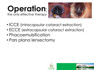
4. Operation:
the only effective therapy
ICCE (intracapsular cataract extraction)
ECCE (extracapsular cataract extraction)
Phacoemulsification
Pars plana lensectomy
6. In extracapsular cataract extraction, an incision is made in the eye
just beneath the iris, or colored part (A). The diseased lens is pulled out (B).
A prosthetic intraocular lens is placed through the incision (D), and is
opened to replace the old lens (E). (Illustration by GGS Inc.)
ECCE:
Extracapsular cataract extraction
Contraindication: Inadequate zonular integrity
14. New Frontiers
Researchers are continuing
to explore new ways to
prevent and treat cataracts,
such as developing
medications that would
reduce or eliminate the need
for surgery
Until then, cataract surgery is
the method to restore vision