MicroT1 myocarditis and pericarditis modified - Copy.pptx
- 1. Module Title Cardiovascular module ’é¦ Course code: IMP-07-203 17 ’é¦ Phase: I ’é¦ Year/ semester: 2nd year / Semester (3) ’é¦ Credit hours: 8 Hr. ’é¦ Course duration: 7 weeks. 2019-2020
- 2. 2019-2020 Micro T1 Prepared by: Dr. Azza Ali Lecturer of microbiology Date of tutorial 30/9/2019 - 1/ 10/2019 Pericarditis and myocarditis
- 3. 2019-2020 Micro T1 Pericarditis and myocarditis Intended Learning Outcomes (ILOs) ’é¦ On completion of this lecture, the student will be able to: 1- Define Pericarditis and myocarditis. 2- Mention causes, viral, bacterial or fungal. 3- Describe pathogenesis, clinical picture and lab. diagnosis. 4- Outline prevention and treatment.
- 4. 2019-2020 Micro T1 Pericarditis and myocarditis ’é¦ Myocarditis ’é¦ Definition: ’é¦ It is inflammation of the heart muscle. ’é¦ The etiology is thought to be caused by a variety of infectious and non-infectious causes.
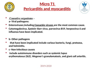
- 6. 2019-2020 Micro T1 Pericarditis and myocarditis ’é¦ Causative organisms: - a- Viral pathogens: ’é¦ Enteroviruses including Coxsackie viruses are the most common cause. ’é¦ Cytomegalovirus, EpsteinŌĆōBarr virus, parvovirus B19, herpesvirus 6 and influenza have been implicated. ’é¦ b- Other pathogens ’é¦ that have been implicated include various bacteria, fungi, protozoa, and helminths. ’é¦ c- Non-infectious causes ’é¦ include autoimmune disorders such as systemic lupus erythematosus (SLE), WegenerŌĆÖs granulomatosis, and giant cell arteritis.
- 7. 2019-2020 Micro T1 Pericarditis and myocarditis ’é¦ Pathogenesis: It occurs most commonly following hematogenous spread of virus or other pathogen to the heart muscle, although direct spread from adjacent structures can occur. ’é¦ It may result in cardiac dysfunction leading to heart failure.
- 8. 2019-2020 Micro T1 Pericarditis and myocarditis ’é¦ Clinical manifestations: Patients with myocarditis present with signs and symptoms of heart failure. ’é¦ Patients may have signs and symptoms of a systemic infection as well (fever, constitutional symptoms ) ’é¦ Those with associated pericarditis often have chest pain.
- 9. 2019-2020 Micro T1 Pericarditis and myocarditis ’é¦ Diagnosis: A definitive diagnosis requires cardiac muscle biopsy revealing myocardial inflammation and necrosis. ’é¦ Most cases are diagnosed in a patient presenting with heart failure, cardiac dysfunction on echocardiogram and elevated cardiac enzymes. ’é¦ Cardiac markers, such as troponin, may be elevated, but during which course of the disease process is mostly unknown. Higher levels of troponin likely correlate with more myocardial damage as it is indicative of myonecrosis, but negative values do not rule out the diagnosis. ’é¦ Other tests that should be ordered include complete blood count (CBC), erythrocyte sedimentation rate (ESR), and c-reactive protein (CRP). The white count, ESR, and CRP may be elevated but are not diagnostic in any way.
- 10. 2019-2020 Micro T1 Pericarditis and myocarditis ’é¦ Viral antibody titers should also be ordered and should include coxsackievirus group B, HIV, CMV, Ebstein-Barr virus, hepatitis and influenza viruses. Titers will typically increase by four-fold during the acute phase with gradual fall with the progression of the disease process. Serial titers may be helpful. ’é¦ Cardiac ECHO should be ordered and may show nonspecific findings such as reduced left ventricular function, global hypokinesis, and even regional wall motion abnormalities. ’é¦ Contrast MRI or nuclear studies can show the extent of inflammation and cellular edema, although this may still be non-specific. Treatment: Treatment of the cause of myocarditis if possible, and supportive care is most often given.
- 11. 2019-2020 Micro T1 Pericarditis and myocarditis 2-Pericarditis ’é¦ Definition: ’é¦ It is inflammation of the pericardium, which can be due to infection, autoimmune diseases, trauma, or malignancy.
- 12. 2019-2020 Micro T1 Pericarditis and myocarditis ’é¦ Causative organisms: ’é¦ Viral infections, Coxsackie virus and echovirus are most common. ’é¦ Non pecific bacteria: Staph. aureus and Strept . pneumoniae are most common. ’é¦ Specific bacterial infection: Mycobacterium tuberculosis is one of the most common infectious causes of pericarditis worldwide. ’é¦ Fungi such as Histoplasma capsulatum and Coccidioides immitis can cause pericarditis, which clinically presents similarly to tuberculous pericarditis
- 13. 2019-2020 Micro T1 Pericarditis and myocarditis ’é¦ Pathogenesis ’é¦ Pathogens reach the pericardium by either : ’é¦ Hematogenous spread through the blood or ’é¦ Direct spread from adjacent intrathoracic structures or, rarely ’é¦ Directly from infected myocardium. ’é¦ Inflammation of the pericardium can result in the formation of pericardial effusion.
- 14. 2019-2020 Micro T1 Pericarditis and myocarditis ’é¦ Clinical Manifestations: ’é¦ Chest pain is the most common manifestation of pericarditis. Pain often worsens with inspiration or coughing Diagnosis: ’é¦ Culture of pericardial fluid or pericardial tissue may reveal causative bacteria. ’é¦ Viruses are rarely isolated.
- 15. 2019-2020 Micro T1 Pericarditis and myocarditis ’é¦ Treatment: ’é¦ It is dependent on the pathogen. ’é¦ Most viral etiologies are treated with symptomatic management and supportive care ’é¦ Bacterial, mycobacterial, and fungal infections will require directed antimicrobial therapy. Prevention: ’é¦ Immunization against Strept . pneumoniae may be effective. ’é¦ Treatment of early or latent stages of infections (e.g., tuberculosis) may prevent development of pericarditis in some cases.
- 16. 2019-2020 Micro T1 Pericarditis and myocarditis Non Specific bacterial infection : Common bacterial infection include: Staph. aureus and Strept. Pnuemonae Laboratory diagnosis: Specimen: sputum, pleural fluid, pericardial effusion aspirate, bloodŌĆ” Microscopic examination: Gram stained film: shows pus cells, Gram positive cocci arranged in clusters (Staph), or Gram positive diplococci surrounded by hallo (Pnuemo). Culture: on blood agar, blood culture, the resulting colonies are identified morphologically and by biochemical reactions. Antibiotic sensitivity testing to choose the proper antibiotic for treatment. Prevention: Vaccination: pnuemococcal conjugate vaccine.
- 17. Catalase test
- 18. Pneumococci in tissue, stained by Gram stain. (Gram-positive capsulated diplococci ) Growth of Strept. viridans or Strept. pneumo on blood agar showing partial or alpha-haemolysis Fig. : Blood culture for diagnosis of : ŌĆó Acute bacterial endocarditis ŌĆó Puerperal sepsis ŌĆó Subacute bacterial endocarditis
- 19. Fig. 2.7:Pneumococci by Quellung reaction (capsule swelling with specific antisera) Fig. 2.8: Optochin sensitivity test Strept. pneumo is sensitive Strept. viridans is resistant Fig. 2.7:, Inulin fermentation test Strept. pneumo ferment inulinŌĆ”..pink Strept. viridans not ferment inulin....colorless Fig. 2.7:, Bile solubility test Strept. pneumo soluble in bile (transparent tube) Strept. viridans not soluble in bile (turbid tube)
- 20. 2019-2020 Micro T1 Pericarditis and myocarditis ’é¦ Some important fungi 1- Histoplasma capsulatum { both cause Systemic mycosis 2-Coccidioides immitis General characters: ’é» Immunity mainly CMI ’é» Restricted in specific area ( specific areas in american desert ) ’é» Infection is by Inhalation of spores ’é» not transmitted among human ’é» Lung ’é» Acute & chronic pulmonary & disseminated infections ’é» Dimorphic form hyphi at 25o C , and yeast at 37o C ’é» cultured on (inhibitory mold agar or Sabouraud’é×s agar ( chloramphenicol & cyclohexamide) (1 ŌĆō 3 days). ’é» TTT: systemic antifungal
- 21. 2019-2020 Micro T1 Pericarditis and myocarditis ’é¦ (1) H. capsulatum (Not capsulated) ’Ć┤ Path.: Inhalation ’āĀ conidia ’āĀ yeast ’āĀ lung MQ ’āĀ RES(reticulo-endothelial system) ’āĀgranuloma In 95% of cases CMI ’āĀ cytokine’āĀ (-) intracellular growth. ’Ć┤Disease: (1) acute pulmonary: flu like (2) chronic pulmonary histoplasmosis (3) Disseminated (fatal form): in immuno suppressed (hepatosplenomegally), enlareged LN & anemia) Diagnosis Direct 1- Specimen: Sputum, BM (bone marrow) 2- Film stained with a)Giemsa (for BM) b)Gomori methenamine silver (GMS) [histological section] Oval cell in side MQ 3-Isolation and Identification on fungal mediaŌĆ”. See above 4- detect Ag 5-DNA probe Indirect 1) Serology: detect IgG by CFT ( titre > 1: 32 = dissemination) 2) ID skin test ( histoplasmin )
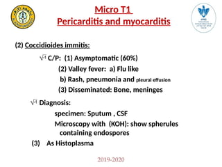
- 22. 2019-2020 Micro T1 Pericarditis and myocarditis (2) Coccidioides immitis: ’āü C/P: (1) Asymptomatic (60%) (2) Valley fever: a) Flu like b) Rash, pneumonia and pleural effusion (3) Disseminated: Bone, meninges ’āü Diagnosis: specimen: Sputum , CSF Microscopy with (KOH): show spherules containing endospores (3) As Histoplasma
- 24. 2019-2020 Micro T1 Pericarditis and myocarditis ’é¦ Some important viruses ’é¦ -EBV ’é¦ It infects B-lymphocytes and reticuloendothelial system (liver, spleen). ’é¦ It causes Latent infection in B-lymphocytes and or pharyngeal epithelial cells. ’é¦ It causes Burkett's lymphoma. ’é¦ -CMV ’é¦ It remains dominant in mononuclear cells to be reactivated in immune compromised patients causing serious disease.
- 25. 2019-2020 Micro T1 Pericarditis and myocarditis Coxsackie viruses It belonges to Picorna viridae family, Enteroviruses genera ’é¦ Capsid’āĀIcosahedral ’é¦ Core’āĀ SS RNA [+ve] sense ’é¦ Envelop’āĀ Naked ’é¦ Coxsackie virus A ’āĀ 23 Serotypes (1 ŌĆō 22 , 24 serotypes) ’é¦ Coxsackie virus B ’āĀ 6 Serotypes
- 26. 2019-2020 Micro T1 Pericarditis and myocarditis Group [A] 23 serotypes Group [B] 6 serotype Fecal -oral ’ā¤ Mode ’āĀ Fecal ŌĆōoral ’āŗ Acute hemorrhagic conjunctivitis ’ā¤ Disease ’āĀ ’āŗ Pleurodynia (Bornholm disease ’āŗ Herpangina Pharynx (the Devil's Grippe): sudden Sharp ’ü╗ Vesicle in Palate pain on one side of chest (self limited) ’ü╗ Fever Tonsil ’āŗ Myocarditis: arrhythmia ’ü╗ Children & self limited ’āŗ generalized disease in infants ’āŗ Hand, foot & mouth disease [Vesicles] ’āŗ Type 1 DM Both causes: ’ü╗ Aseptic meningitis ’ü╗ Common colds
- 27. 2019-2020 Micro T1 Pericarditis and myocarditis ’é¦ Diagnosis ’āŗ Specimen: Throat swab (1st few days), Stool (for weeks), CSF...) 1- Direct demonstration of the virus by E.M. 2-Tissue culture :[C P E] appear in group A within (3 ŌĆō 8 days) while grpup B within (5 ŌĆō 14 days) 3- Serology detection of specific antibody by ELISA ’āŗ PCR is the rapid method for diagnosis (2 ŌĆō 5 hr) Prevention ’āĀ No vaccine
- 28. Fixed cell culture slide showing CPE of enterovirus e.g. coxsackie, and polio (Cell rounding & shrinking, nuclear pyknosis and cell destruction)
- 29. 2019-2020 Micro T1 Pericarditis and myocarditis ECHO viruses [Enteric Cytopathic Human Orphan] ’é¦ Morph. ’āĀ as Coxsackie It belonges to Picorna viridae family, Enteroviruses genera ’é¦ C’āĀ SS RNA [+ve] sense ’é¦ C’āĀ Icosahedral ’é¦ E’āĀ Naked 34 serotypes ’é¦ Mode ’āĀ Feco-oral Disease’āĀ ’é¦ A septic meningitis ’é¦ Febrile illness with or without rash. ’é¦ Diagnosis ’āĀ ................................... ’é¦ Prevention ’āĀ No vaccine
- 30. 2019-2020 Micro T1 Pericarditis and myocarditis Parvovirus B19 ’é¦ Mode of Transmission ’é¦ Transmission is by respiratory secretions, blood, and blood products of infected patients. ’é¦ The virus can be also transmitted vertically from mother to fetus. ’é¦ Pathogenesis ’é¦ It infects primarily two types of cells: ’é¦ -Red blood cell precursors (erythroblasts) in the bone marrow, which accounts for the aplastic anemia, ’é¦ -Endothelial cells in the blood vessels, which accounts, in part, for the ’é¦ rash associated with erythema infectiosum. ’é¦ Clinical manifestation: ’é¦ Transient Aplastic Crisis (TAC): ’é¦ There is temporary arrest of RBCs production. This becomes apparent only in patients with chronic hemolytic anemia.
- 31. 2019-2020 Micro T1 Pericarditis and myocarditis ’é¦ Pure Red Cell Aplasia (PRCA): ’é¦ B19 may establish a persistent infection in immunocompromised patients causing severe chronic aplastic anemia and the patients are dependent on blood transfusions. ’é¦ Laboratory Diagnosis: ’é¦ Specimens: Serum, blood cells. ’é¦ Direct detection: ’é¦ ELISA: for direct detection of viral antigen. ELISA is used to detect B 19 IgM antibodies, which indicates recent infection. ’é¦ PCR: for detection of viral DNA. It is the most sensitive assay. ’é¦ Treatment ’é¦ Symptomatic treatment.
- 32. 2019-2020 Micro T1 Pericarditis and myocarditis ’é¦ Prevention and Control: Screening of blood donors. ’é¦ Standard infection control precautions should be followed to prevent transmission of B19 to healthcare workers from patients with TAC or immunodeficient patients with chronic B 19 infection. ’é¦ There is recombinant vaccine available under evaluation against human parvovirsus for people with chronic anaemia.
- 33. 2019-2020 Micro T1 Pericarditis and myocarditis Intended Learning Outcomes (ILOs) ’é¦ On completion of this lecture, the student abled to: 1- Define Pericarditis and myocarditis. 2- Mention causes, viral, bacterial or fungal. 3- Describe pathogenesis, clinical picture and lab. diagnosis. 4- Outline prevention and treatment.
- 34. 2019-2020 Micro T1 Pericarditis and myocarditis Questions: 1-What is the most common cause of myocarditis in adolescents? a. Drugs b. Coxsackie virus c. Tuberculosis d. Post myocardial infarction 2-Which of the following family of viruses are the most common cause of viral cardiomyopathy? a. Adenoviridae b. Hepadnaviridae c.Picornaviridae d. Reoviridae
- 35. 2019-2020 Micro T1 Pericarditis and myocarditis 3-A 17-year-old male, previously healthy, presents with an acute flu-like illness, malaise, fever and chest pain. During his work up he is found to have sinus tachycardia on ECG and an elevated troponin. He is admitted to the hospital for suspected acute myocarditis. Which clinical finding is most concerning for having a poor prognosis in this patient? ’é¦ Fever ’é¦ Tachypnea ’é¦ Elevated troponin ’é¦ Congestive heart failure 4-Which of the following is most likely to have purulent pericarditis as a possible complication? ’é¦ Coxsackievirus infection ’é¦ Sepsis ’é¦ Myocardial infarction ’é¦ Tuberculosis
- 36. 2019-2020 Micro T1 Pericarditis and myocarditis 5-Select the virus that most often causes acute viral pericarditis. a. Rhinovirus b. Epstein-Barr virus c. Coxsackie B virus d. Adenovirus
- 37. 2019-2020 Micro T1 Pericarditis and myocarditis Learning Resources: References 1- https://www.ncbi.nlm.nih.gov/books/NBK431080/ 2- https://www.ncbi.nlm.nih.gov/books/NBK459259/ 3- Medical microbiology (department book), Ragaa Awad, Laila Saleh, Azza-Elsalakawi. 4- Medical microbiology (Ain Shams department book).


![2019-2020
Micro T1
Pericarditis and myocarditis
’é¦ (1) H. capsulatum (Not capsulated)
’Ć┤ Path.: Inhalation ’āĀ conidia ’āĀ yeast ’āĀ lung MQ ’āĀ RES(reticulo-endothelial system) ’āĀgranuloma
In 95% of cases CMI ’āĀ cytokine’āĀ (-) intracellular growth.
’Ć┤Disease: (1) acute pulmonary: flu like (2) chronic pulmonary histoplasmosis
(3) Disseminated (fatal form): in immuno suppressed (hepatosplenomegally), enlareged LN &
anemia)
Diagnosis
Direct
1- Specimen: Sputum, BM (bone marrow)
2- Film stained with a)Giemsa (for BM)
b)Gomori methenamine silver (GMS) [histological section]
Oval cell in side MQ
3-Isolation and Identification on fungal mediaŌĆ”. See above
4- detect Ag
5-DNA probe
Indirect
1) Serology: detect IgG by CFT ( titre > 1: 32 = dissemination)
2) ID skin test ( histoplasmin )](https://image.slidesharecdn.com/microt1myocarditisandpericarditismodified-copy-241114161935-d84fbfb5/85/MicroT1-myocarditis-and-pericarditis-modified-Copy-pptx-21-320.jpg)


![2019-2020
Micro T1
Pericarditis and myocarditis
Coxsackie viruses
It belonges to Picorna viridae family, Enteroviruses genera
’é¦ Capsid’āĀIcosahedral
’é¦ Core’āĀ SS RNA [+ve] sense
’é¦ Envelop’āĀ Naked
’é¦ Coxsackie virus A ’āĀ 23 Serotypes (1 ŌĆō 22 , 24 serotypes)
’é¦ Coxsackie virus B ’āĀ 6 Serotypes](https://image.slidesharecdn.com/microt1myocarditisandpericarditismodified-copy-241114161935-d84fbfb5/85/MicroT1-myocarditis-and-pericarditis-modified-Copy-pptx-25-320.jpg)
![2019-2020
Micro T1
Pericarditis and myocarditis
Group [A] 23 serotypes Group [B] 6 serotype
Fecal -oral ’ā¤ Mode ’āĀ Fecal ŌĆōoral
’āŗ Acute hemorrhagic conjunctivitis ’ā¤ Disease ’āĀ ’āŗ Pleurodynia (Bornholm disease
’āŗ Herpangina Pharynx (the Devil's Grippe): sudden Sharp
’ü╗ Vesicle in Palate pain on one side of chest (self
limited)
’ü╗ Fever Tonsil ’āŗ Myocarditis: arrhythmia
’ü╗ Children & self limited ’āŗ generalized disease in infants
’āŗ Hand, foot & mouth disease [Vesicles] ’āŗ Type 1 DM
Both causes: ’ü╗ Aseptic meningitis
’ü╗ Common colds](https://image.slidesharecdn.com/microt1myocarditisandpericarditismodified-copy-241114161935-d84fbfb5/85/MicroT1-myocarditis-and-pericarditis-modified-Copy-pptx-26-320.jpg)
![2019-2020
Micro T1
Pericarditis and myocarditis
’é¦ Diagnosis
’āŗ Specimen: Throat swab (1st
few days), Stool (for weeks), CSF...)
1- Direct demonstration of the virus by E.M.
2-Tissue culture :[C P E] appear in group A within (3 ŌĆō 8 days) while grpup B within
(5 ŌĆō 14 days)
3- Serology detection of specific antibody by ELISA
’āŗ PCR is the rapid method for diagnosis (2 ŌĆō 5 hr)
Prevention ’āĀ No vaccine](https://image.slidesharecdn.com/microt1myocarditisandpericarditismodified-copy-241114161935-d84fbfb5/85/MicroT1-myocarditis-and-pericarditis-modified-Copy-pptx-27-320.jpg)

![2019-2020
Micro T1
Pericarditis and myocarditis
ECHO viruses
[Enteric Cytopathic Human Orphan]
’é¦ Morph. ’āĀ as Coxsackie
It belonges to Picorna viridae family, Enteroviruses genera
’é¦ C’āĀ SS RNA [+ve] sense
’é¦ C’āĀ Icosahedral
’é¦ E’āĀ Naked
34 serotypes
’é¦ Mode ’āĀ Feco-oral
Disease’āĀ
’é¦ A septic meningitis
’é¦ Febrile illness with or without rash.
’é¦ Diagnosis ’āĀ ...................................
’é¦ Prevention ’āĀ No vaccine](https://image.slidesharecdn.com/microt1myocarditisandpericarditismodified-copy-241114161935-d84fbfb5/85/MicroT1-myocarditis-and-pericarditis-modified-Copy-pptx-29-320.jpg)


