
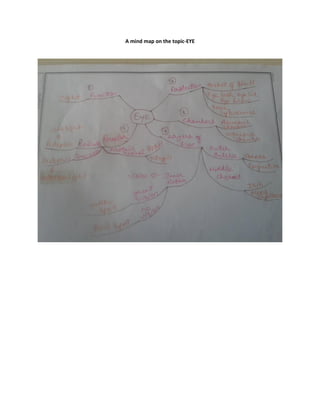
Mind mapping on the topic - EYE
- 1. Name of the teacher: Benson Abraham Standard: X Name of the School: St. Thomas Strength: Subject: Biology Date Unit: Beyond the Senses Period Topic: Eye Time: 40 mins Curricular Statement 1. To obtain Knowledge about eye. 2. To attain Conceptual idea regarding the overall structure and function of eye. 3. To develop creativity skills in developing mind maps. Learning Outcome Factual Knowledge The learners recalls the layers of eye and receptors of eye. Conceptual Knowledge The learner understands the structure, function etc of the eye. Procedural Knowledge The learner compares rod and cone cells Meta cognition The learner evaluates the fucnction of rod cells and cone cells Process Skills The learner develops skill in observing and interpreting mind maps. Content Analysis Terms Socket of Skull, eyelash, eyelid eyebrow, tear, lysosomes, aqueous and vitreous chamber, Sclera, cornea, conjuctiva, choroid, Iris, Yellow spot, blind spot, receptors, rod and cone cells Facts 1. The function of eye is sight 2. The eye is protected by the socket of skull, eyelash, eye lid, eye brow, tear, and lysosome. 3. The eye has two chambers, aqueous chamber and vitreous chamber. 4. The eye contains three layers sclera, choroid and retina. 5. The image is formed in retina 6. There are two types of receptors- Rod cells (dim light), cone cells( intense light).
- 2. Teaching Learning Interaction Pupil Response As part of awakening process the teacher conducts a discussion with the students. How do we see things? Pupil says through eyes So today we are going to study about Eye (BB to the center) To the upper left side of the black board the teacher write Function. Can anyone say the fuction of Eye? Sight (BB Under Function ) Pupil Says Sight To the upper right of the BB teacher writes Protection and asks students to say the parts that help in protecting eye Pupil says Skull (BB), Eyelash, Eyelid, eyebrow(BB), Tears(BB) The Teacher write the pupils response under protection. The eye has two Chambers (BB) (under Eye) ie. Aqueous Chamber and Vitreous Chamber (BB under chamber). Aqueous Chamber is filled with a watery fluid called Aqueous humor that supplies nutrients and oxygen to the cells of lens and cornea. Pupils listens carefully Concept Major Concept Eye Minor Concept Protection, Chambers, layers and receptors of eye. Pre-requistites The learner already knows the function of eye and how the eye is protected by the eye. Teaching- Learning Resources The diagram of the structure of eye. Eye-Mind Map. Reference Textbook of Biology X, Concise Biology, Teacher Plus Magazine February 2011.
- 3. Vitreous Chamber is filled with a jelly like substance which helps to maintain the shape of eyeball. The teacher then show a picture of the structure of the eye and points aqueous chamber and vitreous chamber. Now let us see the different layers of eye. Can you name the Layers of eye(BB) that you know? Pupil says retina ThatŌĆÖs right but there are 3 layers in the Eye, The outer Sclera, middle choroid, and Inner Retine(BB under the layers of eye). The transparent front portion of sclera is called cornea(BB under sclera) and the anterior pair of cornea is protected by a membrane called Conjuctiva (BB). The Teacher with the help of chart points of cornea and conjunctiva The middle layer is called Choroid. It consists of Iris and blood capillaries (BB) Have you seen Iris? Pupil listens and Observes carefully Pupils says Yes, It is the dark color on Eye. The inner most layer is the retina . It helps in vision. The image is formed in Retina, that is sent to brain as the signal. The place in Retina where there is great vision is called Yellow Spot (BB under retina), and a place where there is no vision is called Blind spot (BB under retina). The teacher shows Yellow Spot and Blind Spot on chart. Pupil Listens Carefully Receptor of Eye (BB under Eye). The receptors consists of a protein Retinal(BB) which consists of Opsin and Vitamin A (BB). The receptors are present in 2 type of cells Rod cells and Cone cells (BB under receptors). Rod cells contains pigment called Rodopsin (BB) that helps vision in dim light and cone cells contains pigment called Photopsin (BB)- intense light. Pupils listens carefully The teacher then makes the students to read the mind map created on the board. Pupils learns actively
- 4. A mind map on the topic-EYE