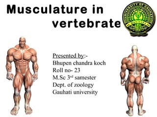
Musculature in vertebrates
- 1. Musculature in vertebrates Presented by:- Bhupen chandra koch Roll no- 23 M.Sc 3rd samester Dept. of zoology Gauhati university
- 2. The Muscular System ŌĆó Muscles are responsible for all movement of the body ŌĆó There are three basic types of muscle ŌĆō Skeletal ŌĆō Cardiac ŌĆō Smooth
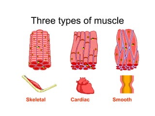
- 3. 3 Types of Muscles
- 4. Three types of muscle Skeletal Cardiac Smooth
- 5. Classification of Muscle Skeletal- found in limbs Cardiac- found in heart Smooth- Found in viscera Striated, multi- nucleated Striated, 1 nucleus Not striated, 1 nucleus voluntary involuntary involuntary
- 6. Characteristics of Muscle ŌĆó Skeletal and smooth muscle are elongated ŌĆó Muscle cell = muscle fiber ŌĆó Contraction of a muscle is due to movement of microfilaments (protein fibers) ŌĆó All muscles share some terminology ŌĆō Prefixes myo and mys refer to muscle ŌĆō Prefix sarco refers to flesh
- 7. Shapes of Muscles ŌĆó Triangular- shoulder, neck ŌĆó Spindle- arms, legs ŌĆó Flat- diaphragm, forehead ŌĆó Circular- mouth, anus
- 8. Skeletal Muscle ŌĆó Most are attached by tendons to bones ŌĆó Cells have more than one nucleus (multinucleated) ŌĆó Striated- have stripes, banding ŌĆó Voluntary- subject to conscious control ŌĆó Tendons are mostly made of collagen fibers ŌĆó Found in the limbs ŌĆó Produce movement, maintain posture, generate heat, stabilize joints
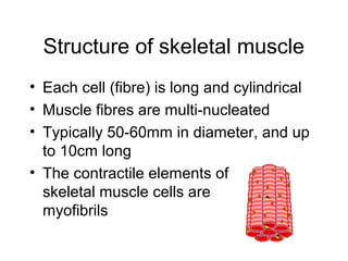
- 9. Structure of skeletal muscle ŌĆó Each cell (fibre) is long and cylindrical ŌĆó Muscle fibres are multi-nucleated ŌĆó Typically 50-60mm in diameter, and up to 10cm long ŌĆó The contractile elements of skeletal muscle cells are myofibrils
- 10. Skeletal muscle - Summary ŌĆó Voluntary movement of skeletal parts ŌĆó Spans joints and attached to skeleton ŌĆó Multi-nucleated, striated, cylindrical fibres
- 11. Smooth Muscle ŌĆó No striations ŌĆó Spindle shaped ŌĆó Single nucleus ŌĆó Involuntary- no conscious control ŌĆó Found mainly in the walls of hollow organs
- 12. Smooth muscle ŌĆó Lines walls of viscera ŌĆó Found in longitudinal or circular arrangement ŌĆó Alternate contraction of circular & longitudinal muscle in the intestine leads to peristalsis
- 13. Structure of smooth muscle ŌĆó Spindle shaped uni-nucleated cells ŌĆó Striations not observed ŌĆó Actin and myosin filaments are present( protein fibers)
- 14. Smooth muscle - Summary ŌĆó Found in walls of hollow internal organs ŌĆó Involuntary movement of internal organs ŌĆó Elongated, spindle shaped fibre with single nucleus
- 15. Cardiac Muscle ŌĆó Striations ŌĆó Branching cells ŌĆó Involuntary ŌĆó Found only in the heart ŌĆó Usually has a single nucleus, but can have more than one
- 16. Cardiac muscle ŌĆó Main muscle of heart ŌĆó Pumping mass of heart ŌĆó Critical in humans ŌĆó Heart muscle cells behave as one unit ŌĆó Heart always contracts to itŌĆÖs full extent
- 17. Structure of cardiac muscle ŌĆó Cardiac muscle cells (fibres) are short, branched and interconnected ŌĆó Cells are striated & usually have 1 nucleus ŌĆó Adjacent cardiac cells are joined via electrical synapses (gap junctions) ŌĆó These gap junctions appear as dark lines and are called
- 18. Cardiac muscle - Summary ŌĆó Found in the heart ŌĆó Involuntary rhythmic contraction ŌĆó Branched, striated fibre with single nucleus and intercalated discs
- 19. Muscle Control Type of muscle Nervous control Type of control Example SkeletalSkeletal Controlled by CNS Voluntary Lifting a glass Cardiac Regulated by ANS Involuntary Heart beating Smooth Controlled by ANS Involuntary Peristalsis
- 20. Types of Responses ŌĆó Twitch- ŌĆō A single brief contraction ŌĆō Not a normal muscle function ŌĆó Tetanus ŌĆō One contraction immediately followed by another ŌĆō Muscle never completely returns to a relaxed state ŌĆō Effects are compounded
- 21. Where Does the Energy Come From? ŌĆó Energy is stored in the muscles in the form of ATP ŌĆó ATP comes from the breakdown of glucose during Cellular Respiration ŌĆó This all happens in the Mitochondria of the cell ŌĆó When a muscle is fatigued (tired) it is unable to contract because of lack of Oxygen
- 22. How are Muscles Attached to Bone? ŌĆó Origin-attachment to a movable bone ŌĆó Insertion- attachment to an immovable bone ŌĆó Muscles are always attached to at least 2 points ŌĆó Movement is attained due to a muscle moving an attached bone
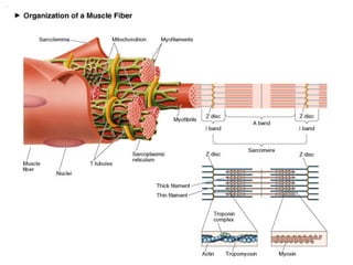
- 23. Structure of Skeletal Muscle: Microstructure ŌĆó Sarcolemma ŌĆō Muscle cell membrane ŌĆó Myofibrils ŌĆō Threadlike strands within muscle fibers ŌĆō Actin (thin filament) ŌĆó Troponin ŌĆó Tropomyosin ŌĆō Myosin (thick filament)
- 25. Structure of Skeletal Muscle: The Sarcomere ŌĆó Further divisions of myofibrils ŌĆō Z-line ŌĆō A-band ŌĆō I-band ŌĆó Within the sarcoplasm ŌĆō Sarcoplasmic reticulum ŌĆó Storage sites for calcium ŌĆō Transverse tubules ŌĆō Terminal cisternae
- 26. Sliding Filament Theory ŌĆó Rest ŌĆō uncharged ATP cross-bridge complex ŌĆó Excitation-coupling ŌĆō charged ATP cross- bridge complex, ŌĆ£turned onŌĆØ ŌĆó Contraction ŌĆō actomyosin ŌĆō ATP > ADP & Pi + energy ŌĆó Recharging ŌĆō reload cross-bridge with ATP ŌĆó Relaxation ŌĆō cross-bridges ŌĆ£turned offŌĆØ
- 27. The Sliding Filament Model of Muscle Contraction
- 29. THANK YOU
