Muscle Physiology A level Biology Powerpoint Edexcel A
- 1. Muscle Physiology Dynamics of Muscle Contraction MMHS Anatomy
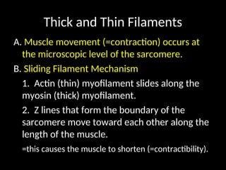
- 2. Thick and Thin Filaments A. Muscle movement (=contraction) occurs at the microscopic level of the sarcomere. B. Sliding Filament Mechanism 1. Actin (thin) myofilament slides along the myosin (thick) myofilament. 2. Z lines that form the boundary of the sarcomere move toward each other along the length of the muscle. =this causes the muscle to shorten (=contractibility).
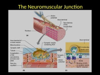
- 3. The Neuromuscular Junction The Neuromuscular Junction
- 4. Muscle Cell Parts 1. Sarcolemma = the muscle membrane 2. Sarcoplasm = the muscle cytoplasm 3. 3. Sarcoplasmic reticulum Sarcoplasmic reticulum = organelle responsible for protein production. ’āĀ This contains high amounts of Ca+2 ions.
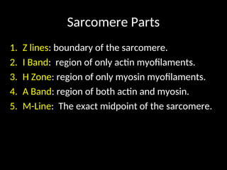
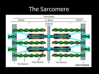
- 5. Sarcomere Parts 1. Z lines: boundary of the sarcomere. 2. I Band: region of only actin myofilaments. 3. H Zone: region of only myosin myofilaments. 4. A Band: region of both actin and myosin. 5. M-Line: The exact midpoint of the sarcomere.
- 7. Muscle ContractionŌĆö10 Steps 1. A nerve impulse enters the presynaptic terminal (nerve) of the neuromuscular junction. 2. The impulse causes Ach to be released from the synaptic vessicles in the axon terminal. 3. Ach diffuses across the synaptic cleft and opens Na+ channels in muscle membranes. 4. Na+ enters the muscle cell and depolarizes it. 5. ŌĆ£TŌĆØ tubules carry impulses into the sarcoplasmic reticulum and releases Ca2+ ions.
- 8. 10 steps of Muscle Contraction 6. Ca +2 enters the individual muscle fibrils and binds to troponin molecules on tropomyosin strands moving the strand and exposing the binding sites. 7. Myosin binds to actin forming crossbridges that ATP can bind to. 8. ATP breaks down, releasing energy, causing cross bridges to pull actin strand.
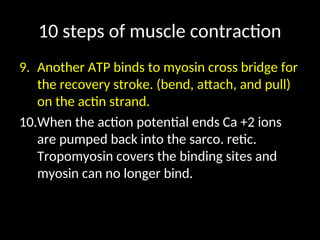
- 9. 10 steps of muscle contraction 9. Another ATP binds to myosin cross bridge for the recovery stroke. (bend, attach, and pull) on the actin strand. 10.When the action potential ends Ca +2 ions are pumped back into the sarco. retic. Tropomyosin covers the binding sites and myosin can no longer bind.
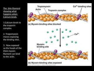
- 10. The thin filament showing what happens when Calcium binds. 1.Calcium binds to the troponin complex. 2. Tropomyosin moves exposing the binding sites. 3. Now exposed so the heads of the thick myosin filament can bind to the actin.
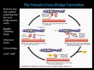
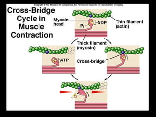
- 11. The Myosin Cross-Bridge Formation The Myosin Cross-Bridge Formation Read the step- wise captions explaining how the cross- bridge process works. Identify: 1.Working stroke 2.Recovery stroke 3.Cross Bridge. 4.ATP + ADP