NECROSIS FOR MBBS FIRST YEAR STUDENTS MADE EASY.pptx
- 2. OVERVIEW • Review of cell injury • Definition of Necrosis • Pathophysiology • Morphology • Patterns/Types of Tissue necrosis – Coagulative Necrosis – Liquefactive Necrosis – Gangrenous Necrosis – Caseous Necrosis – Fat Necrosis – Fibrinoid Necrosis
- 3. CELL INJURY : REVIEW NORMAL CELL CELLULAR ADAPTATION REVERSIBLE CELL INJURY IRREVERSIBLE CELL INJURY NECROSIS APOPTOSIS altered physiological stimuli, non injurious stimuli injurious stimuli (acute/transient) progressive cell injury
- 4. NECROSIS DEFINITION • Necrosis refers to a series of changes that accompany cell death, resulting from degradative action of enzymes on lethally injured cells. • Irreversible cell injury • Causes of Necrosis – Ischemia – Physical agents – Chemical agents – Immunological injury – Microbial infection
- 5. NECROSIS • Characteristics of Necrosis – Denaturation of cellular proteins – Leakage of cellular contents through damaged membranes, – Local inflammation, and – Enzymatic digestion of lethally injured cells
- 6. PATHOPHYSIOLOGY Normal cells Severe damage to cell membrane Entry of lysosomes to cytoplasm Digestion of the cell Leakage of cellular contents to extracellular space Host inflammatory response Severe and progressive injury
- 7. PATHOPHYSIOLOGY Specific substances released from injured cells:k/a Damage associated molecular patterns (DAMP) - ATP (from damaged mitochondria) - Uric acid ( DNA breakdown product) - Other molecules confined to healthy cells *Indicators of severe cell injury Leaked DAMP recognized by receptors in macrophages Trigger phagocytosis of debris and production of cytokines -->induces inflammation Accumulation of inflammatory cells Production of Proteolytic enzymes Leads to clearance of necrotic cells (due to combination of phagocytosis and enzymatic digestion
- 9. CLINICAL APPLICATION Clinical Application of Leakage of intracellular proteins • Basis of blood tests that detect tissue specific cellular injury • Examples – Injury of cardiac muscles cells : Increased cardiac specific protein (troponin) – Bile duct epithelium injury - Specific isoform of alkaline phosphatase – Hepatocyte injury - Transaminases
- 10. MORPHOLOGY • Cytoplasmic Changes – Increased eosinophilia in H&E stain --> Due to loss of cytoplasmic RNA and accumulation of denatured cytoplasmic proteins --> binds to eosin – Glassy homogenous appearance (more compared to normal cells) --> due to loss of glycogen – Vacuolated, moth eaten appearing cytoplasm --> due to enzymatic digestion of the organelles
- 11. MORPHOLOGY
- 12. MORPHOLOGY – Precipitation of Myelin Figures --> Whorled phospholipid precipitates which replaces the dead cells – Myelin figures are phagocytosed by macrophages or further degraded to fatty acids – Calcification of these fatty acids residues --> Deposition of calcium rich precipitates
- 13. MORPHOLOGY Electron microscopic finding – Discontinuities in the plasma membrane and organelle membranes – Marked dilation of mitochondria – Deposition of large amorphous densities in the mitochondria – Intracytoplasmic myelin figures – Amorphous debris in the cytoplasm – Fluffy material representing denatured protein
- 14. MORPHOLOGY • Nuclear Changes – Appears in one of three patterns (Due to break down of DNA) – Karyolysis : Fading of the basophilic chromatin due to loss of DNA as a result of enzymatic degradation by endonucleases – Pyknosis : Characterized by nuclear shrinkage and increased basophilia. Also seen in apoptosis. Chromatin is condensed to a dense shrunken basophilic mass – Karyorrhexis : Pyknotic nucleas undergoes fragmentation – Necrotic cells disappear within one to two days
- 15. • Characteristics of Irreversible injury – Inability to reverse mitochondrial dysfunction (lack of oxidative phosphorylation and ATP generation) – Profound disturbances in in membrane function
- 16. TYPES OF NECROSIS • Necrosis of tissues have some distinct pattern which gives the clue to the underlying cause • TYPES – Coagulative necrosis – Liquifactive necrosis – Gangrenous necrosis – Caseous necrosis – Fat necrosis – Fibrinoid necrosis
- 17. COAGULATIVE NECROSIS • Definition :Form of necrosis in which the architecture of the dead tissue is preserved for the span of at least few days • Morphology – Tissue affected has firm texture --> Injury denatures both structural proteins and enzymes --> Blocks proteolysis of dead cells --> Results in intensely eosinophilic cells with indistinct/reddish nuclei which persists for days to week -->Finally, necrotic cells are broken down by lysosomal enzymes derived from infiltrating leucocytes
- 18. COAGULATIVE NECROSIS -->Infiltrating leucocytes remove necrotic debris by process of phagocytosis – Cause: Ischemia caused by obstruction of blood vessel leads to necrosis of tissue supplied by the vessel – Seen in all the solid organs except Brain – Localized area of coagulative necrosis is called an infarct • Eg. Necrosis of heart, kidney spleen
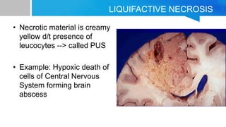
- 20. LIQUIFACTIVE NECROSIS • Definition : Necrosis characterized by digestion of the dead cells resulting in the transformation of tissue into viscous fluid • Seen in focal bacterial and occasionally fungal infection • Bacterial/ fungal infection --> stimulates accumulation of leucocytes--> release of enzymes --> Liquifactive necrosis
- 21. LIQUIFACTIVE NECROSIS • Necrotic material is creamy yellow d/t presence of leucocytes --> called PUS • Example: Hypoxic death of cells of Central Nervous System forming brain abscess
- 22. GANGRENOUS NECROSIS • Not a specific pattern of cell death but term is used in clinical practice • Usually applied to limb, esp. lower limb that has lost its blood supply and has gone necrosis (coagulative necrosis) involving multiple tissue planes. • Bacterial infection is superimposed leading to liquifactive necrosis because of degradative action of bacterial and leucocytic enzymes • Also known as WET GANGRENE
- 24. CASEOUS NECROSIS • Type of coagulative necrosis • Caseous : Cheese-like; derived from friable white appearance of necrosis; Tissue architecture is preserved • Seen in foci of Tuberculosis • Microscopic Features: Appears as structureless collection of fragmented or lysed cells and amorphous granular debris enclosed in a cuff of inflammatory cells --> Granuloma
- 25. CASEOUS NECROSIS
- 26. FAT NECROSIS • Definition : Refers to focal areas of fat destruction, typically resulting from release of activated pancreatic lipases into the substance of pancreas and peritoneal cavity • Occurs in pancreatitis--> pancreatic enzymes leak out of damaged acinar cells --> liquifies the membranes of fat cells in the peritoneum --> releases triglyceride esters split by pancreatic lipases • Generates fatty acids that combines with calcium to produce chalky white areas
- 27. FAT NECROSIS • Histology – Necrotic areas show shadowy outlines of necrotic fat cells – Basophliic calcium deposits – Inflammatory reaction
- 28. FAT NECROSIS
- 29. FIBRINOID NECROSIS • Definition: Special form of vascular damage usually seen in immune reactions involving blood vessels. • Antigen antibody complexes are deposited in the arterial wall --> leaking of plasma proteins out of the vessels • Results in pink amorphous appearance in H&E stain • Example: immunologically mediated vasculitis syndromes


